Research Article
The Effect of Vascular Endothelial Growth Factor in the Improvement of Microcirculation Disturbance in Rats with Severe Acute Pancreatitis
Palikhe M1, Zhan J1*, Jha RK2 and Zhang X2
1Department of Gastroenterology, The Second Affiliated Hospital of Xi’an Jiaotong University, China
2Department of Gastroenterology, Xi’an Medical University, China
*Corresponding author: Zhang Jun, Department of Gastroenterology, The Second Affiliated Hospital of Xi’an Jiaotong University, No.157, Xi Wu Road, Xi’an, China
Published: 23 Oct, 2018
Cite this article as: Palikhe M, Zhan J, Jha RK, Zhang
X. The Effect of Vascular Endothelial
Growth Factor in the Improvement of
Microcirculation Disturbance in Rats
with Severe Acute Pancreatitis. Clin
Surg. 2018; 3: 2175.
Abstract
Objective: To observe the function of Vascular Endothelial Growth Factor (VEGF) in the
improvement of microcirculation disturbance in rats with Severe Acute Pancreatitis (SAP).
Methods: 120 adult male Sprague-Dawley (SD) rats were randomly divided into four groups, the
Sham Operation (SO) SAP VEGF treatment or VEGF antibody (anti-VEGF) treatment group.
The mortality rates were counted at 24 hr; the dry/wet ratios of tissues were measured in the lung,
small intestine, and liver; changes in the ultrastructure of the microcirculation in pancreatic and
lung tissue were observed using a Transmission Electron Microscope (TEM), and the levels of
VEGF, Nitric Oxide (NO), Endothelin (ET), and trypsin were measured using an Enzyme-Linked
Immunosorbent Assay (ELISA).
Results: The mortality in SO group at 24 hr was 0%, SAP group was 90% (9/10), anti-VEGF group
was 80% (8/10), and VEGF group was only 10% (1/10). The dry/wet ratios of all tissues in the VEGF
group were significantly lower than those in the SAP and anti-VEGF groups (P<0.05). Results from
the TEM showed that the SAP and anti-VEGF groups exhibited obvious pathological changes. The
serum ET, NO, and trypsin levels in the SAP and anti-VEGF groups were significantly higher than
those in the SO group (P<0.05).
Conclusion: VEGF could improve microcirculation disturbance in SAP rats through the repair of
the vascular endothelial barrier and maintenance of vascular endothelial function to further reduce
the mortality of rats, which might become a new breakthrough in SAP treatment in the future.
Keywords: Severe acute pancreatitis; Vascular endothelial growth factor; Microcirculation
disturbance; Multiple organ damage
Background
Acute Pancreatitis (AP) is an acute inflammatory process of the pancreas [1]. It is a potentially
life-threatening disease characterized by tissue edema, necrosis, and hemorrhage in the pancreas
[2]. Severe Acute Pancreatitis (SAP) has a poor prognosis and may lead to degradation of the
capillary endothelial basement membrane and alter capillary endothelial cell connections [3,4]. The
presence of microcirculation disturbance is an early event in SAP and penetrates through the whole
process of SAP development [5], finally causes Multiple Organ Dysfunction Syndrome (MODS)
[6]. A large number of clinical observations and animal experiments have confirmed that the major
presentations of microcirculation disturbance in SAP are microvascular permeability changes and
hemorheological changes [7,8]. Therefore, the early pathophysiological change in pancreatitis is the
systemic inflammatory response centered on blood vessels [9]. Currently, based on the above theory,
how to improve microcirculation disturbance to further increase the cure rate of SAP patients has
become a hot research issue in SAP treatment. At present, therapies for SAP, such as corticosteroids
or non steroid anti-inflammatory agents, attempt to reduce the microcirculatory disturbance [10].
Vascular Endothelial Growth Factor VEGF (VEGF) families of proteins are key regulators of
physiological systems [11]. VEGF has the functions of protection of vascular endothelial cells and
maintenance of vascular endothelial functions as well as the promotion of angiogenesis and antiinflammation
and anti-thrombosis activities [12]. It is currently the substance with the strongest
known function in the promotion of angiogenesis and protection of the vascular endothelium
[13,14]. Because of the physiological functions of VEGF, we
considered that using VEGF as a therapeutic drug for SAP could
improve microcirculation disturbance and block the cascade reaction
to further improve the prognosis of SAP patients [15]. Thus, this study
used VEGF as a therapeutic drug and observed its treatment effects
on microcirculation disturbance in SAP rats, in order to provide an
animal experiment basis for seeking novel clinical treatments.
Figure 1
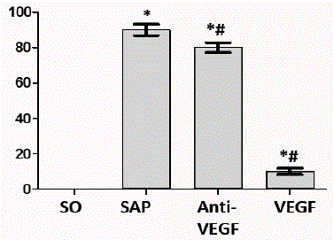
Figure 1
The mortality of rats in the SAP group is very high, by contrast,
there was a significant decrease in the VEGF group (*P<0.05 vs. SO group,
#P<0.05 vs. SAP group).
Figure 2
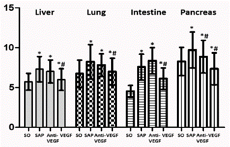
Figure 2
The dry/wet ratios of all tissues in the VEGF group were significantly
lower than those in the SAP and anti-VEGF groups (*P<0.05 vs. SO group,
#P<0.05 vs. SAP group).
Materials and Methods
Materials
A total of 120 healthy adult male Sprague-Dawley (SD) rats with
a body weight of 250 g to 300 g were provided by the Experimental
Animal Center of the College of Medicine of Xi’an Jiaotong
University. Sodium taurocholate was provided by Sigma; VEGF and
anti-VEGF antibodies were provided by Abcam. Enzyme-Linked
Immunosorbent Assay (ELISA) reagent kits were provided by Beijing
Jingmei Biotechnology. This study was approved by the Experimental
Animal Ethics Committee of Xi’an Jiaotong University.
Experimental animals and model establishment
A total of 120 healthy, adult male SD rats were randomly divided
into either the Sham Operation (SO) group, SAP group, VEGF
treatment group, or VEGF antibody (anti-VEGF) treatment group.
Each group had 10 rats. Rats were fasted for 12 hr for food and for
4 hr for water and then anesthetized by intraperitoneal injection of
25 g/L pentobarbital sodium (1.2 mg/kg). A median incision in the
upper abdomen was made at a length of approximately 2 cm. The
pancreaticobiliary duct was located, and its proximal and distal
ends were occluded using an arterial clamp. The SAP model was
established by a retrograde injection of 40 g/L sodium taurocholate
into the pancreaticobiliary duct, and the arterial clamp was then
removed. For the rats in the SO group, the abdomen was opened and
the duodenum was turned, and the abdominal wall was then sutured.
After establishment of the SAP model, VEGF (10 μg/kg) and VEGF
antibodies (10 μg/kg) were immediately injected through the dorsal
vein of the penis in the VEGF and anti-VEGF groups, respectively.
Rat specimens were collected at 6 hr, 12 hr, and 24 hr after model
establishment.
Comparison of mortality of rats at 24 hr
The mortality of rats was calculated using the ratio between the
number of deaths of rats within 24 hr and the total number of rats in
each group. This study only compared mortality at 24 hr after model
establishment.
Measurement of the water content (dry/wet ratio) in
tissues of the liver, lung, and small intestine of rats
Rats were sacrificed and dissected, and 1 cm3 of tissues in the
same locations of the liver, lung, pancreas, and small intestine were
collected. The wet weight was measured. Tissues were baked at 70° for
72 hr, and the dry weight was measured. The water content (%) = (wet
weight-dry weight)/wet weight.
Detection of serum VEGF, nitric oxide (NO), endothelin
(ET), and trypsin using ELISA
Before the rats were sacrificed and dissected, 4 mL venous
blood was collected from the portal vein and centrifuged for 2 min
to collect serum samples. Some specimens were stored at -80°C for
ELISA detection. The ELISA reagent kits provided by Beijing Jingmei
Biotechnology were used to detect the levels of VEGF, NO, ET, and
trypsin in plasma. The detection was performed strictly according to
the procedures in the reagent kit instructions.
Observation of ultra structure of microcirculation in lung
and pancreatic tissues
A volume of 1 mm3 of each aforementioned tissue was fixed in
25 mL/L glutaraldehyde at 4°C for 8 hr. Tissues were dehydrated in
gradient alcohol, immersed in epoxy resin Epon 812, and used for
ultra-thin sections in an LKB-V ultra-thin microtome (50 nm to 70
nm). Changes in the ultra structure of the microcirculation in tissues
were observed under an electron microscope.
Statistical analyses
Data analyses were performed using SPSS 19.0. The comparison
within the same group was performed using a one-way analysis of
variance. The mortality of rats was examined using the χ2 test. The
results are expressed as the mean ± standard deviation (x̅ ± s). P<0.05
indicates the difference had statistical significance.
Figure 3
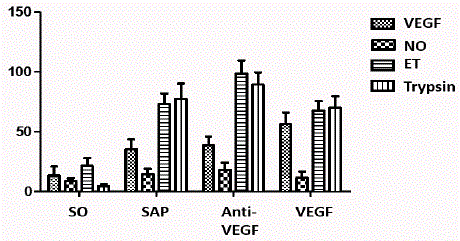
Figure 3
The levels of VEGF, NO, ET, and Trypsin at all time points in the
SAP and anti-VEGF groups were significantly higher than those in the SO
groups (*P<0.05 vs. SO group, #P<0.05 vs. SAP group).
Table 1

Table 1
Results of mortality after 24 h in each group.
Figure 4
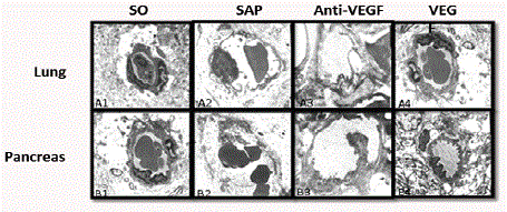
Figure 4
Ultra structural alterations in the lung and pancreas at 24 h. (TEM
× 10,000).
Results
The mortality of rats at 24 hr
The mortality of rats at 24 hr was 0.0% (0/8) in the SO group, 90%
(9/10) in the SAP group, 10.0% (1/10) in the VEGF group, and 80%
(8/10) in the anti-VEGF group (Figure 1). The mortality of rats in
the VEGF group was significantly lower than that in the SAP group
(P<0.05) (Table 1).
The dry/wet ratios of tissues in liver, lung, small intestine,
and pancreas
The dry/wet ratios of all tissues in the anti-VEGF group were the
highest among all groups. The dry/wet ratios of all tissues in the VEGF
group was significantly lower than those in the SAP and anti-VEGF
groups (P<0.05) (Figure 2). These results suggest that VEGF plays an
obvious role in the reduction of tissue edema in SAP rats (Table 2).
ELISA detection results of serum VEGF, NO, ET, and
trypsin
The levels of VEGF, NO, ET, and trypsin at all time points in the
SAP and anti-VEGF groups were significantly higher than those in
the SO group (Figure 3). In the VEGF group, the VEGF level was the
highest among all groups and the NO and ET levels were significantly
lower than those in the SAP and anti-VEGF groups (P<0.05). The
trypsin levels in the three groups other than the SO group did not
have significant differences (Table 3).
Changes in the ultra structure of the microcirculation in
lung and pancreatic tissues
No tissues in the SO group had obvious pathological changes
under a Transmission Electron Microscope (TEM). The typical
pathological changes, such as vascular endothelial cell apoptosis,
mitochondrial swelling, cell edema, capillary congestion, thrombosis,
destruction or disappearance of vascular endothelial integrity in the
microcirculation, extra vascular bleeding, and edema, were observed
in the SAP and anti-VEGF groups under a TEM and became gradually
aggravated over time. However, those pathological changes in the
VEGF group were significantly attenuated (Figure 4).
Table 2
Table 2
Results of mortality after 24 hr in each group and the dry/wet ratio of tissues in the liver, lung, small intestine, and pancreas.
Table 3
Table 3
Results of VEGF, NO, ET and trypsin in serum at 24 hr.
Discussion
We designed this study according to the hypothesis that protease
is the culprit in SAP [16,17]. The results indicated that the serum
trypsin levels in pancreatitis in all groups significantly increased after
the establishment of the SAP model, suggesting that trypsin indeed
participated in the SAP pathological damage process. To further
validate that the destructive function of trypsin on the vascular wall
was the core link in the SAP pathophysiological process, VEGF was
applied, which has obvious vascular repair functions [18]. The results
showed that over time, the mortality of rats in the VEGF group was
significant lower than that in the SAP group. The mortality of rats at
24 hr, the gold standard for treatment effects, was significantly lower
in the VEGF group, indicating that the treatment effect of VEGF
was ideal. To further investigate its mechanisms of action, the water
content of rat organs was measured using the dry/wet ratio of tissues in
these organs. The results indicated that the water content of all tissues
was significantly higher in SAP, suggesting that obvious vascular
leakage was indeed present in SAP. To further confirm the reason
for vascular leakage, a detailed observation of the ultra structure of
tissues in the pancreas and lung was performed. The results showed
destruction of the vascular wall integrity in the microcirculation
in SAP. The major presentations were endothelial lysis, defect of
the vascular wall, endothelial cell apoptosis, and even necrosis. In
addition, the phenomenon of leakage of the visible components of
blood, such as erythrocytes, to extra vascular tissue spaces was also
observed. This was a very special pathological change. Any type of
inflammation is centered on the vascular response, and the general
presentation is the increase in vascular wall permeability to cause
the extravasation of plasma components into the interstitial spaces.
However, the presentations of SAP are vascular wall destruction and
tissue bleeding, which strongly confirms our previous hypothesis
that activated trypsin affects vascular wall substrates to destroy
the vascular wall integrity, thus inducing this special pathological
phenomenon. Previous reports suggested that early stages of SAP are
accompanied by the increase in plasma levels of ET and NO levels,
and altered systolic and diastolic function of local pancreatic blood
vessels. Plasma levels of NO are likely to be increased due to increased
activity of NO synthase [19]. Increased levels of ET upregulate
intracellular calcium levels and promote activation of inflammatory
cells [20]. To confirm the role of inflammatory factors in vascular
leakage in SAP, the levels of serum NO and ET was detected. The
levels of serum NO and ET significantly increased in SAP; however,
their levels did not particularly change in the VEGF group when
the VEGF treatment effect was very obvious. These results suggest
that inflammatory factors do not pay a very critical role in the SAP
pathophysiological process.
Furthermore, we also showed the phenomenon of the increase
in serum VEGF in SAP. It has been reported that VEGF is involved
in the process of organ dysfunction in SAP [21,22]. However, the
conclusion was only based on the phenomenon of the increase
in VEGF in SAP. Our study showed that, with the increase in
the VEGF level, pathological damage of organs in all groups was
significantly attenuated; in particular, the vascular wall integrity in
microcirculation was significantly improved. These results suggested
that VEGF indeed had the effect of a significant improvement of
microcirculation injury in SAP. The increase in VEGF in SAP might
be completely due to the increase in the self-protective responses in
the body.
Overall, our application of VEGF as a therapeutic drug had
unexpected excellent results. Its specific mechanism might be the
rapid repair of the damaged vascular intima and maintenance of
the vascular endothelial integrity to prevent systemic multiple organ
dysfunction induced by obvious vascular leakage in SAP. Whether
there are other mechanisms still remains to be further studied.
Acknowledgment
The authors express their gratitude to the Nature Natural Science
Foundation of China for financial support (Grant NO 81550110256).
The author will like to thank Dr. Sha Huanchen for providing
new ideas while conducting this Research.
References
- Sand J, Nordback I. Acute pancreatitis: Risk of recurrence and late consequences of the disease. Nat Rev Gastroenterol Hepatol. 2009;6(8):470-7.
- Whitcomb DC. Clinical practice. Acute pancreatitis. N Engl J Med. 2006;354(20):2142-50.
- Lu F, Huang H, Wang F, Chen Y. Intestinal capillary endothelial barrier changes in severe acute pancreatitis. Hepatogastroenterology. 2011;58(107-108):1009-17.
- Nathens AB, Curtis JR, Beale RJ, Cook DJ, Moreno RP, Romand JA, et al. Management of the critically ill patient with severe acute pancreatitis. Crit Care Med. 2004;32(12):2524-36.
- Landahl P, Ansari D, Andersson R. Severe Acute Pancreatitis: Gut Barrier Failure, Systemic Inflammatory Response, Acute Lung Injury, and the Role of the Mesenteric Lymph. Surg Infect. 2015;16(6):651-6.
- Sha H, Ma Q, Jha RK. Trypsin is the culprit of multiple organ injury with severe acute pancreatitis. Med Hypotheses. 2009;72(2):180-2.
- Zhou Y, Wang W X. Wang. Advances of microcirculatory disturbance in acute pancreatitis. Chinese J General Practice. 2017;15(9):1559-62.
- Bonnardel-Phu E, Wautier JL, Vicaut E. Advanced glycation end products are involved in microvascular permeability changes observed in microcirculation of diabetic rats in vivo. J Mal Vasc. 2000;25(2):122-7.
- De-madaria E. Latest advances in acute pancreatitis. Gastroenterol Hepatol. 2015;38(1):100-5.
- Leach SD, Gorelick FS, Modlin IM. New Perspectives on Acute Pancreatitis. Scand J Gastroenterol. 1992;192(sup192):29.
- Bates DO, Beazley-Long N, Benest AV, Ye X, Ved N, Hulse RP, et al. Physiological Role of Vascular Endothelial Growth Factors as Homeostatic Regulators. Compr Physiol. 2018;8(3):955-79.
- Hang TC, Tedford NC, Reddy RJ, Rimchala T, Wells A, White FM, et al. Vascular Endothelial Growth Factor (VEGF) and Platelet (PF-4) Factor 4 Inputs Modulate Human Microvascular Endothelial Signaling in a Three-Dimensional Matrix Migration Context. Mol Cell Proteomics. 2013;12(12):3704-18.
- Pandey AK, Singhi EK, Arroyo JP, Ikizler TA, Gould ER, Brown J, et al. Mechanisms of VEGF (Vascular Endothelial Growth Factor) Inhibitor-Associated Hypertension and Vascular Disease. Hypertension. 2017;117:10271.
- Li S, Chen X, Wu T, Zhang M, Zhang X, Ji Z. Role of heparin on serum VEGF levels and local VEGF contents in reducing the severity of experimental severe acute pancreatitis in rats. Scand J Gastroenterol. 2012;47(2):237-44.
- Zhao Y, Adjei AA. Targeting angiogenesis in cancer therapy: Moving beyond vascular endothelial growth factor. Oncologist. 2015;20(6):660-73.
- Chvanov M, De Faveri F, Moore D, Sherwood MW, Awais M, Voronina S, et al. Intracellular rupture, exocytosis and actin interaction of endocytic vacuoles in pancreatic acinar cells: Initiating events in acute pancreatitis. J Physiol. 2018;596(13):2547-64.
- Sha H, Ma Q, Jha RK. Trypsin is the culprit of multiple organ injury with severe acute pancreatitis. Med Hypotheses. 2009;72(2):180-2.
- Watanabe K, Karimpour-Fard A, Michael A, Miyamoto SD, Nakano SJ. Elevated Serum Vascular Endothelial Growth Factor and Development of Cardiac Allograft Vasculopathy in Children. Send to J Heart Lung Transplant. 2018;37(9):1075-82.
- Rahman SH, Ammori BJ, Larvin M, McMahon MJ. Increased nitric oxide excretion in patients with severe acute pancreatitis: evidence of an endotoxin mediated inflammatory response? Gut. 2003;52(2):270-4.
- Jaworek J, Jachimczak B, Tomaszewska R, Konturek PC, Pawlik WW, Sendur R, et al. Protective action of lipopolysaccharides in rat caerulein-induced pancreatitis: role of nitric oxide. Digestion. 2000;62(1):1-13.
- Ye J, Dai JY. Expression and significance of vascular endothelial growth factor in the course of increasing microcirculation disturbance of severe acute pancreatities. Chinese J General Practice. 2012;15(9):679-82.
- Greco M, Palumbo C, Sicuro F, Lobreglio G. Soluble Fms-Like Tyrosine Kinase-1 Is A Marker of Endothelial Dysfunction During Sepsis. J Clin Med Res. 2018;10(9):700-6.