Case Report
Treatment of Facial Neuralgia Developed after Inferior Meatal Antrostomy by Narrowing of the Inlet with Endoscopic Cartilage Graft
Jong Sook Yi and Hong Ryul Jin*
Department of Otorhinolarygnology-Head and Neck Surgery, Seoul National University, Boramae Medical Center, South Korea
*Corresponding author: Hong RyulJin, Department of Otorhinolarygnology-Head and Neck Surgery, Seoul National University, Boramae Medical Center, South Korea
Published: 14 Sep, 2018
Cite this article as: Yi JS, Jin HR. Treatment of Facial
Neuralgia Developed after Inferior
Meatal Antrostomy by Narrowing of the
Inlet with Endoscopic Cartilage Graft.
Clin Surg. 2018; 3: 2103.
Abstract
Inferior Meatal Antrostomy (IMA) is a widely performed surgical technique to treat postoperative
maxillary mucocele. It is safe and easy to perform, without major complications, compared with
other approaches. Facial pain developing after IMA is a rare clinical entity that can be challenging
to diagnose and treat. The authors present an unusual case of acute facial neuralgia triggered by cold
air that developed after IMA. The antrostomy was located at the anterior-most part of the inferior
meatus, and the inlet size was relatively large compared to the size of the remaining sinus. Surgical
narrowing of the antrostomy inlet with endoscopy dramatically reduced symptoms, and symptom
relief was maintained a year after surgery.
Keywords: Facial pain; Caldwel-Luc operation; Inferior meatal antrostomy; inferior orbital nerve dehiscence
Introduction
Transnasal endoscopic marsupialization via Inferior Meatal Antrostomy (IMA) and middle
meatal antrostomy (MMA) is gaining popularity as a standard surgical method to treat postoperative
maxillary mucocele. Although MMA is more physiologic, IMA is preferred because the mucocele is
mostly located near the lower part of the lateral nasal wall, with bulging into the nasal cavity. Even
with contracted and fibrotic sinus, which is common after the Caldwell-Luc operation, IMA can be
performed easily and effectively without major complications, such as bleeding, facial numbness,
and nasolacrimal duct injury [1-5].
The authors experienced a rare case of facial neuralgia that developed after IMA to treat
postoperative maxillary mucocele. The patient was successfully treated by surgically narrowing the
IMA ostium. The diagnosis, surgical method, results, and possible pathophysiologic mechanism are
discussed.
Case Presentation
A 52-year-old woman reported a 6-year history of left-sided facial and ocular pain that developed after left-sided IMA to treat postoperative mucocele. She had undergone a Caldwell-Luc (C-L) operation 20 years earlier. The pain was sharp and electric aggravated in cold air and relieved when the nostril was partially blocked with cotton pledget. She did not report other rhinologic symptoms, such as nasal obstruction, rhinorrhea, orhyposmia. Endoscopic examination showed a patent antrostomy inlet, about 1.0 cm in diameter, at the anterior-most part of the left inferior meatus. The inferior turbinate was intact, and the nasal cavity was normal. OMU Computed Tomography (CT) scan showed a contracted, small maxillary sinus due to new bone formation and fibrosis and a relatively large antrostomy (Figure 1). The Infra Orbital Nerve (ION) canal was sagging downward in the contracted sinus. Because she demonstrated pain relief with nasal blockage, we planned to narrow the IMA inlet to prevent cold air from directly entering and stimulating the sinus wall.
Operation
Figure 2 an inferior vestibular incision was made in the left nostril from floor to lateral wall. The submucosal flap was elevated to the antrostomy inlet while preserving mucosa and skin integrity. Meticulous dissection was done around the antrostomy anteriorly, inferiorly, and superiorly. Harvested septal cartilage and conchal cartilage pieces were inserted into the submucosal pocket to obstruct the antrostomy inlet, with approximately 80% obstruction achieved. The patient immediately noticed improvement of symptoms after the operation, and this improvement persisted thereafter. She could breathe painlessly even in cold weather without nostril blockage. On endoscopic examination, well-maintained IMA inlet narrowing was observed at 6 months after operation (Figure 3). We believe this is the first case report of successful treatment of facial neuralgia that developed after IMA.
Figure 1
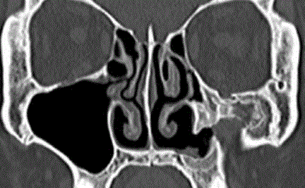
Figure 1
OMU CT scan showed small, contracted left maxillary sinus with
fibrosis and new bone formation. The antrostomy inlet is relatively large
compared to the small sinus.
Figure 2
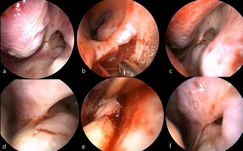
Figure 2
IMA site reconstruction. Yellow dotted line indicates IMA size.
a: About 1.0 cm sized, patent antrostomy at the anterior part of the left inferior
meatus
b: Endoscopic vestibular incision
c-d: Dissection of pocket
e: Insertion of cartilage chips
f: Ostium narrowing after surgery
Discussion
Considering the character of the patient’s pain-sharp, electric,
and sudden-we could assume that the pain is neuropathic in origin.
We also supposed that cold air directly entering the sinus may have
triggered neuralgic pain in the left side of the face. Although cold air
is known as a common triggering factor for trigeminal neuralgia,
the exact pathophysiologic mechanism of how the cold ambient air
caused neuralgic pain in the left side of the patient’s face is not clear.
CT scan of this patient showed the ION sagging into the sinus
during its course on the orbital floor, suggesting a possible clue to
the neuralgic origin of the patient’s pain. The ION travels anteriorly
on the orbit floor through the thin infra orbital canal and exits from
the infra orbital foramen into the face, reaching the skin. Not all
sinus mucosa exposed to cold air would induce facial pain, but nerve
irritation may happen, especially when the nerve is exposed from the
canal. According to anatomic study, ION canal thickness is only 0.2
mm, and the incidence of partial or complete dehiscence of the infra
orbital canal is between 12% and 20% in dried skull specimens [6-8].
Moreover, unexpected variations of the nerve course in the maxillary
sinus or canal dehiscence can increase the possibility of iatrogenic
injury during C-L surgery. The mechanism of pain development by
the dehiscent infra orbital canal is still unclear. Facial pain in the
distribution of the ION is often labelled “vacuum maxillay sinusitis”
and treated empirically by intranasal antrostomy to enlarge the inlet
[9]. Some authors have suggested alterations in atmospheric pressure
as a barotrauma, and others have suggested a vasodilation effect of the
antral mucosal blood vessel [10,11]. However, these assumptions are
mostly related to small antral ostium size and do not correlate to our
patient, who has a large IMA inlet. Sessle et al. [12] suggested that the
trigeminal primary afferent fibers terminated in the nasal mucosa as
a free nerve ending and were activated by noxious stimuli, including
mechanical and chemical irritants.
Another possible factor contributing to neuralgic pain
development in our case is the location and size of the IMA opening.
IMA is usually performed at the inferior portion of the inferior
meatus where the maxillary cyst that developed after C-L operation
bulges most. Thus, the neo-ostium location depends on the location
of the cyst in general. The IMA opening tends to become smaller
with fibrotic changes of the opening, although the opening is large
as created initially. Lund et al5 reported that 53% of the IMA opening
had closed with a mean follow-up time of 26 months, and adequate
initial hole size should be as large as 1 cm × 2 cm to prevent its
closure. In this case, the neo-ostium had remained mostly open at
6 years after surgery. In addition, it was located too anteriorly. This
situation allowed non-humidified, cold air to easily enter the sinus
and irritate exposed nerve. Our treatment of blocking the IMA inlet
was based on these theories and was successful.
Blocking the IMA inlet with conchalcartilage graft via an
endonasal approach is not technically challenging. However, the
mucosa needs to be protected, and the ostium should not be destroyed
to prevent recurrence of mucocele. An endoscope helped us to dissect
precisely around the ostium and to place grafts exactly to surround
the ostium. The initial narrowing of up to 80% of the ostium size
was maintained at 6 months after surgery. Slight resorption of the
cartilage with widening of the narrowed ostium is expected with time,
and long-term follow-up is warranted.
Figure 3
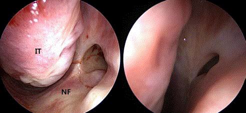
Figure 3
Pre (left) and postoperative 6months (right) endoscopic view of the
IMA opening. Narrowed antrostomy inlet was well maintained.
IT: Inferior Turbinate; NF: Nasal Floor
Conclusion
We report a case of successful treatment of facial neuralgia that developed after IMA by endoscopically narrowing the ostium with cartilage graft. The neuralgia was triggered by cold air directly entering the maxillary sinus, and we believe that the exposed ION was a contributing factor in the neuralgia.
References
- Basu MK, Rout PG, Rippin JW, Smith AJ. The post-operative maxillary cyst. Experience with 23 cases. Int J Oral Maxillofac Surg.1988;17(5):282-4.
- Saito T, Ikeda T, Kono Y, Ohtsubo T, Noda I, Saito H. Implications of endoscopic endonasal surgery for the treatment of postoperative maxillary mucoceles. ORL J Otorhinolaryngol Relat Spec. 2000;62(1):43-8.
- Huang CC, Chen CW, Lee TJ, Chang PH, Chen YW, Chen YL, et al. Transnasal endoscopic marsupialization of postoperative maxillary mucoceles: middle meatal antrostomy versus inferior meatal antrostomy. Eur Arch Otorhinolaryngol. 2011;268(11):1583-7.
- Lee JY, Baek BJ, Byun JY, Shin JM. Long-term efficacy of inferior meatal antrostomy for treatment of postoperative maxillary mucoceles. Am J Oto Surg. 2014;35(6):727-30.
- Lund VJ. Fundamental considerations of the design and function of intranasal antrostomies. Rhinology 1985;23:231-6.
- Sharma N, De M, Pracy P. Recurrent facial paresthesia secondary to maxillary antral cyst and dehiscent infraorbital canal: case report. J Laryngol Otol. 2007;121(6):e6.
- Hollinshead WH. Anatomy for region v.1: the head and neck. New York: Harper & Row. 1968;6.
- Gray HGCM. Anatomy of the human body. Philadelphia, PA: Lea &Febiger. 1966.
- Whittet HB. Infraorbital nerve dehiscence: the anatomic cause of maxillary sinus "vacuum headache"? Otolaryngol Head Neck Surg. 1992;107(1):21-8.
- Mirza S, Richardson H. Otic barotrauma from air travel. J Laryngol Otol. 2005;119(5):366-70.
- Falck B, Svanholm H, Aust R, Bäcklund L. The relationship between body posture and pressure in occluded maxillary sinus of man. Rhinology. 1989;27(3):161-7.
- Sessle BJ. Peripheral and central mechanisms of orofacial inflammatory pain. Int Rev Neurobiol. 2011;97:179–206.