Clinical Image
Tracheal Stenosis by Brachiocephalic Artery Compression
Toshiyuki Mukai, Yujiro Hoshi, Toshihito Sahara and Rumi Ueha*
Department of Otolaryngology, The University of Tokyo, Tokyo, Japan
*Corresponding author: Rumi Ueha, Department of Otolaryngology, The University of Tokyo, Tokyo, Japan
Published: 10 Aug, 2018
Cite this article as: Mukai T, Hoshi Y, Sahara T, Ueha R.
Tracheal Stenosis by Brachiocephalic
Artery Compression. Clin Surg. 2018;
3: 2066.
Keywords
Tracheal stenosis; Brachiocephalic artery compression; Tracheotomy
Clinical Image
A 77-year-old woman had been intubated for about two weeks following a stroke caused
by right middle cerebral artery infarction. She was re-intubated owing to respiratory disorder
after extubation, so that tracheotomy was required. Since her body mass index was 34.2, the
tracheal structure was checked by computed tomography, and the tracheal compression by the
brachiocephalic artery on the right side about 3.5 cm below the lower edge of the cricoid cartilage
was revealed (Figure 1 and 2).
After tracheotomy, flexible fibers copy revealed the right anterolateral tracheal wall
recessed inward by the brachiocephalic artery (Figure 3). To avoid the danger of developing a
tracheoinnominate fistula following the use of a general tracheal
cannula, we prepared and used a slender spiral-wire-reinforced
silicone tracheostomy tube with an adjustable neck flange to ensure
better fit in the proper position. Preoperative evaluation of the
tracheal structure should be considered to avoid complications after
tracheotomy.
Figure 1
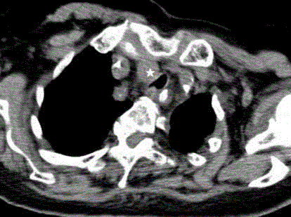
Figure 1
Axial view of computed tomography. A white star shows the brachiocephalic artery.
Figure 2
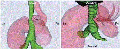
Figure 2
Three-dimensional reconstruction computed tomographic views. Black arrows show the part of
tracheal stenosis by brachiocephalic artery compression.
Figure 3
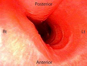
Figure 3
Intratracheal view from a flexible fiberscope. The right anterolateral tracheal wall recessed inward by
the brachiocephalic artery.