Clinical Image
What become of the Changes of the Stuck Guide Wire during Cardiac Percutaneous Coronary Intervention?
Jun-Ted Chong1, Se-Yuan Liu2 and Huai-Min Chen3,4*
*Corresponding author: Huai-Min Chen, Department of Surgery, College of Medicine, Kaohsiung Medical University, Kaohsiung, Taiwan
Published: 31 Jul, 2018
Cite this article as: Chong J-T, Liu S-Y, Chen H-M.
What become of the Changes of the
Stuck Guide Wire during Cardiac
Percutaneous Coronary Intervention?.
Clin Surg. 2018; 3: 2054.
Clinical Course Description
The patient was a 71-year-old man suffered from Complete Total Occlusion (CTO) of Right
Coronary Artery (RCA) for years. Because repeated chest tightness bothered him, so he decided to
receive coronary intervention (PCI) on 15’-07-10. The PCI was approached via right femoral artery,
and the guide wire was put into the left anterior descending artery (LAD) and to take the route of
retrograde into the Right Coronary Artery (RCA). The distal guidewire was snared through the
RCA ostium from right-armed puncture (Figure 1). Unfortunately, the whole lengths of guidewires
(SionTM & Fielder FCTM) were stuck after the distal part of wire was pulled back to the right brachial
artery. Then the cardiologist wanted to pull the both sides of guidewire out but the wire was
unmovable. After repeated procedures the distal segments of wire were curled. At that moment we
both decided not to take any surgical approach because there were no clinical pictures noted and
indeed the wire was stuck in spite of open remove.
The patient attacked a shape pain over anterior left chest and he found a piece of wire was
protruded outside the skin four days later (15’-07-14). The wire was pulsatile along with the heart
beats. We surgeon were asked to remove the wire again, but there was only a short piece could
be pulled out and it was resected (Video 1 & Figure 2A). For the guidewire protrusion outside
the skin, the cardiologist was afraid of the cardiac penetrating injury with pericardial effusion, so
they performed the echocardiographic examination and moderate amount of blood was found. The
pericardiocentesis was done and totally 430 ml of blood was drained
out. Another four days later (15’-07-18) a new piece of wire appeared
from the previous operative wound (Figure 2B). We tried to pull
the wire out again and luckily a long segment of wire was removed
bit by bit (Figure 2C). The followed-up roentgenograms after the
two episodes revealed that the whole length of wire was retained
from right arm to RCA, intracoronary, ascending aorta, and to the
descending aorta in the film of Figure 3A (filmed at 07-16). But the
later film demonstrated there still was a residual wire in Figure 3B
(filmed at 07-19) retained from aortic root to descending aorta. Few
days later the patient discharged without any clinical discomfort.
But the things were not ended. In the following month he felt
epigastralgia with general abdominal discomfort, and one episode
of tarry stool also noted. The GI man took the gastro-endoscopic
examination and a shallow gastric ulcer was told. In this period the
patient still felt abdominal irritable, especially when he changed the
body positions. He went back to the CV OPD for help on 15’-09-
07. The abdominal plain film revealed the retained wire was moved
into the abdominal cavity (Figure 3C). The abdominal Computed
Tomography (CT) was also arranged. The CT scan revealed the same
pictures of foreign body retention (Video 2.1 & Video 2.2). Finally
he received diagnostic laparoscopy to survey the possibilities of
abdominal organs’ injuries by the wire and the most important was
to remove the retained wire (Figure 4). But to my surprise, the long
guide wire traveling in the abdominal cavity got no further injuries.
Conclusion and Discussion
1. Open surgical remove of the guidewire may be the best
choice of treatment in such condition, but we should consider the
sites of surgical approach. If we approach the wire from the peripheral
arteries, the problems of stuck of the wire were persisted in the
beginning; if we approached from the more central part, sternotomy
and more aggressive procedures such as aortic root manipulation or
cardiac apex dissection might be unavoidable.
2. Whether the episodes of UGI bleeding with tarry stool were
caused by penetration of GI tract was unprovable, but the penetration
of wire from diaphragm into abdomen was confirmed.
3. The fracture site of the guidewire was considered at the
region of apex because of constant cardiac contraction, the right half
one of wire was protruded out from the left anterior chest wall, and
the left half one of wire was penetrated into the abdominal cavity.
4. The rare seen experience gave us another opinion for the
treatment of such an awkward condition. So the best practice is not
always the most aggressive one.
Figure 1
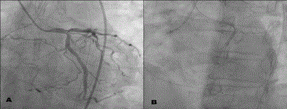
Figure 1
A) The left coronary angiography revealed the guide wire was retrograded into the right coronary artery
with the wire extending from ascending-left ostium-LAD-RCA-right ostium-to ascending. B) The same picture as
Figure 1A without contrast.
Figure 2
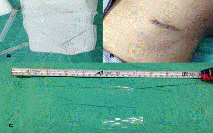
Figure 2
A) The first resected short piece of guide wire. B) Re-appearance of a stump of the wire from previous
surgical wound on 4 days later. C) The long wire directly pulled out bit by bit.
Video 1
Video 2
Figure 3
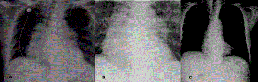
Figure 3
A) X-ray taken on 15’-07-16, white arrows indicate the right half of
wire, and the red arrows indicate the left half of wire. B) X-ray taken on 15’-
07-19, the white arrows indicate the residual left half part of wire, and the wire
had no change of position. C) X-ray taken on 15’-09-07, the residual wire in
abdomen was marked by the arrows.
Figure 4
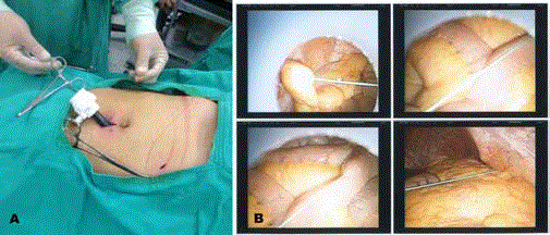
Figure 4
A) Laparoscopy removes of intra-abdominal retained wire. B)
Photo pictures taken during laparoscopic examination, wire could be seen.