Case Report
Cardiac Tamponade: A Case Series
Chiu-Yang Lee1,2*
1Department of Surgery, Division of Cardiovascular Surgery, Taipei Veterans General Hospital, Taiwan
2Department of Senior Citizen Service Management, Chia Nan University of Pharmacy and Science, Taiwan
*Corresponding author: Chiu-Yang Lee, Department of Surgery, Taipei Veterans General Hospital, 201, Section 2, Shih-Pai Road, Taipei 112, Taiwan
Published: 27 Jul, 2018
Cite this article as: Lee C-Y. Cardiac Tamponade: A Case
Series. Clin Surg. 2018; 3: 2051.
Abstract
Cardiac tamponade is a condition in which the heart is compressed by excess fluid in the pericardial
space, which can result in diastolic filling impairment, subsequent cardiac dysfunction, and even
cardiac collapse. Cardiac tamponade is an uncommon sequela of chest contusions from blunt chest
trauma that brings with it a severe risk of sudden death. We present a small series of cases with
successful treatment for cardiac injuries: a young man who was struck by a bull cart and an old man
with chronic kidney disease receiving stent graft placement for superior vena cava syndrome.
This report highlights the need to remain alert for cases of tamponade, and measures such as
emergent pericardiocentesis should first be administered to maintain the hemodynamics of vital
organs such as the heart.
Keywords: Chest contusion; Cardiac tamponade; Hemodynamics
Introduction
Cardiac tamponade is a condition in which the heart is compressed by excess fluid in the
pericardial space, which can result in impaired cardiac filling, subsequent cardiac dysfunction, and
even cardiac collapse. Cardiac tamponade is an uncommon and fatal sequela of chest contusions
from blunt chest trauma that can frequently lead to death if left undiagnosed. Common causes of
cardiac tamponade vary, but they include acute pericarditis, post-myocardial infarction, cardiac
surgery, sharp or blunt chest trauma, aortic dissection, and malignancy.
Pericardial effusions may develop rapidly (acute) or more gradually (subacute or chronic).
When intrapericardial pressure develops quickly and becomes high enough to impede cardiac
filling, cardiac function quickly becomes impaired, and cardiac tamponade can be considered
present and acute.
The true incidence of cardiac rupture following blunt chest contusion is not well documented,
with current records primarily dependent on geography and patient population. Herein, we present
successful treatment for cardiac tamponade following cardiac injuries in a young man who was
struck by a bull cart and an old man with chronic kidney disease receiving stent graft placement for
superior vena cava syndrome.
This report highlights the need to remain alert for cases of tamponade, and life-saving measures
such as emergent pericardiocentesis should first be administered to maintain the hemodynamics of
vital organs such as the heart.
Case 1
An 18-year-old man was involved in a high-speed frontal collision in which his motorcycle struck a bull cart. Approximately 30 minutes later, he was admitted to the emergency room. At presentation, he was confused, violent, and complaining of thoracic and abdominal pain. He developed hypotension with a systolic blood pressure ranging between 60 mmHg and 80 mmHg, with a pulse rate of 120 bpm and a respiratory rate of 35 breaths per minute. Bruise marks were found on his right flank as well as on his chest. His extremities were clammy with marked peripheral hypoperfusion. Notably, his external jugular veins were distended. Cardiovascular examination revealed auscultated and muffled dual heart sounds with no cardiac murmur. Plain X-ray suggested a widened mediastinum. After chest and abdominal Computed Tomography (CT) scans were finished, he underwent circulatory collapse. The CT scans revealed massive pericardial effusion and confirmed cardiac tamponade. After emergency pericardiocentesis with echocardiography, the patient was quickly transferred to the operating room, undergoing a median sternotomy incision. After cardioplegic arrest under assistance of a heart-lung machine, a 1.5 cm tear was found at the junction of the right atrium and superior vena cava (Figure 1). The tear was repaired with Teflon-buttressed sutures. The patient’s postoperative course was uneventful, and he was discharged on postoperative day 15.
Figure 1
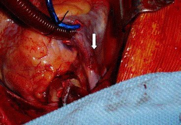
Figure 1
A 1.5 cm tear was found at the junction of the right atrium and
superior vena cava (see arrow).
Figure 2
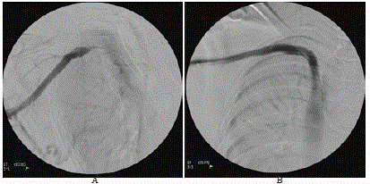
Figure 2
A) Obstruction of right brachiocephalic vein. B) Placement of gore
extension limb stents from the proximal subclavian vein to the proximal
superior vena cava.
Case 2
A 78-year-old man with a history of diabetes mellitus, cerebrovascular accident, superior vena cava syndrome, and chronic kidney disease with regular hemodialysis underwent placement of gore extension limb stents to treat his obstruction of right brachiocephalic vein (Figure 2). After the procedure, he presented with newly onset dyspnea and clinical signs of hypotension. An emergency twodimensional echocardiogram confirmed a diagnosis of cardiac tamponade. Therapeutic pericardiocentesis resulted in prompt cardiac relief, and his hemodynamics developed stably with a systolic blood pressure up to 90 mmHg. Later, the patient was transferred to the intensive care unit for observation. Simultaneously, bleeding tendency including prolonged activated Partial Thromboplastin Time (aPTT) and Activated Clotting Time (ACT) were corrected through transfusion of plasmapheresis and fresh frozen plasma. The patient’s postoperative course was uneventful and he was discharged on day 8.
Conclusion
Incidence of myocardial rupture has decreased with the prevalence
of urgent revascularization and aggressive pharmacological
therapy for the treatment of acute myocardial infarction. The real
occurrence of cardiac rupture following blunt chest trauma is not well
documented, but reports have indicated that it occurs in less than 1%
of patients with such trauma [1]. The mortality rate varies, ranging
from 75% to 81.3% because of asymptomatic presentation, delayed
occurrence, and delayed diagnosis [2,3].
Diagnosis of cardiac tamponade is challenging. The three
principal features of tamponade (Beck’s triad) are soft or absent
heart sounds, hypotension, and jugular venous distension with a
prominent “x” descent but absent “y” descent [4]. Because cardiac
tamponade occurs only after myocardial injuries, prompt diagnosis
with echocardiography and emergent pericardiocentesis should be
undertaken to save patients’ lives.
In Case 1, the young man experienced a high-speed frontal
collision with a bull cart, which is an unusual but nonetheless
traumatic circumstance. Indeed, approximately 80% to 90% of
patients with cardiac rupture die almost immediately at the scene
or before hospital admission [5]. Reports have stated that the right
ventricle is the chamber most frequently ruptured, followed by the
right atrium and left ventricle [6,7].
In cases of cardiac injuries, previous history of heart disease and
blunt mechanical forces must be considered [8]. Mechanical forces
associated with blunt chest trauma include deceleration, acceleration,
compression, and shearing. Cardiac injuries caused by blunt chest
trauma are more difficult to detect than penetrating injuries are,
because the extent of damage caused by blunt trauma is less obvious,
making actual diagnosis difficult. Therefore, patients with blunt chest
trauma must be observed closely to detect any injuries that may not
be initially apparent. In patients with blunt trauma and presenting
with hemodynamic change, the diagnosis of cardiac rupture requires
a high degree of clinical suspicion. Additionally, echocardiography
is a useful tool for rapid detection of blood volume in the pericardial
space before any signs of cardiac tamponade develop. However, if the
patient is hemodynamically unstable, emergent pericardiocentesis
should be performed immediately.
In most patients, immediate surgery is necessary and should not
be delayed by attempts to stabilize the patient. The success rate in
managing cardiac rupture depends on early recognition of its severity
through careful observation and timely diagnosis. Although atrial
tears have been managed without Cardiopulmonary Bypass (CPB),
instituting CPB during surgery is effective and vital for such lifethreatening
situations. CPB can stabilize the hemodynamic state and
allow surgeons to easily locate the site of bleeding after opening the
pericardium, facilitating secure repair under an empty and relaxed
ventricular condition [9]. In our cases, after removal of clots, bright
red blood emanating from the right side of the pericardium was
noted.
In summary, suspicion of blunt cardiac rupture, timely diagnosis,
and proper management create an environment for life-saving
treatment and effectively reduce subsequent mortality in patients
with devastating cardiac injuries.
Acknowledgement
This manuscript was edited by Wallace Academic Editing.
References
- Martin TD, Flynn TC, Rowlands BJ, Ward RE, Fischer RP. Blunt cardiac rupture. J Trauma. 1984;24(4):287-90.
- Fulda G, Brathwaite CE, Rodriguez A, Turney SZ, Dunham CM, Cowley RA. Blunt traumatic rupture of the heart and pericardium: a ten-year experience (1979-1989). J Trauma. 1991;31(2):167-72.
- Brathwaite CE1, Rodriguez A, Turney SZ, Dunham CM, Cowley R. Blunt traumatic cardiac rupture. A 5-year experience. Ann Surg. 1990;212(6):701-4.
- Fauci A, Braunwald E, Kasper D, Hauser S, Longo D, Jameson J, editors. Harrison’s principles of internal medicine. 17th ed. USA: Mcgraw-hill; 2008.
- Got C, editor. Mécanismes lésionnels des traumatismes thoraciques. In: Actualités en réanimation et urgences. SRLF ed. Paris: Elservier; 2000;313-28.
- Türk EE, Tsokos M. Blunt cardiac trauma caused by fatal falls from height: an autopsy-based assessment of the injury pattern. J Trauma. 2004;57(2):301-4.
- Siderys H, Strange PS. Rupture of the heart due to blunt trauma. Successful treatment utilizing cardiopulmonary bypass. J Thorac Cardiovasc Surg. 1971;62(1):84-6.
- Utter GH, Scherer LA, Wisner DH. Blunt cardiac rupture in a patient with prior ventricular septal defect repair: a case report. J Trauma. 2004;57(3):635-7.
- Yaku H, Fermanis G, Horton DA, Guy D, Lvoff R. Successful repair of a ruptured post infarct pseudoaneurysm of the left ventricle. Ann Thorac Surg. 1995;60(4):1097-8.