Case Report
Primary Mixed Adeno - Neuroendocrine Carcinoma(MANEC) of the Appendix Presenting with Small - Bowel Obstruction and Requiring Emergency Laparotomy
Shen Ann Eugene Yeo*
Department of Colorectal Surgery, Singapore General Hospital, Singapore
*Corresponding author: Shen Ann Eugene Yeo, Department of Colorectal Surgery, Singapore General Hospital, Singapore
Published: 18 Jul, 2018
Cite this article as: Ann Eugene Yeo S. Primary Mixed
Adeno - Neuroendocrine Carcinoma
(MANEC) of the Appendix Presenting
with Small - Bowel Obstruction and
Requiring Emergency Laparotomy. Clin
Surg. 2018; 3: 2047.
Abstract
A case of a 57 year old male presenting with abdominal pain, abdominal distension and vomiting. Computed tomography of the abdomen and pelvis showed small bowel obstruction. He underwent an emergency open right hemicolectomy which showed a stenotic mass in ileocaecal junction. Histopathology was that of Mixed Adeno - Neuroendocrine Carcinoma of the appendix (MANEC), a very rare histological subtype of primary appendiceal cancer with a poor prognosis.
Backgrounds
Primary cancer of the appendix is a rare clinical entity and accounts for about 0.5% of the tumours of the gastrointestinal tract. Of all primary appendiceal tumours, carcinoid tumours are the most common and account for about 50% [1]. Uncommon histological subtypes include mucinous cystadenomas and adenocarcinomas. Mixed Adeno - Neuroendocrine Carcinoma (MANEC) of the appendix is a very rarely encountered histological diagnosis. Common presenting signs and symptoms of appendiceal cancer include abdominal pain, a palpable abdominal mass or as an incidental finding on histology post appendicectomy for acute appendicitis. Another common presentation of an appendiceal neoplasm or cancer is the clinical picture of pseudomyxoma peritonei, which is the intra peritoneal accumulation of mucinous material due to rupture of a mucinous appendiceal neoplasm or cancer. Rarely, the patient with primary appendiceal cancer presents with intestinal obstruction [2]. In this case report, we present a case of primary appendiceal MANEC complicated by small bowel intestinal obstruction, and subsequently requiring emergency laparotomy.
Case Presentation
The patient is a 57 - year - old male of Chinese ethnicity who presented with a 2 - month history of central colicky abdominal pain associated with nausea, vomiting and weight loss of 10kg over a duration of 4 months. Apart from this, the patient did not report any diarrhoea, constipation or change in bowel habits. Physical examination showed that while the patient’s vital parameters were stable, he was dehydrated. His abdomen was distended but there was no peritonism and no masses were palpable. Inflammatory markers were normal while serum Carcinoembryonic Antigen (CEA) was normal at 2.9 ug/L. Computed Tomography scan of the Abdomen and Pelvis (CTAP) showed a long continuous segment of dilated bowel with mural thickening of the terminal ileum with surrounding fat stranding. The patient underwent a colonoscopy which showed an ulcerative appearance of the ulcerative ileo - caecal valve (Figure 1) with biopsies showing mild lymphoid hyperplasia with patchy active inflammation. No obvious tumour was visualised on colonoscopy and endoscopic biopsies did not reveal any malignancy. The patient was treated for severe enterocolitis and was given intravenous antibiotics (ceftriaxone and metronidazole). His symptoms improved after one week - the patient’s abdominal distension resolved and he was able to resume an oral diet. He was subsequently discharged well from hospital. Three weeks later, the patient represented to the emergency department with intestinal obstruction, complaining of generalised colicky abdominal pain, associated with abdominal distension and vomiting. He could still open his bowels. His abdomen was distended on examination. Inflammatory markers remained normal. Repeat CTAP showed severe small dilatation with the transition point in the terminal ileum associated with mural thickening of the ileocaecal valve, consistent with Small Bowel Obstruction (SBO). In view of recurrent intestinal obstruction, the patient underwent an emergency exploratory laparotomy. Intra - operatively, there was serous ascites on entry into the abdomen. The small bowel was grossly dilated with the transition point at a 2 x 2 x 2.3 cm stenotic mass in the ileo - caecal junction adherent to the right pelvic side wall. Multiple peritoneal nodules were studded along the small bowel mesentery, pelvis and paracolic gutters (Figures 2 and 3). In view of the above findings, the patient underwent a right hemicolectomy with creation of a double barrelled ileo-colostomy. Final histopathology of the mass was that of MANEC of the appendix (Figure 4). The tumour was noted to have originated from the appendix and invaded directly into the ileocaecal junction. Extensive perintraneural invasion was seen. There were multiple tumour deposits in the small bowel serosa and small and large bowel mesentery. Fourteen lymph nodes were negative for metastatic carcinoma. The ascitic fluid drained was positive for malignancy. Pathologic staging was consistent with pT4bN0M1 in keeping with Stage IV appendiceal cancer [3]. Immunohistochemistry of the tumour cells showed immunoreactivity to synaptophysin and CD56 (<1%) and it stained negative for chromogranin (Figure 5). The Ki - 67 proliferative indexes was approximately 5% at the solid area. Post operatively, the patient made an uneventful recovery and was discharged from hospital 2 weeks after the operation. The patient was subsequently discussed at a multidisciplinary meeting whereby the recommendation was to offer him Cytoreductive Surgery (CRS) with Hyperthermic Intraperitoneal Chemotherapy (HIPEC).
Figure 1
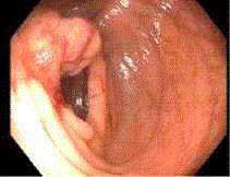
Figure 1
Friable, firm and ulcerative ileo - caecals valve seen on colonoscopy.
Figure 2
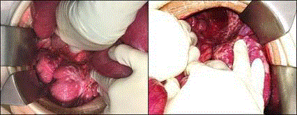
Figure 2
Intra operative images showing multiple peritoneal nodules
along the small bowel mesentery, pelvis and paracolic gutters found
intraoperatively. A stenotic mass is at the terminal ileum was found to be the
cause of obstruction, it was later noted on final histology that this mass arose
from the appendix.
Figure 3
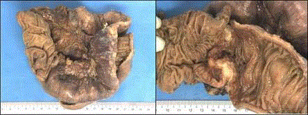
Figure 3
2 cm x 2 cm x 2.3 cm stenotic mass in the ileo-caecal junction.
Figure 4
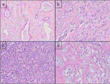
Figure 4
Histopathology of appendiceal tumour showing various
characteristics of MANEC. a) Well-differentiated adenocarcinoma. b)
Infiltrative nests of cells with no lumen formation. The cells are composed
of relatively monomorphic medium sized cells with eosinophilic cytoplasm
as well as cells that contain intracytoplasmic mucin resemble goblet cells.
c) Solid growth pattern with closely packed nests of cells with eosinophilic
cytoplasm and vesicular nuclei. The cells are monomorphic with mild to
moderate atypia. d) Extracellular dissecting mucin pools with nests of tumour
cells within.
Figure 5
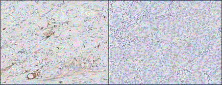
Figure 5
Immunostaining of cells showing immunoreactivity to CD56
(left) and dimmunoreactivity to synaptophysin (right) in keeping with the
histological diagnosis of appendiceal MANEC.
Discussions
The appendix is often the site in the gastrointestinal tract where distinctive benign and malignant neoplasms can occur. Tumours of the appendix can be classified into epithelial or non-epithelial tumours. Epithelial tumours of the appendix range from benign lesions like adenomas to malignant lesions such as carcinoid tumours, adenocarcinomas, mucinous adenocarcinomas and signet ring cell carcinomas. Non epithelial tumours of the appendix include lesions such as appendiceal neuromas, leiomyomas or gastrointestinal stromal tumours. The majority of patients with appendiceal tumours are diagnosed incidentally during appendicectomies. Other usual presenting signs and symptoms of appendiceal tumours include abdominal pain or a palpable abdominal mass. It is exceedingly rare that the patient with primary appendiceal cancer present with intestinal obstruction. In this case report, the stenotic lesion resulting in the patient’s presentation of small bowel intestinal obstruction originated from the appendix with direct tumour extension into the ileo - caecal junction. MANEC of the appendix represents a rare histological subtype of malignant appendiceal neoplasms whereby features of adenocarcinomas and carcinoids are both present. It is defined by the World Health Organisation (WHO) as neoplasms comprising of at least 30% each of epithelial and neuroendocrine components, containing least two out of the three neuroendocrine markers, namely, synaptophysin, CD56 and chromogranin [4]. Apart from the appendix, MANEC has also been reported to develop in the other regions of the gastrointestinal system such as colon, rectum, stomach and biliary tract [5,6]. Prior to the 2010 WHO definition, neoplasms with mixed epithelial and neuroendocrine characteristics were given varied pathological designations including goblet cell carcinoid, adenocarcinoma ex goblet cell carcinoid, and mixed endocrine-exocrine tumours [7]; this lack of standardised diagnostic criteria made characterisation of the disease challenging. In fact, earliest reports of this tumour date back as early as 1969 by Gagne et al [8]. The histological subtype of appendiceal neoplasms and stage of tumour at diagnosis greatly influences its biological behaviour and overall survival. Being only defined recently by WHO, the true biological behaviour and incidence of MANEC is largely unclear but has been reported to be an aggressive clinical entity [7]. In terms of prognosis, in a 2016 study of a patient database that analysed clinicopathological characteristics of gastrointestinal MANEC cases, Brathwaite et al [9] found a large majority (87%) of MANEC arose from the appendix and patients had an overall survival of about 4 years. 49% of MANEC cases had nodal metastases and 67% had stage IV disease at the time of diagnosis. Due to its rarity, the optimal management of appendiceal MANEC requires multidisciplinary and multimodal oncologic management. This includes right hemicolectomy with adjuvant chemotherapy. In selected patients with peritoneal metastasis, cytoreductive surgery with Hyperthermic Intraperitoneal Chemotherapy (HIPEC) constitutes optimal treatment. This is a complex procedure that requires a multidisciplinary team comprising specialists such as surgical oncologists, anaesthesiologists, oncologists and perfusionists. In this group of patients, overall survival varies from 20% to 90% at 5 years [10,11]. Notably, peritoneal surface malignancies of appendiceal origin treated with CRS and HIPEC is associated with better prognosis [12].
Outcome and Follow-up
The patient developed recurrent intestinal obstruction secondary to progressive and extensive peritoneal metastases one month after his MANEC diagnosis. Palliative chemotherapy was trialled with no clinical response. Chemotherapy was eventually with - drawn and he was managed with best supportive cares. The patient died about four months post the MANEC diagnosis.
Conclusion
Primary MANEC of the appendix is a rare clinical entity that carries a poor prognosis; in our case report, our patient with MANEC had the very uncommon presentation of small bowel intestinal obstruction due to invasion into the ileocecals junction by the primary tumour.
References
- American Society of Clinical Oncology (ASCO).
- Connor SJ, Hanna GB, Frizelle FA. Appendiceal tumors: retrospective clini-copathologic analysis of appendiceal tumors from 7,970 appendectomies. Dis Colon Rectum. 1998;41(1):75-80.
- American Joint Committee on Cancer (AJCC) Cancer Staging Manual. In: Edge SB, Byrd DR, Compton CC, editors. 7th ed. New York: Springer; 2010;133.
- World Health Organisation (WHO) Classification of Tumours of the Digestive System.In: Lyon, editor. 4th ed. France: IARC Press; 2010:122-5.
- Zheng SL, Yip VS, Pedica F, Prachalias A, Quaglia A. Intrahepatic bile duct mixed adenoneuroendocrine carcinoma: a case report and review of the literature. Diagn Pathol. 2015;10:204.
- Gagné F, Fortin P, Dufour V, Delage C. [Tumors of the appendix associating histologic features of carcinoid and adenocarcinoma]. Ann Anat Pathol (Paris). 1969;14(4):393-406.
- Turaga KK, Pappas SG, Gamblin T. Importance of histologic subtype in the staging of appendiceal tumors. Ann Surg Oncol. 2012;19(5):1379-85.
- Brathwaite S, Jonathan R, Martha MY, Tanios BS, Wei L, Wendy LF, et al. Mixed adeno - neuroendocrine carcinoma: an aggressive clinical entity. Annals of surgical oncology. 2016;23(7):2281-6.
- Verwaal VJ, Zoetmulder FA. Follow-up of patients treated by cytoreduction and chemotherapy for peritoneal carcinomatosis of colorectal origin. Eur J Surg Oncol. 2004;30(3):280-5.
- Haslinger M, Francescutti V, Attwood K, McCart JA, Fakih M, Kane JM 3rd, et al. A contemporary analysis of morbidity and outcomes in cytoreduction/hyperthermic intraperitoneal chemoperfusion. Cancer Med. 2013;2(3):334-42.
- Verwaal VJ, Bruin S, Boot H, van Slooten G, van Tinteren H. 8 - Year follow-up of randomized trial: cytoreduction and hyperthermic intraperitoneal chemotherapy versus systemic chemotherapy in patients with peritoneal carcinomatosis of colorectal cancer. Ann Surg Oncol. 2008;15(9):2426-32.