Review Article
Cadaveric Atlas for Orthoplastic Lower Limb and Foot Reconstruction of Soft Tissue Defects
Kaitlyn L Ward1, Anthony Romano1 and Edgardo R Rodriguez-Collazo2*
1Franciscan Foot & Ankle Institute, Federal Way, WA, USA
2Presence Saint Joseph Hospital, Chicago, IL, USA
*Corresponding author: Edgardo Rodriguez-Collazo, Department of Surgery, Director Chicago Foot & Ankle Deformity Correction Center, Illizarov Correction & Microsurgical Limb Reconstruction Presence Saint Joseph Hospital, Chicago, USA
Published: 28 Jun, 2018
Cite this article as: Ward KL, Romano A, Rodriguez-
Collazo ER. Cadaveric Atlas for
Orthoplastic Lower Limb and Foot
Reconstruction of Soft Tissue Defects.
Clin Surg. 2018; 3: 2001.
Abstract
Soft tissue deficits or non-healing wounds are a common and challenging problem faced by the
lower extremity reconstructive surgeon. These cases often end in proximal amputation, especially
in those with co-morbidities, compromised angiosomes, or following significant trauma. This
atlas provides a guide for surgeons to understand and treat soft tissue lower extremity defects and
complications. We discuss basic orthoplastic reconstructive principles and patient work-up; thus,
alleviating the need to refer to a plastic or microsurgical specialist. Additionally, incision placement,
anatomy of perforators, axial flow and arc of rotation for flaps are shown for medial, lateral and
anterior compartments of the lower leg as well as the foot. The muscular and fascio cutaneous flaps
in this atlas can be used to cover almost all areas of the lower extremity from the knee distally to the
digits. The purpose of this atlas is to serve as a guide for surgeons to more effectively treat these soft
tissue defects without the need for amputation.
Keywords: Orthoplastic; Reconstruction; Soft tissue defects; Flaps; Lower extremity
Introduction and Preoperative Planning
The first step in preparation for performing any flap is precise preoperative planning. Anatomic
landmarks should be utilized to map out major neurovascular structures and perforating vessels.
Locations and patency of said vessels can be further confirmed with the use of Doppler ultrasound
and/or angiography if necessary. A typical map of such structures in the lower extremity is shown in
the figures above. Figure 1a shows the major landmarks that should be drawn out pre- operatively.
The green lines represent the anterior and medial borders of the tibia. The dashed blue lines
represent proper incision placement. They are drawn 1cm posterior and 1cm anterior to the medial
and anterior tibial borders respectively. Perforating vessels lie along these blue lines and are marked
by the red X’s. They correlate with the anatomic map of perforating vessels at approximately 5 cm,
10 cm, and 15 cm proximal to the ankle joint as shown in Figure 1b. Figure 1c shows a contrast
angiogram confirming adequate blood flow to the perforators stemming from the major vascular
structures of the lower extremity.
Section I: Medial approach to the superficial and deep compartment of the lower leg
• Tibial Exposure
• Medial Gastrocnemius Flap
• Medial Soleal Flap
• Proximal cutaneous sural perforator flap
• Distal cutaneous sural perforator flap
Tibial exposure (for osseous, muscle flap or compartment fasciotomy): In order to avoid
any further damage to the cutaneous perforators, tibial exposure should be obtained 1 cm from the
medial tibial border. This is noted by the dashed red line in Figure 2a. Locations of the perforators
are noted by red X’s.
Medial gastrocnemius and medial soleal flap
Incision Planning: As previously stated, a thorough under-standing of the relevant anatomical
structures and preoperative mapping should be performed. Figure 2b is a cross-section demonstrating
important structures and the incision placement when performing the medial gastroc- nemius
and medial soleal muscle flaps. Perforators and axial flow should be
marked on the posterior border of the tibia at approximately 5 cm,
10 cm, and 15 cm proximal to the ankle joint. An incision is made
approximately 1cm posterior to the medial tibial border on the medial
aspect of the leg as pictured below in Figure 2c. The perforator 15cm
proximal to the ankle is also known as the “C-Point” coined by Dr.
Pedro Rodriguez MD. This is where the division of the medial hemi
soleus should be performed.
Anatomy of perforators & axial flow: It is important to take care
when dissecting down to the fascial planes. The gastrocnemius and
soleus muscles lie in the superficial posterior compartment (Figure
3a). The posterior tibial artery supplies this entire compartment with
the exception of the medial head of the gastrocnemius (supplied by
the medial sural artery). Again, these perforators should be identified
at 5 cm, 10 cm, and 15 cm proximal to the ankle joint in the posterior
medial compartment of the leg (Figure 3b). Posterior tibial perforators
arise from the deep fascia into the superficial compartment (Figure
3c,3d). It is paramount to avoid dissection over the septum because
they carry the blood supply.
Arc of Rotation: The division of the medial hemi soleus should
be performed to protect the distal perforator at 15cm proximal to
the ankle joint, the “C-Point” (Figure 4a). Segmental fascia resection
for additional gastrocnemius and soleus length may be performed if
more coverage is needed. The arc of rotation for these flaps allows
for adequate coverage of the posterior proximal portion of the leg
(proximally based medial gastrocnemius), the anterior leg (distally
based medial hemi-soleus) as seen in Figure 4a. The medial hemisoleus
may also provide coverage for the medial malleolar area,
posterior heel, distal tibia, and anterior ankle (Figure 4b,4c).
Proximal & distal cutaneous sural perforator flap: Two
additional flaps with relevant anatomy to the medial aspect of the
lower extremity are the proximal and distal cutaneous adipose fascia
sural artery perforator flaps (Figure 4d).
Incision planning and anatomy of perforators & axial flow: The
medial aspect of the tibial should be palpated. The perforators for the
proximal cutaneous adipose fascia sural artery flap are demonstrated
in Figure 4d and 4e. Incision placement is key and needs to include
the perforators for vital blood supply.
Arc of Rotation: The Proximally Based Sural Perforator Flap is
excellent for coverage of proximal medial tibial defects (Figure 4f)
and medial popliteal area (Figure 4g). The Proximally Based Sural
Perforator Flap is excellent for coverage of the medial malleolus and
calcaneus (Figure 4h), anterior ankle (Figure 4i) and anterior tibia
(Figure 4j).
Section II: Approach to the lateral and anterior compartment of
the lower extremity
• Peroneus Brevis Flap
• Septal Peroneal Perforator Flap
• Lateral Compartment Options
• Common Peroneal Nerve Exposure
• Proximal Based Lateral Gastrocnemius Muscle Flap
• Anterior Compartment
Peroneus brevis flap
Incision planning: When dissecting the peroneus brevis flap, the
fibular head and lateral malleolus should be palpated and marked. The
incision should start approximately 4 cm distal to the fibular head in
order to avoid common peroneal nerve damage and is made directly
over the fibula as shown in Figure 5b. It is important to take care to
avoid incisions over the anterior and posterior borders of the fibula in
order to preserve septal perforators. The posterior border and septum
of the fibula should also be marked. The perforators should also be
marked out and typically lie approximately 5 cm, 10 cm, and 15 cm
proximal to the lateral malleolus. The viability of the flap is completely
dependent on maintaining the distal most perforator. Dissection is
sharply carried down through cutaneous, adipose and superficial
fascia tissue layers approaching midline (Figure 5c). It is important
to carry dissection towards midline in order to avoid the Superficial
Peroneal Nerve (SPN) (Figure 5d). The SPN travels anterior to the
peroneus brevis muscle over the roof of the anterior compartment or
anterior aspect of the floor of the lateral compartment.
Anatomy of perforators & axial flow: As previously mentioned,
the perforating vessels from this flap, stem from the peroneal artery
and consistently lie approximately 5 cm, 10 cm, and 15 cm proximal
to the lateral malleolus; however, they can be much more numerous
(Figure 5e).
The peroneus brevis muscle belly is mobilized beginning
proximal to distal from the floor of the compartment (superior
surface of the flat surface of the middle 1/3 of the fibula) (Figure 5f).
The muscle is carefully teased off the fibula until the level of the distal
most perforator approximately 5cm to 7 cm proximal to the lateral
malleolus (Figure 5g).
Arc of rotation: The peroneus brevis muscle flap has been termed
the “work horse flap” of the lower extremity due to its large arc of
rotation and many applications. These include: lateral malleolus
(Figure 5f), retrocalcaneal and Achilles insertion (posterior and
plantar calcaneus). Additionally, it is adequate for anterior tibia and
defects (Figure 5g).
Septal peroneal perforator flap
Incision planning: When dissecting the septal peroneal
perforator flap, the posterior border of the fibula should be palpated
and marked as shown in Figure 6a with a blue line. It is vital to
keep dissection away from this line as it also corresponds with the
posterior-lateral septum, the perforators and all the necessary blood
flow to this flap. The lateral malleolus should also be marked out. The
distal perforator for this flap is typically 10cm proximal to ankle joint
and is marked by the red X in Figure 6a. Dissection is sharply carried
down through cutaneous, adipose and superficial fascia tissue (Figure
6c). It is important to keep a paddle of 2 cm (at minimum) on either
side of the posterior septum in order to ensure adequate blood supply
to the flap (Figure 6b and 6d).
Anatomy of perforators & axial flow: As previously mentioned
the distal septal perforator for this flap is typically located
approximately 10 cm proximal to the lateral malleolus (Figures 6e-
6g).
Arc of rotation: This flap has similar indications as the peroneus
brevis flaps and includes: anterior tibia, lateral foot (Figure 6h and
6i), lateral malleolus (Figure 6h and 6i), retrocalcaneal and Achilles
insertion.
Lateral compartment exposure: It should be noted that the
lateral compartment dissection technique also allows for exposure to
the lateral gastrocnemius, lateral soleus, peroneus longus muscle flaps
as well (Figure 7a).
Common peroneal nerve exposure: One key structure in this
area is the common peroneal nerve. It can be accessed effectively
through the lateral compartment (Figure 8a).
Proximal based lateral gastrocnemius muscle flap: for those
practitioners involved with microsurgical techniques and treating
revisional common peroneal nerve injuries or proximal tibia
defects, it is important to be aware that the proximal based lateral
gastrocnemius is a flap that can be harvested in this area (Figure 8b).
This flap receives its blood supply from the sural artery.
Anterior compartment (Figure 9a)
Section III: Medial arch approach for the foot
• Medial Plantar Artery Cutaneous Adipofascia Flap
• Abductor Hallucis Muscle Flap
• Flexor Digitorum Brevis Muscle Flap
• Flexor Hallucis Brevis Muscle Flap
• Plantar Fasciocutaneous Flap
Medial plantar artery cutaneous adipofascia flap
Incision planning: When performing the medial plantar artery
cutaneous adipofascia flap the following structures are important to
identify and mark: calcaneus, metatarsal heads and 3rd ray (Figure
10a). The dissection for this flap will take place medial to the 3rd
ray. Dissection begins laterally and is carried full thickness down to
muscle of the medial instep of the foot.
Anatomy of perforators & axial flow: Blood flow for this flap
is dependent upon the medial plantar artery and its corresponding
perforators (Figure 10b). It is crucial to keep the septum intact.
Arc of rotation: This flap is excellent for coverage of tarsal tunnel
or medial malleolus, dorsal foot and plantar foot.
Abductor hallucis muscle flap
Incision planning: The same landmarks, structures and incision
as the Medial Plantar Artery Cutaneous Adipofascia are utilized
for the Abductor Hallucis Muscle flap (Figure 10a). The dissection
for this flap will take place medial to the 3rd ray. Dissection begins
laterally and is carried full thickness down to muscle of the medial
instep of the foot. The Abductor Hallucis Muscle is then identified
and transected distally.
Anatomy of perforators & axial flow: Blood flow for this flap
is dependent upon the medial plantar artery and its corresponding
perforators (Figure 10b).
Arc of rotation: This flap is excellent for coverage of plantar
calcaneal and plantar midfoot defects (Figure 10c).
Flexor digitorum brevis flap
Incision planning & anatomy of perforators & axial flow: The
flexor digitorum brevis flap is typically harvested through a plantar
midline incision extending from the metatarsal heads and carried
towards the calcaneus (Figure 10d). It is important to note that the
incision placement preserves the medial plantar skin for medial
plantar artery adipofascial flaps. Branches from the lateral plantar
artery mainly supply the muscle, but there is contribution from the
medial plantar artery as well. These arteries should be identified and
marked with a Doppler preoperatively. Dissection is taken through
the plantar fascia which is retracted medially and laterally to expose
the flexor brevis muscle belly. Once the muscle is identified, the
tendinous portion to the toes is transected at the metatarsal neck
(Figure 10d). Dissection is then carried from distal to proximal on
the plantar foot. Dissection is then taken as proximal as needed while
preserving the proximal as needed while preserving the proximal
perforators that have been identified.
Arc of rotation: Coverage of plantar calcaneal and medial foot
defects are possible with this muscle as seen in Figure 10d.
Flexor hallucis brevis flap
Incision planning & anatomy of perforators & axial flow: The
flexor hallucis brevis may be harvested deep to the abductor hallucis
muscle belly. This flap is mainly supplied by the medial plantar artery
(Figure 10e). Preoperative mapping should be performed with a
Doppler.
Arc of rotation: The main advantage of using this flap is that it
may be distally based to provide coverage for the plantar aspect of the
first metatarsophalangeal joint as seen in Figure 10e,10f.
Plantar fasciocutaneous flap
Incision planning & anatomy of perforators & axial flow: The
Plantar Fasciocutaneous Flap incision is performed on the plantar
arch and is demarcated by the dashed yellow lines in Figure 10g.
Blood supply to this flap comes from both medial and lateral plantar
arteries.
Arc of rotation: Coverage of medial arch defects can be obtained
with this flap (Figure 10h).
Figure 1
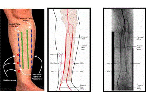
Figure 1
Figure 2
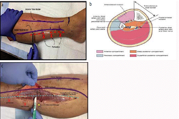
Figure 2
a: Tibial exposure and incision placement. b: Cross-section
demonstrating important structures and the incision placement when
performing the medial gastrocnemius and medial soleal muscle flaps. c:
Incision placement for dissection into the posterior compartment of the leg
approximately 1 cm posterior to the medial border of the tibia. The “C-Point”
is identified from the 15cm perforating artery and pierces the muscle belly
into the superficial fascia.
Figure 3
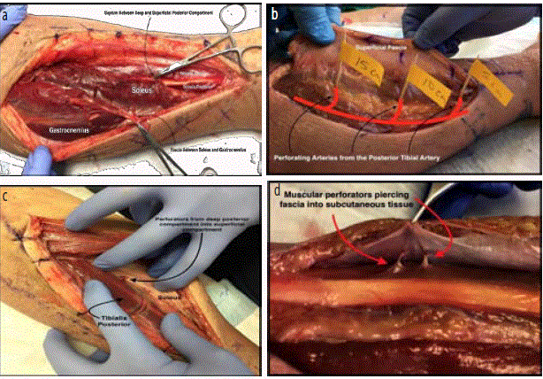
Figure 3
a: Medial incision exposing the posterior compartment of the leg
with fascial and septal divisions. b: Perforators from the posterior tibial artery
at 5, 10, and 15 cm piercing through the muscle bellies the triceps surae
into the subcutaneous tissue. Identified with Doppler preoperatively. c:
Identification of the posterior tibial perforating arteries from the deep posterior
muscle compartment to the superficial posterior muscle compartment. d:
Muscular perforators piercing the fascia into subcutaneous tissue. These
must be preserved for adipofascial and Fasciocutaneous flaps.
Figure 4
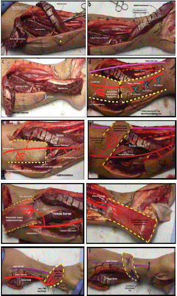
Figure 4
a: After incision and dissection into the deep posterior compartment,
the gastrocnemius and soleus are exposed. The distally based medial hemisoleus
is flapped onto the anterior leg and the proximally based medial
gastrocnemius is flapped over the popliteal fossa. Note the location of the
C-Point, which is crucial to the arc of rotation for the hemi-soleus muscle
flap. b: Medial hemi- soleus muscle flap over distal ankle. Facial resections
may be performed as shown to gain additional length of the muscle flap
for more distal coverage if necessary. c: Distally based medial hemi-soleal
flap turned down over medial malleolus. Additionally, the proximally based
medial head of the gastrocnemius is flapped proximally and posteriorly over
the popliteal fascia and proximal leg. d: Adipofascial flaps from the posterior
medial compartment may be proximally or distally based. The perforator
anatomy, as shown, is exactly the same as the muscular flaps. e: Proximally
Based Sural Perforator Flap and pertinent structures. f: Proximally based
adipofascial/Fasciocutaneous flap from the posterior medial leg. Blood
supply from the medial sural arteries. Coverage of the anterior tibia is
possible with this flap. g: Proximally based adipofascial flap from the poster
medial leg covering the medial popliteal area. Blood supply from medial sural
arteries. h: Distally based posterior tibial artery flap coverage turned down
over the medial malleolus. i: Distally based medial adipofascial flap turned
down showing coverage over the anterior ankle. j: Distally based poster
medial Fasciocutaneous flap covering the anterior tibia. Blood supply from
posteriortibial perforators.
Figure 5
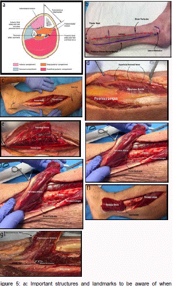
Figure 5
a: Important structures and landmarks to be aware of when
performing flaps along the lateral and anterior compartments of the lower
extremity. b: Incision Planning for Peroneus Brevis Flap. c: Peroneus
Brevis Muscle Flap Incision 1cm posterior to posterior fibular border. d:
Superficial peroneal nerve from lateral compartment dissection. e: Peroneal
brevis muscle flap perforating vessels branching from the peroneal artery.
f: Peroneus Brevis is dissected from the middle 1/3 of the fibula. g: Distal
perforator of Peroneal Brevis Flap found 5 cm proximal to lateral malleolus. f1:
The Peroneus Brevis muscle flap is excellent for lateral malleolar coverage.
g1: The Peroneus Brevis muscle flap can also be used for coverage over
anterior tibial defects.
Figure 6
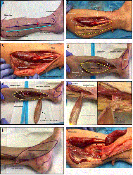
Figure 6
a: Incision planning for the Septal Peroneal Perforator Flap.
b: Septal Peroneal Perforator Flap. c: Septal Peroneal Perforator Flap
demonstrating the septal, fascia, adipose and cutaneous. d: Variation of the
Septal Peroneal Perforator Flap. e: The distal septal perforator is located
10 cm proximal to the lateral malleolus. f: Another look at the distal septal
perforator. g: Close up of the distal septal perforator. h: Septal Peroneal
Perforator Flap providing lateral malleolar and foot coverage. i: Septal
Peroneal Perforator Flap providing lateral malleolar and foot coverage.
Peroneal brevis flap providing anterior ankle joint coverage.
Figure 7
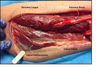
Figure 7
Other lateral compartment flap options.
Figure 8
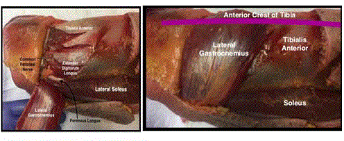
Figure 8
a: Common Peroneal Nerve Exposure. b: Proximal Based Lateral
Gastrocnemius Muscle Flap for CPN injuries of proximal tibia defects.
Figure 9
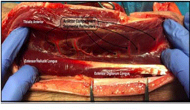
Figure 9
Segmental Perforators over the Anterior Tibial Muscle.
Figure 10
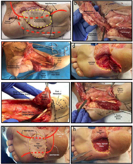
Figure 10
a: Medial plantar artery flap with incision placement. Blood supply
mainly from medial plantar artery.
Blood supply is mapped preoperatively with a Doppler. b: Medial plantar
artery Fasciocutaneous flap with blood supply from medial plantar artery
(proximally based) with dissection at the level of the tarsal tunnel. c: Medial
foot flaps depicting the versatility of intrinsic and fasciocutaneous flaps
coverage of foot defects as shown. d: Incision placement and dissection of
proximally based flexor digitorum brevis flap. Exposed portion of the FDB is
transected at the level of the tendinous portion. Note the incision placement
preserves the medial plantar skin for medial plantar artery adipofascial flaps.
The flap is excellent for coverage of proximal plantar calcaneus. e: Flexor
hallucis brevis flap and the distally based perforator off of the medial plantar
artery. Note the distal coverage sub first metatarsal head. f: Dissection of the
medial and plantar foot at the level of the tarsal tunnel and distal showing
proximally and distally bases muscle flaps with blood supply from the medial
and lateral plantar arteries. g: Plantar Fasciocutaneous flap with perforating
arteries from both medial and lateral plantar arteries. h: Exposure of the flexor
digitorum brevis and dissection of plantar Fasciocutaneous flap. Coverage of
medial arch defects can be obtained with this flap.