Case Report
Irreducible Dislocation of the Metacarpophalangeal Joint
Katsumi Takase*
Department of Orthopedic Surgery, Tokyo Medical University, Japan
*Corresponding author: Katsumi Takase, Department of Orthopedic Surgery, Tokyo Medical University, 6-7-1 Nishishinjuku Shinjuku-ku Tokyo, 160-0023 Japan
Published: 28 Jun, 2018
Cite this article as: Takase K. Irreducible Dislocation of the
Metacarpophalangeal Joint. Clin Surg.
2018; 3: 1997.
Abstract
Dorsal dislocation of the Metacarpophalangeal (MP) joint is less common in the thumb. Simple MP
joint dislocations are described that a closed reduction is expected to be relatively easy. However,
complex MP joint dislocations are, by definition, irreducible by closed means and require open
reduction, as the volar plate becomes entrapped between the metacarpal head and proximal phalanx.
We present a case of dorsal complex MP joint dislocation of the thumb. A 20-year-old right-hand
dominant young lady presented to our hospital with complaints of pain and deformity of the right
thumb. The plain radiographs demonstrated dorsal complete dislocation of MP joint, and that a
sesamoid bone was trapped within the joint space. We performed a surgical procedure using dorsal
approach on this patient. A curvilinear incision was made overlying the MP joint. We confirmed the
proximal end of the phalanx lying completely dorsal to the metacarpal head. The inspection of the
joint revealed that the volar plate and sesamoid bones were completely dorsally translocated over
the metacarpal head. A freer elevator was passed over the metacarpal head to distract open the MP
joint space, and gentle, distally directed pressure was applied to the end of the proximal phalanx to
reduce the MP joint. We attempted several time this maneuver to maintain the continuity of the
volar plate. Finally, we confirmed a stable reduction through full range of motion.
Keywords: Irreducible dislocation; Metacarpophalangeal joint; Thumb; Volar plate
Introduction
Dorsal dislocation of the Metacarpophalangeal (MP) joint is less common in the thumb. Simple MP joint dislocations are described that a closed reduction is expected to be relatively easy. In comparison, Johnson et al. [1] reported that complex MP joint dislocations are, by definition, irreducible by closed means and require open reduction, as the volar plate becomes entrapped between the metacarpal head and proximal phalanx. For the thumb MP joint, anatomical structures that may become trapped include the volar plate, sesamoid bones or the flexor pollicis longus tendon. The pathology of this complex MP joint dislocation is easily reflected to radiographic findings. The radiographs reveal an increased distance between the palmar edge of the base of the proximal phalanx and the metacarpal head, indicating an interposition of the volar plate. We present a case of dorsal complex MP joint dislocation of the thumb. The patient was informed that her data would be submitted for publication, and gave consent.
Case Presentation
A 20-year-old right-hand dominant young lady presented to our hospital with complaints of pain and deformity of the right thumb. At the time of injury, she fell to backward, and the right palm landed on the ground with pronation of the forearm. Then, her right thumb suffered an extreme hyperextension position. On examination, she had a hyperextended deformity at the MP joint and absence of active and passive movements of this joint. There was no neurovascular damage. The plain radiographs demonstrated dorsal complete dislocation of MP joint, and that a sesamoid bone was trapped within the joint space (Figure 1A and 1B). Also, this radiographs showed an increased distance between the palmar edge of the base of the proximal phalamx and the metacarpal head. MR images revealed interposition of the volar plate between the palmar edge of the base of the proximal phalanx and the metacarpal head, and the deep transverse ligament was also involved in the entrapment mechanism (Figure 1C). However, the flexor pollicis longus tendon is intact. Finally, we diagnosed this injury as isolated complex dislocation of the MP joint of the thumb. At first, we treated her by a closed reduction under regional anesthesia. However, we could not accomplish to reduce the dislocated MP joint of the thumb. Then, we changed to perform a surgical procedure using dorsal approach on this patient. A curvilinear incision was made overlying the MP joint. We confirmed the proximal end of the phalanx lying completely dorsal to the metacarpal head. The inspection of the joints revealed that the volar plate and sesamoid bones were completely dorsally translocated over the metacarpal head (Figure 2). A Freer elevator was passed over the metacarpal head to distract open the MP joint space, and gentle, distally directed pressure was applied to the end of the proximal phalanx to reduce the MP joint. We attempted several time this maneuver to maintain the continuity of the volar plate. Finally, we confirmed a stable reduction through full range of motion. After operations, an immobilization cast was applied for 20 days. At 3 years postoperatively (Figure 3), range of motion on interphalangeal joint was o degree to 60 degrees and MP joint was 0 degree to 30 degrees, and grip strength was 21.5 kg (91.2% compared to non-affected side).
Figure 1
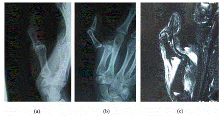
Figure 1
Anteroposterior (A) and lateral (B) radiographs demonstrated
complex dorsal dislocation of the thumb MP joint. Also, they revealed
possible signs of entrapped structures within the joint, and that a sesamoid
bone was trapped within the joint space. MR images (C) revealed interposition
of the volar plate between the proximal end of the phalanx and the head of
the metacarpal, and the deep transverse ligament was also involved in the
entrapment mechanism.
Figure 2
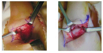
Figure 2
The dislocated MP joint is visualized through a dorsal approach.
Rt side: The inspection of the joint revealed that the volar plate was
completely dorsally translocated over the metacarpal head. Lt Side: After a
stable reduction, there was no injury of the articular surface of the metacarpal
head.
Discussion
Bohart et al. [2] explained Farabeuf’s classification divided into three types for MP joint dislocations. Incomplete dislocations are those where the collateral ligaments are intact. Simple dislocations are those where the collateral ligaments and the volar plate rupture but the latter is not interposed in the joint. Complex dislocations are classically described as complete, irreducible dislocations, and require surgical approach for reduction and proper ligament. Patterson et al. [3] suggested that dorsal MP joint dislocations tended to occur most frequently among the exposed border digits, with the index finger most commonly affected, followed by the small finger. They are relatively rare in the thumb and exceedingly uncommon in the middle or ring fingers. Afifi et al. [4] studied the structures around the MP joint and their contribution to irreducibility of the dislocation. They concluded that the volar plate was the primary structure preventing reduction of the dislocation. Division of the deep transverse metacarpal ligament is not effective in reducing the dislocation. The flexor tendons, lumbricals, superficial transverse ligament, and natatory ligaments do not contribute to irreducibility. In surgical procedure, there is some controversy regarding the preferred approach. Durakbasa et al. [5] described that the strangulated metacarpal head can be directly visualized and the volar plate, which is longitudinally split for reduction, can be repaired using the volar approach. However, Bohart et al. suggested that the dorsal approach to complex dislocations offered several advantages over the volar approach. The dorsal approach allows full visualization of the primary impediment to reduction, and is easy to require vertical splitting of volar plate to reduce the MP joint. Also, Patterson et al. described three advantages concerning about the dorsal approach: reduced risk to palmar displaced neurovascular structures, facilitated management of osteochondral fractures, and full exposure of the volar plate. Postoperatively, there is some debate over the period of immobilization. Eglseder et al. [6] recommend an early mobilization protocol, while Green et al. [7] prefer immobilization three to four weeks. Patterson et al. said that the splitting of the volar plate may delay recovery. In present study, we performed the dorsal approach without longitudinal split of volar plate for reduction. At this time, the time to the reduction of MP joint without damages of the joint cartilage was taken about ten minutes. However, at present, the contractures of MP joint are remained, after an immobilization cast was applied for 20 days.
Figure 3
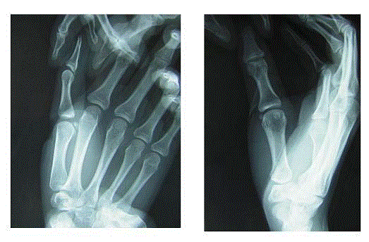
Figure 3
Anteroposterior and lateral radiographs demonstrated the reduction
of the thumb MP joint.
References
- Johnson AE, Bagg MR. Ipsilateral complex dorsal dislocations of the index and long finger metacarpophalangeal joint. Am J Orthop (Belle Mead NJ). 2005;34(5):241-5.
- Bohart PG, Gelberman RH, Vandell RF, Salamon PB. Complex dislocations of the metacarpophalangeal joint. Clin Orthop Relat Res. 1982;(164):208-10.
- Patterson RW, Maschke SD, Evans PJ, Lawton JN. Dorsal approach for open reduction of complex metacarpophalangeal joint dislocations. Orthopedics. 2008;31(11):1099.
- Afifi AM, Medoro A, Salas C, Taha MR, Cheema T. A cadaver model that investigates irreducible metacarpophalangeal joint dislocation. J Hand Surg Am. 2009;34(8):1506-11.
- Durakbasa O, Guneri B. The volar surgical approach in complex dorsal metacarpophalangeal dislocations. Injury. 2009;40(6):657-9.
- Eglseder WA Jr, Gens DR, Burgess AR. Multiple ipsilateral dorsal metacarpophalangeal and interphalangeal joint dislocations: a case report. J Trauma. 1995;38(6):955-7.
- Green DP, Terry GC. Complex dislocation of the metacarpophalangeal joint. Correlative pathological anatomy. J Bone Joint Surg Am. 1973; 55(7): 1480-1486.