Clinical Image
Thumb Malignant Melanoma
Majid Akrami1 and Mohammad Yasin Karami2*
1Breast Diseases Research Center, Shiraz University of Medical Sciences, Iran
2Department of Surgery, Shiraz University of Medical Sciences, Iran
*Corresponding author: Yasin Karami, Department of Surgery, Student Research Committee, Shiraz University of Medical Sciences, Shiraz, Iran
Published: 06 Jun, 2018
Cite this article as: Akrami M, Karami MY. Thumb
Malignant Melanoma. Clin Surg. 2018;
3: 1984.
Clinical Image
A 54-year-old man presented with a 9-month history of blackish discoloration on his right thumb. He had previously been well. On examination, diffuse black pigmentation was seen on palmar side of right thumb (Figure 1). There was not clinical evidence of axillary lymphadenopathy. Incisional biopsy and histologic analysis revealed malignant melanoma (Clark's level 2). Unfortunately, diagnosis may be delayed. Diffuse malignant melanoma of palmar side of thumb was painless and dermatologist physician missed it due to asymptomatic long-standing presentation. The lesion was completely resected and full thickness skin grafting was performed (Figure 2). Right axillary Lymph node Sentinel lymph node biopsy was negative for malignancy. All margins (especially deep margin) were free from the tumor. The patient received systemic treatment with Interferon. At the last follow-up, 12 months after the initial diagnosis, he had no metastases and recurrence.
Figure 1
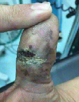
Figure 1
Black pigmentation was seen on palmar side of right thumb.
Figure 2
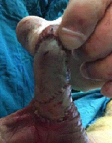
Figure 2
The lesion was completely resected and full thickness skin grafting.