Case Report
Multiple Surgical Treatment of Recurrent Fifth Metatarsal Stress Fracture
Miroslav Haspl1*, Damir Starcevic1, Nenad Medancic1 and Marko Pecina2
1Department of Orthopaedic Surgery, Akromion, Special Hospital for Orthopaedic Surgery, Croatia
2Department of Orthopaedic Surgery, School of Medicine, University of Zagreb, Croatia
*Corresponding author: Miroslav Haspl, Akromion, Department of Orthopaedic Surgery, Special Hospital for Orthopeadic Surgery
Published: 26 Feb, 2018
Cite this article as: Haspl M, Starcevic D, Medancic N,
Pecina M. Multiple Surgical Treatment
of Recurrent Fifth Metatarsal Stress
Fracture. Clin Surg. 2018; 3: 1925.
Abstract
Jones’ refracture after surgical treatment represents great challenge for surgeon. Purpose of this
article is to review surgical options for treating this injury and analyze extrinsic factors which predict
possible refracture. We are presenting the case of a young professional soccer player with repeated
Jones’ fracture of the fifth metatarsal who was treated surgically two times with malleolar screw
fixation. After second refracture authors performed new surgical refixation with malleolar screw
and autologous cancellous bone transplantation. Seven months after surgery patient returned to
professional soccer and remains symptom free for next 7 years. In case of a repeated Jones’ fracture
of the fifth metatarsal bone, refixation should be performed with refreshing the site of fracture
and utilizing bone grafting with autologous cancellous bone. Premature return to sport activity,
cavovarus feet, curved fifth metatarsal bone, weak toe-grip strength, increased body mass index
(BMI) and lower vitamin D levels are risk factors for refracture and they should be considered and
corrected if possible.
Keywords: Jones fracture; Fifth metatarsal stress fracture; Refracture; Professional sport;
Surgical treatment; Bone grafting
Introduction
Jones fracture is now defined as an acute fracture of the fifth metatarsal bone, at the junction between the proximal diaphysis and metaphysis, without dislocation, and at the level of the joint between the fourth and fifth metatarsal bone [1,2]. It was first described by Sir Robert Jones in year 1902 [3] which led many to name all fractures of the metatarsal base a Jones fracture. In 1984, Torg [4] divided fractures at the base of the fifth metatarsal bone into acute fractures, fractures with delayed coalescence and fractures that do not coalesce (non-union). Lawrence and Botte in year 1993 [5] divided fractures in base of the fifth metatarsal bone into tuberosity avulsion, real Jones fracture and fracture of the proximal diaphysis of the fifth metatarsal bone. Stress fracture of the diaphysis of the metatarsal bone is defined as a stress fracture in the zone of the proximal part of the fifth metatarsal bone distally from the zone of Jones fracture [1,5-8]. DeLee et al. [9] defined the characteristics of this type of fracture: (a) present a medical history of previous pain from the lateral side of the foot prior to the acute event that led to the patient requesting medical attention, (b) present radiological evidence of stress reaction in the bone, (c) there is no data on previous treatment for fractures of the fifth metatarsal bone. Conservative treatment has an unacceptably high percentage of non-coalescence and re-fracture, as well as a long and unpredictable return to sporting activities, therefore, many authors today prefer primary, early fixation of fractures, especially with and intramedullary screw, particularly when treating athletes [10,11]. Treatment of acute Jones fractures and stress fractures of the proximal fifth metatarsal bone is well described in literature [2,4,8-22]. Re-fractures after a fixation with an intramedullary screw and finished healing are significantly less mentioned in literature [23,24]. In this paper we will present a professional soccer player, who has been treated surgically three times, on the case of Jones fractures, by using an intramedullary screw, and after the second re-fracture treated again by applying a malleolar screw with autologous cancellous bone graft.
Case Presentation
M.Š., born in 1988, a professional football player, at the age of 19 - in October 2007, suffered
after a prodromal symptomsa stress fracture of the fifth metatarsal bone in his right foot. Two weeks
after the injury, a surgical procedure was done, in terms of osteosynthesis with a malleolar screw in the fifth metatarsal bone. After two months he started with jogging
and after three and a half months he returned to full sport activity.
Throughout that entire period, he experienced some difficulties,
discomfort in the foot accompanied by swollen feet after a workout,
with swelling withdrawing the next day. In January 2008 after 10 days
of intensive training, the patient felt pain and discomfort in the foot
with a sense of insecurity. A new fracture on the plantar side of the
fifth metatarsal bone was found on X-ray. In March 2008 a repeated
osteosynthesis was performed, this time with a longer malleolar screw
(Figure 1). After the operation he was immobilized with brace and
for two months he walking with crutches, and the following month
the load on the foot was increased gradually, using brace. Four and a
half months after surgery, he began with jogging, and the full return
to sport activity was allowed after 7 months. For the next year and a
half, he was playing football with a full load, until April 2010, when he
played a match on an extremely hard field lining and pain occurred on
the outside of the operated foot, with the feeling that something has
snapped. He could not stand on his foot and a swelling appeared. The
X-ray showed that new re-fracture of the base of the fifth metatarsal
bone occurred, accompanied by a radiolucency round intramedullary
screw (Figure 2), which is better seen on CT images of the foot
(Figure 3). A few days later, he was admitted to our Department and
he underwent a new osteosynthesis of the fifth metatarsal bone of
the right foot with a malleolar screw of a 4.5 mm diameter, with an
autologous cancellous bone graft, taken from iliac crest.
The surgical procedure was performed under regional anaesthesia
with the use of Esmarch tourniquet. Using the old operative incision,
we gained access to the base of the fifth metatarsal bone and the screw
was removed. Under the control of an X-ray amplifier a Kirschner
wire is set intramedullary in a new direction. A cannulated bur is used
to make a new ring for the screw in a way that it engages the medial
cortex. A malleolar screw is introduced, with partial thread of 4.5 mm
diameter so that the screw thread passes over the fracture line. Under
the control of X-ray the screw is introduced into the medullary canal, until the distal part of the medial cortices, in which it is screwed in
with two threads. In this way, it is ensure to have a good compression
on the place of the fracture cracks and excellent stability of the screw.
At the fracture site, the periosteum is cut through and the bone is
coated with an autologous cancellous bone from the iliac crest. After
surgery immobilization is set with a walking brace. Load on the
operated leg from 10 kg to 15 kg allowed. The patient starts with early
physiotherapy. After six weeks it is allowed to walk with one crutch
for 30 days and after that with full weight bearing. Three months
after surgery, the patient experienced no pain, and control X-ray
showed healing of the re-fracture site (Figure 4). He started with
strength training after a pedobarographic analysis and preparation
of individual orthopaedic insoles to prevent re-occurrence of
fracture. Seven months after the surgery control X-ray showed visible
complete remodeling of the location of re-fracture of the base of the
fifth metatarsal bone (Figure 5), so he was allowed to perform a full
physical exercise and play soccer. For the next seven years, during
which we followed the patient, while he played professional soccer,
there were no new subjective complaints on the operated foot, and
the X-ray of the foot done at the last check (five and a half years after
the last surgery), was normal with a visible functional adaptation of
the fifth metatarsal bone in his right foot (Figure 6).
Figure 1
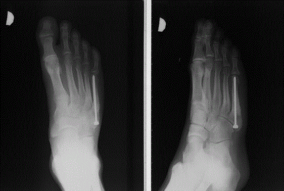
Figure 1
X-ray after first refracture and reoperation at August 8, 2008 (four
months after reoperation) performed with a longer malleolar screw.
Figure 2
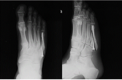
Figure 2
Second refracture at April 11, 2010. Notice screw instability and
don't passing medial corticalis. Good, preferable position of the screw is
marked with line.
Figure 3
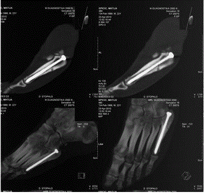
Figure 3
CT scan showing refractures of the fifth metatarsal bone at April
11, 2010
Figure 4
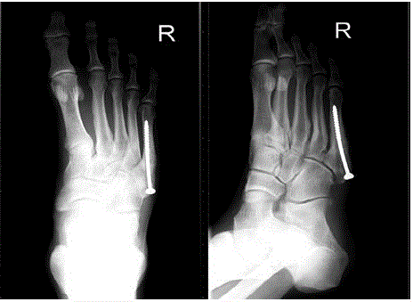
Figure 4
Three months after second reosteosynthesis with bone grafting at
July 21, 2010, Good bone healing.
Figure 5
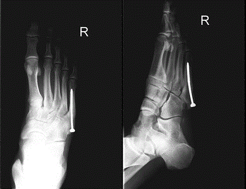
Figure 5
Seven months after second reosteosynthesis shoving finished
bone healing at November 24, 2010. Starting full sport activity.
Figure 6
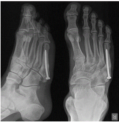
Figure 6
Last X-ray at February 2016 (FU 5.5 years after second
reosteosynthesis and bone grafting).
Discussion and Literature Review
Surgical treatment of acute (real) Jones fractures and stress fractures of the diaphysis of the fifth metatarsal bone is proven to be a safe and effective method of treatment [1,3,5-14]. Chuykpaiwong and associates [1] have analysed 32 real Jones fractures and 29 stress fractures of the proximal diaphysis of the fifth metatarsal bone. They found no difference in outcome between the two anatomical locations, and have therefore suggested that the fractures of the base of the fifth metatarsal bone (except avulsion) should be named Jones fractures. Treatment of non-healing and/or re-fracture require surgical treatment. The question is why did our patient require repeated surgery? Our patient came back too soon into a full physical exercise. Reoperation was done by re-osteosynthesis with a longer screw, but the screw was too long, reaching subcortical bone of fifth metatarsal bone following intramedullary canal, which prevented maximal compression. In our patient a new re-fracture occurred after two years. X-ray and CT investigations indicated the refracture, but also a loosening of the osteosynthetic material, i.e. the screw. This suggests that the osteosynthetic material (screw) was unstable, and was not acting as an additional reinforce for the bone. The patient was reoperated in such a way that we changed the direction of the screw and the screw was introduced from the base of the metatarsal bone in the medial cortex of the distal diaphysis. A. Thus, a stable osteosynthesis and rapid healing of the refracture was achieved. The screw now serves as an additional reinforcement for the bone. Today, the most used are cannulated screws, usually without a head, for easy and accurate insertion, with a lower risk of bad positioning of the screw, and the option of insertion through smaller incisions [22]. Biomechanical data suggest that the initial strength of the force required to move the fragments of the simulated fracture of the fifth metatarsal bone fixed with a cannulated screw of 4.5 mm, is the same as in fractures fixed with a full 4.5 mm malleolar screw [25].The results of another biomechanical study showed strength reduction of cannulated screws in comparison to full screws, but the difference is gradually reduced as the size of the screw increases. Based on these results, Reese and associates [26] suggested the use of the largest cannulated screws that can fit in the medullary canal. Kelly and associates also proposed the use of larger screws in diameter, but Shah and associates [27] emphasize that inserting a screw with largest possible diameter is not critical to achieve the rigidity of fixation, yet it can increase the risk of intraoperative fracture. Although the description of the different methods of surgical treatment exists (intramedullary fixation screw with or without heads, cannulated or full, use corticocancellous bone graft, method of comprehensive bounds, external fixation, application of tile), we agree with the majority of authors that the recommended method of treatment is fixation with an intramedullar screw but inserted in right position. Treatments of non-healing and re-fractures of Jones fractures bear a lot of controversy. Thomas and associates [28] recommend treatment by intramedullary fixation, without showing and cleaning the location of pseudarthrosis and application of bone graft, in order to preserve the surrounding soft tissues and local circulation. According to Hunt and associates [29] treatment of non-healing and repeated fractures requires removal of osteosynthetic materials, debridement (refreshment) of the fracture location, re-fixation with a screw of larger diameter - the largest that can fit in the channel, and the application of autologous bone from the iliac bone or the application of allogenic bone with bone marrow aspirate from the iliac bone. The authors recommend the application of a screw of 5.5 mm or 6.5 mm diameter, to meet the demands of most athletes. There are several described risk factors of re-fracture of the fifth metatarsal bone. It is well known that a premature return to sport activities presents a high risk of re-fracture, and therefore an athlete must be warned about this complication [29,30]. In case of top athletes, a use of computed tomography or magnetic resonance should be taken in consideration before returning to training in order to avoid the possibility of re-fracture [31]. Athletes with high foot arches have a higher risk of injury to the lateral structure of feet. Popovic and associates [32] have used under scopic tests to find that 14 of the 17 patients treated with surgery had emphasized cavovarus foot, while 3 patients had planovalgus feet. These characteristics can be a predisposing factor for the development of overuse injuries of the fifth metatarsal bone. Fujitaka and associates [13] followed 273 college soccer players between 2005 and 2013 and they tried to find risk factors for proximal fifth metatarsal bone fracture. They found that weak toe-grip strength may lead to an increase in the load applied onto the lateral side of the foot, resulting in stress fracture. Risk factors because of foot deformation need to be identified and it is mandatory to propose correction of shoes or orthotics application or orthopaedic insoles, before returning to sports activities and training after surgical treatment of Jones fractures. Clinical studies of Raikin and associates [33] warned of varus of rear foot part at 16 feet, while radiological imaging showed varus of rear foot part in 18 feet of 21 patients treated surgically for the fracture of the fifth metatarsal bone. They concluded that postoperative orthopaedic insoles that corrected varus feet may be useful in the prevention of re-fracture. Similarly, Darabos and associates [34] proposed application of custom-made foot orthosis for surgically treated patients to facilitate quick recovery and return to normal sports activities. Pećina and associates [35] have used pedobarografy on 20 surgically treated football players and have found varus position of feet in 90% of athletes. They proposed as mandatory preventive measure a pedobarographical analysis of athletes and preparation of individual orthopaedic insoles to prevent re-occurrence of fractures of the fifth metatarsal bone. The appearance of re-fracture of the fifth metatarsal bone, especially recurrent refractures, may be associated with increased body mass index (BMI) and the radiological parameters associated with the protrusion of the head of the fifth metatarsal bone - increased intermetatarsal angle between the fourth and the fifth metatarsal bone, as well as lateral deviation of the fifth metatarsal on the angled X-rays [36]. One of the risk factors indicating a possible worse outcome on healing is the distance between fragments plantar greater than 1 mm, visible on a standard angled X-ray image [37]. In this case, it takes a long time to heal, and often more non-healing and repeated fractures. The topic of metabolic difficulties in healing is another open question, in reference to healing the base of the fifth metatarsal bone. Clutton and Perera [38] showed a study in which they analysed 40 patients with fractures of the fifth metatarsal bone and found a reduced level of vitamin D at 65% patients. Lower vitamin D levels have been noted in patients who have suffered a fracture during the winter months. We can end our discussion with the statement that the problem of the fifth metatarsal bone fracture is actual as it always has been especially in sports medicine [39-42].
Conclusion
Surgical treatment and by an intramedullary fixation of Jones fracture with a screw, is the optimal treatment for athletes, because it enables faster and more predictable healing of fractures and more rapid return to sports activities. It is necessary to bear in mind the risk factors that can lead to re-fracture after fractures, all athletes should be prescribed to have custom made orthopaedic insoles, based on pedobarographic analysis. If there is a new refracture, it is necessary to do a re-fixation of the fracture with a refreshment of fracture site, and by application of autologous cancellous bone. During reoperation it is especially important to pay attention of the direction of the screw that needs to go from the base of metatarsal distal to the medial cortical distal diaphysis.
References
- Chuckpaiwong B, Queen RM, Easley ME, Nunley JA. Distinguishing Jones and proximal diaphyseal fractures of the fifth metatarsal. Clin Orthop Relat Res. 2008;466(8):1966-70.
- Kelly IP, Glisson RR, Fink C, Easley ME, Nunley JA. Intramedullary screw fixation of Jones fractures. Foot Ankle Int. 2001;22(7):585-9.
- Jones R. Fracture of the base of the fifth metatarsal bone by indirect violence. Ann Surg. 1902;35(6):697-700.
- Torg JS, Balduini FC, Zelko RR, Pavlov H, Peff TC, Das M. Fractures of the base of the fifth metatarsal distal to the tuberosity. Classification and guidelines for non-surgical and surgical management. J Bone Joint Surg Am. 1984;66(2):209-14.
- Lawrence SJ, Botte MJ. Jones' fractures and related fractures of the proximal fifth metatarsal. Foot Ankle. 1993;14(6):358-65.
- Dameron TB Jr. Fractures and anatomical variations of the proximal portion of the fifth metatarsal. J Bone Joint Surg Am. 1975;57(6):788-92.
- Landorf KB. Clarifying proximal diaphyseal fifth metatarsal fractures. The acute fracture versus the stress fracture. J Am Podiatr Med Assoc. 1999;89(8):398-404.
- Pecina M, Bojanic I. Stress fractures. In: Pecina M, Bojanic I, editors. Overuse injuries of the musculoskeletal system, 2nd edn. Boca Raton: CRC Press. 2004;315-49.
- DeLee JC, Evans JP, Julian J. Stress fracture of the fifth metatarsal. Am J Sports Med. 1983;11(5):349-53.
- Brown SR, Bennett CH Craig H. Management of proximal fifth metatarsal fractures in the athlete. Curr Opin Orthop. 2005;16(2):95-9.
- Boden BP, Osbahr DC. High-risk stress fractures: evaluation and treatment. J Am Acad Orthop Surg. 2000;8(6):344-53.
- Quill GE Jr. Fractures of the proximal fifth metatarsal. Orthop Clin North Am. 1995;26(2):353-61.
- Fujitaka K, Taniguchi A, Isomoto S, Kumai T, Otuki S, Okubo M, et al. Pathogenesis of Fifth Metatarsal Fractures in College Soccer Players. Orthop J Sports Med. 2015;3(9):1-7.
- Japjec M, Staresinic M, Starjacki M, Zgaljardić I, Stivicic J, Sebecic B. Treatment of proximal fifth metatarsal bone fractures in athletes. Injury. 2015;46(6):S134-6.
- Mallee WH, Weel H, van Dijk CN, van Tulder MW, Kerkhoffs GM, Lin CW. Surgical versus conservative treatment for high-risk stress fractures of the lower leg (anterior tibial cortex, navicular and fifth metatarsal base): a systematic review. Br J Sports Med. 2015;49(6):370-6.
- Roche AJ, Calder JD. Treatment and return to sport following a Jones fracture of the fifth metatarsal: a systematic review. Knee Surg Sports Traumatol Arthrosc. 2013;21(6):1307-15.
- Mologne TS, Lundeen JM, Clapper MF, O’Brien TJ. Early screw fixation versus casting in the treatment of acute Jones fracture. Am J Sports Med. 2005;33(7):970-5.
- Mindrebo N, Shelbourne KD, Van Meter CD, Rettig AC. Outpatient percutaneous screw fixation of the acute Jones fracture. Am J Sports Med. 1993;21(5):720-3.
- Nagao M, Saita Y, Kameda S, Seto H, Sadatsuki R, Takazawa Y, et al. Headless compression screw fixation of jones fractures: an outcomes study in Japanese athletes. Am J Sports Med. 2012;40(11):2578-82.
- Sarimo J, Rantanen J, Orava S, Alanen J. Tension-band wiring for fractures of the fifth metatarsal located in the junction of the proximal metaphysis and diaphysis. Am J Sports Med. 2006;34(3):476-80.
- Lombardi CM, Connolly FG, Silhanek AD. The use of external fixation for treatment of the acute Jones fracture: a retrospective review of 10 cases. J Foot Ankle Surg. 2004;43(3):173-8.
- Porter DA, Duncan M, Meyer SJ. Fifth metatarsal Jones fracture fixation with a 4.5-mm cannulated stainless steel screw in the competitive and recreational athlete: a clinical and radiographic evaluation. Am J Sports Med. 2005;33(5):726-33.
- Glasgow MT, Naranja RJ Jr, Glasgow SG, Torg JS. Analysis of failed surgical management of fractures of the base of the fifth metatarsal distal to the tuberosity: the Jones fracture. Foot Ankle Int. 1996;17(8):449-57.
- Granata JD, Berlet GC, Philbin TM, Jones G, Kaeding CC, Peterson KS. Failed Surgical Management of Acute Proximal Fifth Metatarsal (Jones) Fractures: A Retrospective Case Series and Literature Review. Foot Ankle Spec. 2015;8(6):454-9.
- Pietropaoli MP, Wnorowski DC, Werner FW, Fortino MD. Intramedullary screw fixation of Jones fractures: a biomechanical study. Foot Ankle Int. 1999;20(9):560-3.
- Reese K, Litsky A, Kaeding C, Pedroza A, Shah N. Cannulated screw fixation of Jones fractures: a clinical and biomechanical study. Am J Sports Med. 2004;32(7):1736-42.
- Shah SN, Knoblich GO, Lindsey DP, Kreshak J, Yerby SA, Chou LB. Intramedullary screw fixation of proximal fifth metatarsal fractures: a biomechanical study. Foot Ankle Int. 2001;22(7):581-4.
- Thomas JL, Davis BC. Treatment of Jones fracture non-union with isolated intramedullary screw fixation. J Foot Ankle Surg. 2011;50(5):566-8.
- Hunt KJ, Anderson RB. Treatment of Jones fracture nonunions and refractures in the elite athlete: outcomes of intramedullary screw fixation with bone grafting. Am J Sports Med. 2011;39(9):1948-54.
- Larson CM, Almekinders LC, Taft TN, Garrett WE. Intramedullary screw fixation of Jones fractures. Analysis of failure. Am J Sports Med. 2002;30(1):55-60.
- Wright RW, Fischer DA, Shively RA, Heidt RS Jr, Nuber GW. Refracture of proximal fifth metatarsal (Jones) fracture after intramedullary screw fixation in athletes. Am J Sports Med. 2000;28(5):732-6.
- Popovic N, Jalali A, Georis P, Gillet P. Proximal fifth metatarsal diaphyseal stress fracture in football players. Foot Ankle Surg. 2005;11(3):135-41.
- Raikin SM, Slenker N, Ratigan B. The association of a varus hindfoot and fracture of the fifth metatarsal metaphyseal-diaphyseal junction: the Jones fracture. Am J Sports Med. 2008;36(7):1367-72.
- Darabos N, Obrovac K, Knez N, Darabos A, Hudetz D, Elabjer E. Combined surgical therapy and orthotic management of stress and tuberosity avulsion fracture of the fifth metatarsal bone: a case report. J Am Podiatr Med Assoc. 2009;99(6);529-35.
- Pecina M, Bojanic I, Smoljanovic T, Ivkovic A, Mirkovic M, Jelic M. Surgical Treatment of Dyaphyseal Stress Fractures of the Fifth Metatarsal in Competitive Athletes. Long-term Follow-up and Computerized Pedobarographic Analysis. J Am Podiatr Med Assoc. 2011;101(6):517-22.
- Lee KT, Park YU, Jegal H, Kim KC, Young KW, Kim JS. Factors associated with recurrent fifth metatarsal stress fracture. Foot Ankle Int. 2013;34(12):1645-53.
- Lee KT, Park YU, Young KW, Kim JS, Kim JB. The Plantar Gap: Another Prognostic Factor for Fifth Metatarsal Stress Fracture. Am J Sports Med. 2011;39(10):2206-11.
- Clutton J, Perera A. Vitamin D insufficiency and deficiency in patients with fractures of the fifth metatarsal. Foot(Edinb). 2016;27:50-2.
- Begly JP, Guss M, Ramme AJ, Karia R, Meislin RJ. Return to play and performance after jones fracture in the National Basketball Association Athletes. Spots Health. 2016;8(4):342-6.
- Lareau CR, Hsu AR, Anderson RB. Return to play in National Football League Players after Operative Jones Fracture Treatment. Foot Ankle Int. 2016;37(1):8-16.
- Bowes J, Buckley R. Fifth metatarsal fractures and current treatment. World J Orthop. 2016;7(12):793-800.
- Le M, Anderson R. Zone II and III fifth metatarsal fractures in athletes. Curr Rev Musculoskelet Med. 2017;10(1):86-93.