Case Report
Correction of Simple Complete Syndactyly: Case Report and Operative Technique
Georgeanne G Botek1* and Scott D Walker2
1Department of Orthopaedic and Rheumatologic, Cleveland Clinic, Cleveland, Ohio, USA
2Department of Foot and Ankle Surgery Residency, Mercy Regional Medical Center, Lorain, Ohio, USA
*Corresponding author: Georgeanne G Botek, Department of Orthopaedic Surgery, Orthopaedic and Rheumatologic Institute, Cleveland Clinic, Cleveland, Ohio, USA
Published: 16 Feb, 2018
Cite this article as: Botek GG, Walker SD. Correction of
Simple Complete Syndactyly: Case
Report and Operative Technique. Clin
Surg. 2018; 3: 1912.
Abstract
Syndactyly is abnormal congenital or acquired webbing of the digits. Presented is a case report with
the operative technique used for desyndactylization of webbed second and third toes, highlighting
methods to facilitate anatomic correction of the deformity, adequate skin coverage, and tension-free
closure. In this case, the syndactyly is congenital, simple, and complete and a full-thickness skin
graft from the sinus tarsi of the ipsilateral foot is used.
Keywords: Syndactyly; Syndactylism; Desyndactylization; Full thickness skin graft; Sinus tarsi
Case Presentation
A 40-year-old male presented with complaints of pain at the first metatarsophalangeal joint and conjoined second and third toes of his right foot. He also disliked the appearance of his webbed toes. Past medical history included cystic fibrosis status post remote bilateral lung transplant, wellcontrolled hypertension, stage III chronic kidney disease, and steroid-induced insulin-dependent diabetes mellitus (hemoglobin A1C 7.1%). On physical examination webbing of the second and third toes of both feet was found with extent past the distal interphalangeal joint and without synonychia (Figure 1). Examination of the hands demonstrated no concomitant finger deformity. Hallux abductovalgus deformity of the right foot was also present with clinical findings consistent with degenerative joint disease. Radiographs of the symptomatic foot confirmed the first ray deformity and showed no evidence of any osseous abnormality of the syndactylized toes (Figure 2). The patient elected desyndactylization of the second and third toes and arthrodesis of the first metatarsophalangeal joint.
Operative Technique
Anesthesia for the procedure consisted of a popliteal nerve block and monitored anesthesia care. An ankle tourniquet was applied and inflated to 250mmHg for the first metatarsophalangeal joint arthrodesis. The arthrodesis was performed in standard fashion using conical reamers with plate and screw fixation. After deflating the tourniquet, an 18g1¾” hypodermic needle was placed between the second and third proximal phalanges from proximal dorsal to distal plantar, a previously described technique [1]. This needle serves as a guide both to ensure the planned incision is placed equidistant between the proximal phalanges and to prevent against extending the plantar incision too far proximally, which would deviate from normal web space anatomy. A marking pen was used to connect the plantar and dorsal aspects of the needle, taking care to stay directly between the toes, followed by incision with a No.15 blade full thickness through the skin. As sharp and blunt dissection through the subcutaneous tissues was performed using Littler scissors, care was taken to avoid the neurovascular bundles by staying midline, until the toes were completely separated (Figure 3). Next, a small square was trimmed from an Esmarch bandage, folded in half, and placed between the separated digits (Figure 4) [2]. The toes were then gently brought together to leave a mark upon the bandage, outliningthe area of commissure requiring graft coverage. The outlined area was trimmed out as a template. The maximum dimensions of the template in length and width were used to plan and perform an elliptical full-thickness skin graft at the sinus tarsi, orienting the long axis of the graft parallel to the relaxed skin tension lines of the donor site (Figure 5). After lavage of the recipient site with copious sterile normal saline, the skin graft was secured without tension using 5-0 polypropylene monofilament sutures. Prior to subcutaneous and cutaneous closure of the donor site, wide undermining of the skin was performed to facilitate approximation of the wound edges and minimize scar formation [3]. The operative sites were dressed with bismuth containing non-adherent gauze and gently compressive overlying dry dressing. A padded posterior splint was applied to keep the foot from plantar flexing, thereby minimizing skin tension at the donor site (Figure 6). The patient was instructed to be non-weight-bearing to the surgical site for two weeks as is the surgeon’s protocol for first metatarsophalangeal joint arthrodesis. At six weeks post-operatively, this 40-year-old male was ready for advancement to a stiff-soled surgical shoe or supportive footwear ambulating limitedly without pain and pleased with the resultant foot (Figure 7).
Figure 1
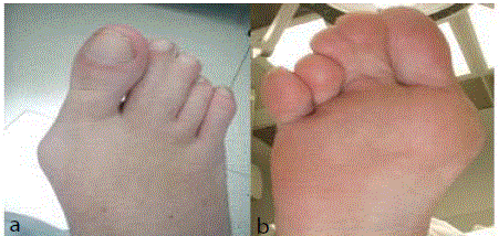
Figure 1
Right dorsal forefoot (A) and plantar forefoot (B).
Figure 2
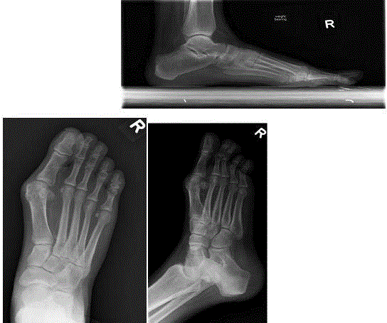
Figure 2
Right foot radiographs, Anteroposterior (A),Medial oblique (B), Lateral (C).
Figure 3
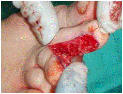
Figure 3
Separation of the second and third toes.
Figure 4
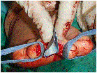
Figure 4
The folded esmarch template.
Figure 5
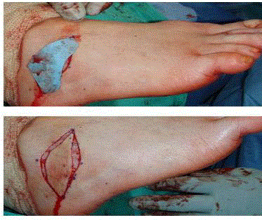
Figure 5
The esmarch template at the donor site (A). Elliptical full thickness
skin graft from the sinus tarsi (B).
Figure 6
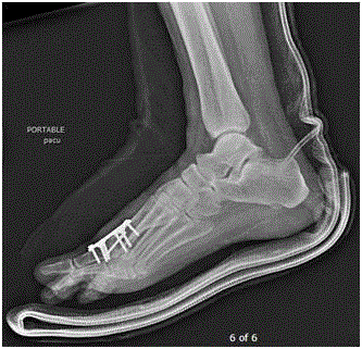
Figure 6
Lateral foot radiograph following posterior splint application,
preventing plantar flexion.
Figure 7
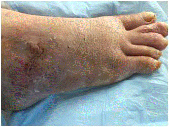
Figure 7
Post-operative appearance of the right foot at six weeks.
Discussion
Congenital syndactyly is the failure of fingers or toes to separate during development, resulting in webbed digits. Syndactyly may also be acquired, such as due to burn injury. The prevalence of congenital syndactyly is estimated between 3-40 per 10,000 births [4]. Davis and German classified syndactylism as complete or incomplete, based on the distal extent of webbing between the digits, and simple or complicated [5]. Syndactyly is simple if it involves normal phalanges, whereas it is complicated if the phalanges are abnormal in size, shape, number, or arrangement [5]. Our case represents simple complete syndactyly, necessitating a larger graft size than in incomplete cases, but requiring no osseous work. If classified according to a modified Temtamy and McKusick scheme, this patient represents type Ia, which is the most common type of congenital syndactyly, accounting for 70% of non-syndromic syndactyly [4]. Myriad procedures are available by which desyndactylization of simple syndactyly can be carried out. Most described techniques involve use of the conjoined digits’ skin and the skin just proximal to the web space to provided coverage for the commissure. For these types of correction, zigzagtype arrangements are most frequent to prevent longitudinal scar contracture, which may lead to digital deformity. Our preferenceis for the described method of using a full thickness skin graft from the sinus tarsi with direct longitudinal separation of the digits, which is preferable in the foot due to less tension on the wound at the site of correction. While donor site morbidity is a concern with this method, this is minimized by the redundant skin normally located at the dorsolateral hind foot and the ease of orienting the graft according to relaxed skin tension lines. Optimal results for healing the donor site can be achieved by keeping the foot in neutral dorsiflexion during healing of the donor site, accomplished via a posterior splint, cast, or properly fitted and applied controlled ankle motion walker.
Conclusion
For correction of simple complete syndactyly of the toes, longitudinal separation of the digits with full thickness skin grafting from the sinus tarsi produces satisfactory results. Proper web space anatomy can be afforded with the described incision placement using a hypodermic needle for guidance. Adequate graft coverage can be ensured by making a template prior to graft harvest. Both the donor and recipient sites can be closed without tension using the described techniques.
References
- Chang TJ. Master techniques in podiatric surgery: The foot and ankle. Philadelphia: Lippincott Williams & Wilkins. 2012.
- Anderson JJ, Spencer LK, Rowe GP, Fowler Z. Pediatric desyndactyly. Poster: Pediatric-desyndactyly. 2017.
- Brodland DG, Pharis DB, Bolognia JL, Jorizzo JL, Schaffer JV. Dermatology. Philadelphia: Saunders. 2012.
- Malik S. Syndactyly: phenotypes, genetics and current classification.Eur J Hum Genet. 2012;20(8):817-24.
- Davis JS, German WJ. Syndactylism coherence of the fingers or toes. JAMA Surg. 1930; 21(1):32-75.