Research Article
Saving the Upper Limb after Mutilating Trauma
Sebe IT1,2, Lascăr I1,2,Cortan S3,2, Plostinariu M R2, Carstea A I2, Pencu DA2 and Hindocha S4*
1Department of Medicine and Pharmacy, "Carol Davila" University, Romania
2Department of Plastic, Aesthetic and Reconstructive Microsurgery, Emergency Clinical Hospital Bucharest,
Romania
3Department of Plastic, Aesthetic and Reconstructive Microsurgery, Country Clinical Emergency Hospital of
Constanta, Romania
4Department of Plastic Surgery and Regional Laser Center, Bedford Hospital, UK
*Corresponding author: Sandip Hindocha, Department of Plastic Surgery and Regional Laser Center, Bedford Hospital, UK
Published: 24 Jan, 2018
Cite this article as: Sebe IT, Lascăr I,Cortan S, Plostinariu
MR, Carstea AI, Pencu DA, et al.
Saving the Upper Limb after Mutilating
Trauma. Clin Surg. 2018; 3: 1885.
Abstract
The full muscular biceps brachial rupture through strong trauma is a rare pathology reported in
the literature, most of the publications displaying closed biceps brachii ruptures. The complexity
of the trauma, per se (the IIIC humeral fracture type and the complete radial nerve section), is
supplemented with Enterococus spp infection, resulting in a muscle defect of the anterior
brachial compartment of approx. 10 cm. In this article we present the case of a 28-year-old, righthanded,
victim of a road accident (car occupant) transferred from a regional hospital for sutured,
superinfected wound, on the internal face of the right arm and forearm 4 days after right humerus
osteosynthesis with external fixator for open type IIIC humeral fracture and radial nerve neuroraphy.
After the admission in our clinic we have instituted antibiotic treatment adapted to the antibiogram.
Over the next 20 days, a series of surgical interventions is being proposed and performed to repair
the infectious outbreak and repair the resulting substance defect. To achieve these goals, we opted
for a seemingly simple surgical technique, but adapted to the condition of the patient-the direct
muscular suture, and immobilisation of the upper limb affected in the hyperflex of the elbow joint
with the hip joint support. The chosen therapeutic attitude, the post-surgical recovery exercises
and the periodic evaluation of the patient's evolution led to a functional result that enabled him to
fully reintegrate the socio-professional and to a satisfying aesthetic result, but with the possibility of
improvement. The complete rupture of muscular brachial biceps is a rare pathology. In our clinic,
a reference center for severe cases of trauma in Romania, registering a number of 4 cases in the last
5 years, all of which are closed trauma, excepting the one presented. Soft tissues infections are a
common complication when complex trauma occurs, especially if associated with open fractures.
The likelihood of the infection spreading in depth depends on the type of trauma, localization, soft
tissue thickness, patient co-morbidity, and treatment prior to immobilization. The radial nerve is
the most commonly injured nerve in the arm, due to its localization in the proximity of the bone
structure, being frequently affected in the case of humerus fractures, especially of the transversal and
spiroids of the middle-distal third of the arm. Covering the substance deffect is a challenge of plastic
surgery. It may be necessary to use the pediculated or freely transfered flaps, if the straight, tension
free suture of the biceps' own muscle heads can not be achieved. Depending on the patient's option,
scar tissue excisions or tissue expansion may be practiced to improve aesthetic appearance with
continued medical recovery to increase the mass and muscle strength of the affected upper limb.
Keywords: Complete rupture body biceps brachial; Nosocomial infection; Recovery; Radial
nerve; Enteroccocus spp; Humerus fracture type IIIC; Neurorrhaphy; Rehabilitation; Limb
saving; Mutilated trauma; Complex injuries of the limbs
Background
Full brachial biceps muscle brawls through strong trauma are rarely seen and reported in the specialty literature, most of the publications displaying closed muscular ruptures. The complexity of the traumatism itself (the IIIC type humerus fracture and the complete radial nerve section) is supplemented with the infectious complication with Enterococus spp, a rare bacterium at this level, resulting in a muscular defect of approx 10 cm. Through this article, we want to propose the "Gordian Knot" of Plastic Surgery solution, covering the soft tissue defect by a simple apparent surgical solution - the direct muscular suture and the immobilization of the upper limb affected in the elbow joint hyperflexion and the fist joint supination. The chosen therapeutic attitude, the postsurgical recovery exercises and the periodic evaluation of the patient's evolution led to a functional result that enabled him to fully reintegrate the socio-professional and to a satisfying aesthetic result, but with the possibility of improvement.
Figure 1
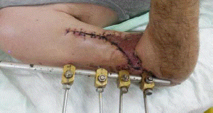
Figure 1
After skin grafting.
Figure 2
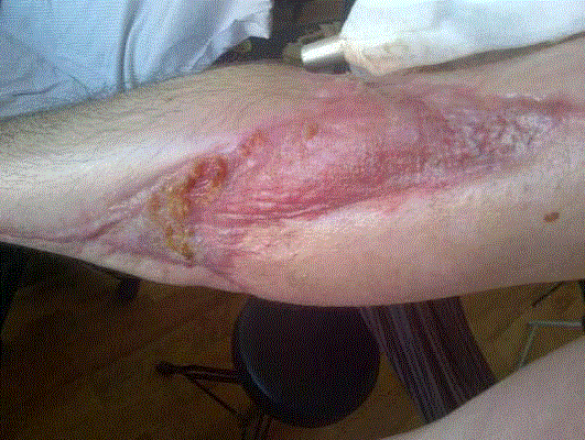
Figure 2
At 3 month post surgery.
Materials and Methods
A 28-year-old victim of a road accident (car occupant) presented himself in our service for the wound, sutured, superinfected, internal face of the right forearm face by transfer from a regional hospital. From the anamnestic-clinical data, we note that the patient is right handed, and from the accompanying medical documents it appears that on the right humerus osteosynthesis has been practiced with an external fixator for open type IIIC humeral fracture, fasciotomy and radial nerve neurorraphy. At 4 days postoperatively, a yellowish purulent secretion appeared, for which antibiotic treatment was established according to the antibiogram with Linezolid 2 mg/day and the suture material was partially removed to favor drainage. After the admission to our clinic we have instituted antibiotic treatment adapted to the antibiogram. Over the next 20 days, a series of surgical interventions is being proposed and performed forrehabilitation of the infectious outbreak and repair the t of the resulting substance. Since the patient was initially operated, urgently, in the general surgery room, we can assume that it is a nosocomial infection. This assumption was based on the duration of onset of clinical signs and on the isolation of Enterococus spp from wound secretions, the bacterium commonly found in the lower limb region, but atypical for soft portions of the upper floor, where the order of frequency of occurrence is isolated: With S Aureus, Pseudomonas aeruginosa, beta-hemolytic streptococci. During 20 days of admission, a series of surgical interventions are being proposed and carried out, aiming at rehabilitation of the infectious outbreak and repairing the defect of the resulting substance. In the first intervention, the suture is removed from the skin, is practiced large excision of the septic transformed tissue, abundant lavage with antibiotic ampicillin solution 1 g/500 ml of physiological serum, according to the antibiogram, haemostasis, the integrity of the radial nerve coaptation was checked, miorraphy, bandage, immobilization with a 45º antebrahio brachial splinter with the hand in supination for tensionfree suture of the muscle ends. The recommended position for the relaxation of the anterior brachial compartment is with the elbow at 90º flexion with supine hand and progressive extension of the extension angle in order to avoid muscle retraction. Our choice allows to avoid laborious reconstruction with autologous tissues in the vicinity or freely transferred by microsurgical techniques in several operating times, but assuming the risk of achieving a higher degree of joint stiffnes at the elbow and the inability of complete recovery. Following the established drug treatment, under the double antibiotic therapy with Linezolid be 2 mg/ml × 2/day 7 days, to which Ampicillin is added, po. 500 mg × 4/day, 14 days, NSAID, IPP, Eubiotic, and supportive Freshubin drink 2 fl/day, resulted a decrease in the dynamics of procalcitonin, fibrinogen (from 623 mg/dl, at 480 after 5 days of treatment), WBC normalization on day 7, remission of inflammatory signs, absence of purulent secretions with negative cultures for Enterococus spp at the end of treatment and appearance of granulation tissue. It is decided the reintervention by completing both the surgical removal of devitalized tissues and the miorraphy in the anatomical planes with U type suture and immobilization in flexion at the same angle of the forearm on the arm to relax the anterior brachial muscle compartment. In the case of soft tissue infections with Enterococus spp, the National Antibiotic Protocol propose antibiotic treatment with trimethoprim-sulfamethoxazole 2 cp × 2/day in combination with rifampicin 300 mg × 2/day. However, the above mentioned treatment was preferred because the isolated culture is resistant to rifampicin and trimethoprim, but is sensitive to Amoxicillin/ Clavulanate, Ampicillin, Penicillin, Linezolid, Erythromycin. Over the next 9 days after the last intervention, hemoglobin is corrected from 8.7 mg/dL to 10.5 mg/dL by administration of MER 2 U in order to cover the remaining skin defect with the PPLD skin graft harvested by on the anterior face of the ipsilateral thigh. The intervention is performed under general anesthesia, the postoperative antebrahiobrachial immobilization angle being increased to 60º. It has been chosen for a medium-thickness PLD on the arm portion due to the uncertainty-quality receptor bed (possible bacterial contamination) and the thick PLD at the elbow level to preserve the mobility of the receptor bed under the overlying tissues. In order to counteract the retraction at the level of the elbow, the boundary between the graft and healthy skin was shaped as a frangible line. After integrating the graft, immobilization on the gypsum plaster for another 6 weeks with the elbow and the antebrahiopalmara nocturnal throat retainer remains on the neutral position, the period of time sufficient for controlled muscular scarring to withstand the stresses exerted during day-to-day efforts. At the same time, the recovery of the joints of the hand and the fist is started by passive extension movements and flexion actives to avoid the installation of the stiffness of joint. After the elbow joint is immobilized, the recovery process with serial weights begins at this level for the progressive increase of the extension angle. Due to the heterogeneity of the types of production mechanisms and the resulting muscle damage, it was not possible to standardize an efficient, unanimously accepted treatment. After obtaining a degree of extension of 150º, it was practiced under the AGIV to remove the external humerus external fixator (one bar and 4 chips), and to reduce fracture under Rx-TV and osteosynthesis with centro-medullary rod, diameter 8 mm, length 60 mm with 3 bolts, lavage, suture in anatomical planes, sterile bandage. Evolution of the patient was favorable, being monitored at 3, 6, 9, 12, 18, 24 months in terms of mobility of the upper limb joints and the evolution of radial nerve neurotrophication.
Figure 3
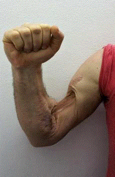
Figure 3
Final aspect.
Figure 4
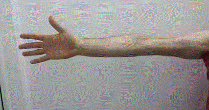
Figure 4
Final result.
Figure 5
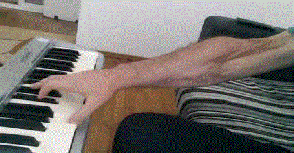
Figure 5
Patient returns to his jobs and hobbies
Figure 6
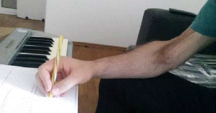
Figure 6
Appearance of wound after the skin grafting (post- operative day
13). All authors declare that they have no conflict of interests.
Results
The therapeutic attitude, followed by a rigorous recovery process, and periodic monitoring of patient evolution led to a functional result appreciated by the visual analogue scale with 9 out of 10 points and a satisfying aesthetic result with the possibility of improvement. At the present time (about 30 months) we can see the integrity of the skin, slight hyperpigmentation in the grafted skin, mobile bands without affecting the mobility of the elbow joint, the complete recovery of the flexural mobility and the extensions of the brahio-antebrahio-digital passive and the sensitivity in normal limits, measured by the Webber test, distal to the site of the lesion affected in the case of humerus fractures. Although, rarely, there is the possibility of its damage by fitting external fixators, often requiring subsequent microsurgical reconstruction. In the case of humeral fractures, especially of the transversal and spiroids of the medial-distal third of the arm, the radial nerve can be trapped in the healing calus, causing an entrapment syndrome. Covering substance flaws at the brachial level is a challenge to plastic surgery. If it were not possible to carry out the direct, tension-free sutures of the biceps' own head muscles, we would have considered the use of pediculous or freely transferred flaps. After vascular integration of the graft, the main concern for recovery of the arm function was to prevent the appearance of scarring bands and adhesions. Depending on the patient's option, for the improvement of the aesthetic appearance, serial scar excisions or tissue expansion can be performed on the external brachial face or on the proximal third of the forearm with the continuation of the medical recovery to increase the mass and muscle strength in the affected upper limb.
Conclusion
The complete rupture of muscular brachial biceps is a rare pathology. In our clinic, a reference center for severe cases of trauma in Romania, registering a number of 4 cases in the last 5 years, all of which are closed trauma, except the one presented. This pathology correlates with a long-term unsatisfactory prognosis, especially if it is associated with soft-tissue infections or radial vascular-nerve or medial nerve damage in the middle or distal third of the arm. Soft skin infections are a common complication when complex trauma occurs, especially if associated with open fractures. The likelihood of the infection spreading in depth depends on the type of trauma, localization, soft tissue thickness, patient co-morbidity, and treatment prior to immobilization. The radial nerve is the most commonly injured nerve in the arm. Due to its placement in the proximity of the bone structure it is frequently.
References
- Ferreira N, Marais LC. Prevention and management of external fixator pin track sepsis. Strategies in Trauma and Limb Reconstr. 2012;7(2):67-72.
- Bibbo C, Brueggeman J. Prevention and management of complications arising from external fixation pin sites. J Foot Ankle Surg. 2010;49(1):87-92.
- Sims M, Saleh M. External fixation - The incidence of pin site infection: A prospective audit. J Ortho Nursing. 2000;4(2):59-63.
- Kazmers NH, Fragomen AT, Rozbruch SR. Prevention of pin site infection in external fixation: a review of the literature. Strategies Trauma Limb Reconstr. 2016;11(2):75-85.
- Bumbasirevic M, Palibrk T, Lesic A. Radial nerve palsy. EFORT Open Rev. 2017;1(8):286-94.
- Chen HW, Chew FS. Complete rupture of both heads of the biceps brachii muscle belly by blunt trauma. Radiology case reports. 2006;1(4):145-8.
- Ki V, Rotstein C. Bacterial skin and soft tissue infections in adults: a review of their epidemiology, pathogenesis, diagnosis, treatment and site of care. Can J Infect Dis Med Microbiol. 2008;19(2):173-84.
- Olawale KO, Fadiora SO, Taiwo SS. Prevalence of hospital acquired enterococci infections in two primary-care hospitals in Osogbo, Southwestern Nigeria. Afr J Infect Dis. 2011;5(2).
- Lascar I. Principii de chirurgie plastica si microchirurgie reconstructiva. Editura National. 2005.
- Ioan Lascar, Sebe IT. Neuropatia compresiva a nervului ulnar la nivelul cotului. editura Printech. 2010;65-83.
- Carstea AI, Sebe IT, Lascar I, Cortan S, Diaconescu BI, Ferariu N, et al. Early rehabilitation in severe forearm injury.
- Joel A. Delisa. Phisical Medicine and Rehabilitation. Editura Lippincot. 2005;63(2).