Clinical Image
Stranger Things: Clinical and Radiological Diagnosis Confirmed by Surgery
Altomare M1* and Dovidio G2
1Department of General Surgery, University of Milan, Italy
2Department of Radiology, Sapienza University of Rome, Rome, Italy
*Corresponding author: Michele Altomare, Department in General Surgery, University of Milan, Milan, Italy
Published: 22 Jan 2018
Cite this article as: Altomare M, Dovidio G. Stranger
Things: Clinical and Radiological
Diagnosis Confirmed by Surgery. Clin
Surg. 2018; 3: 1876.
Clinical Image
A 42-year-old woman transferred from Emergency Room (ER) to General Surgery Unit for acute abdominal pain associated with mild distension of the abdomen, nausea and 2 episodes of vomit. No previous history of ER entrances or abdominal/gynaecological surgery. Laboratory data show a mild impairment of inflammatory parameters (WBC: 11,4 x 10³/ml CRP: 7,89 mg/dl). Abdominal X-Ray revealed a generalized distension of small and large bowel without free air (Figure 1). Considering the stability of vital parameters, patients undergone to CT-scan that demonstrated a centroabdominal region with stratify imagine consisting of mesenteric fat surrounding from bowel’s loop. The remaining colon downstream seems to be collapse and is recognizable a roundish shape mass having the densitometric parameters of lipoma in the contest of splenic flexure (Figure 1). Due to the deterioration of clinical condition the patient has been subjected a surgical exploration that confirmed the presence of intussusception arising from the last loop of ileum until splenic flexure caused by voluminous intestinal lipoma. Right hemicolectomy was performed and the patient had an uneventful post-operative course and was discharged on day 7.
Discussion
Despite representing a common cause of bowel obstruction in the pediatric population, intussusception account for only 1% of all bowel obstructions in the adults [1]. Interestingly the prevalence of enteric intussusception is grater than colonic intussusception (78% vs. 22%) [2], but cases of ileocolic intussusception caused by enteral lipomatosis almost remain anecdotal in literature. Considering the rarity of this clinical feature, a multidisciplinary team is essential to achieve the correct diagnosis and adopt the best management.
Figure 1
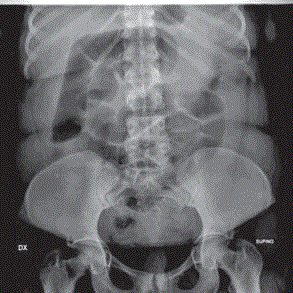
Figure 1
Wound appearance at the time of diagnosis.
Figure 2
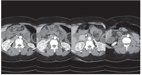
Figure 2
Densitometric parameters of lipoma in the contest of splenic flexure.