Case Report
A Case of Severe Herniation Following a Posterior Component Separation
Danni Lu*, Andrew Bates and Aurora Pryor
Department of Gastroenterological Surgery, Stony Brook University School of Medicine, USA
*Corresponding author: Danni Lu, Department of Gastroenterological Surgery, Stony Brook University School of Medicine, USA
Published: 22 Jan, 2018
Cite this article as: Lu D, Bates A, Pryor A. A Case of
Severe Herniation Following a Posterior
Component Separation. Clin Surg.
2018; 3: 1875.
Abstract
The component separation technique can be used for the management of large or complex midline abdominal wall defects resulting from infection, trauma, or complications of multiple abdominal surgeries. This technique was first described in 1990 as a surgical procedure that would release tension from the circumference of the abdominal wall while restoring function and stability of the abdominal wall muscles. The general technique involves the separation of abdominal muscle layers followed by disconnection of the muscle unit from the fascial sheath envelope and bilateral expansion, allowing for equilibration of pulling forces and centralization of the midline. A supplemental mesh may be used to reinforce the repair, and may help to reduce short-term recurrence rates. Currently, there are two approaches that are used - anterior and posterior - involving the isolation and division of the external abdominal oblique or transversus abdominis muscles, respectively. While the component separation technique yields promising results in restoring abdominal support following large wall defects, further study is required to adequately understand the risks of this procedure. This case report study presents an acute case of severe herniation following the failure of the abdominal wall following a posterior component separation.
Case Presentation
A 62-year old female presented with a history of previous open sigmoid colectomy with end colostomy for perforated diverticulitis. Her colostomy was later reversed, which was complicated by anastomotic leak necessitating a diverting loop ileostomy, which was also later reversed. She presented as an outpatient with a 10cm x 10 cm symptomatic midline incisional hernia. She was a chronic smoker with a 30 pack year history. Treatment options were discussed with the patient for repair of her incisional hernia, and a bilateral component separation was elected. The patient was taken to the operating room. The midline incision was opened and all intra abdominal adhesions to the abdominal wall were taken down (Figure 1). The rectus sheath was incised bilaterally at the medial aspect and the rectus abdominis muscles were elevated off the posterior fascia. As per the operating surgeon’s report, a posterior component separation was performed bilaterally from within the abdominal cavity. The posterior fascia was reapproximated at the midline and a 20 cm x 30 cm biologic mesh was placed in the retrorectus space and secured with multiple prolene sutures. Two months later, the patient presented to a different surgeon with the complaint of a constant “pulling” sensation bilaterally, with mid-abdominal fullness and bilateral swelling that never resolved since the surgery. She also detailed a constant, mild abdominal pain. The patient denied nausea, vomiting, heartburn, dysphagia, or changes in bowel movements. The abdomens were soft upon palpation and non-tender. The midline was also well healed. A CT scan was obtained, which revealed bilateral stable “spigelian” hernias, as well as a midline hernia that had recurred suprapubically. Upon further review, the image showed an absent abdominal fascia at the semilunar lines bilaterally, indicating that the fascia had been completely incised. Given her history of smoking and the combination of suprapubic and bilateral hernia, it was elected not to operate due to complications and a higher baseline recurrence rate. The patient was directed to pursue means of smoking cessation and will return to the office once she has completely discontinued nicotine use.
Figure 1
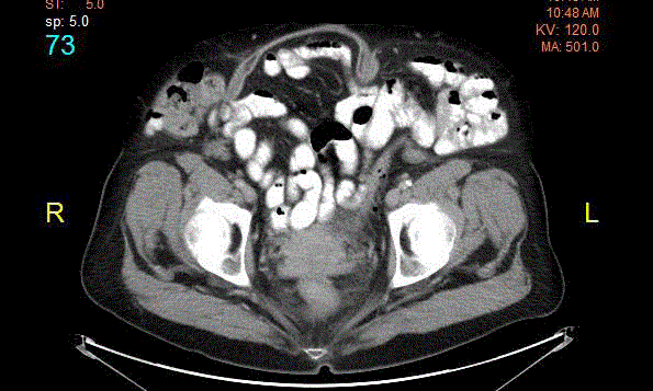
Figure 1
CT scan of the abdomen revealed persistent bilateral Spigelian hernias at the level of the semilunar
lines, in combination with a recurrent suprapubic hernia.
Discussion
While mesh repair is often the ideal treatment for small- or medium-sized midline incisional hernias, there is no standard procedure recommended for larger-sized abdominal defects. The component separation technique is a relatively new approach in treating these larger event rations, and confers excellent reinforcement of abdominal wall stability with restoration of abdominal wall function. The general procedure involves separating the abdominal wall muscle layers, disconnecting the oblique abdominal muscle unit from its fascial sheath, and creating maximal expansion of each muscle, in order to reduce tension and mitigate the chances of fascial strangulation and re-herniation in the reconstruction of larger abdominal wall defects [1]. There are two fundamental approaches to the component separation procedure. In the anterior approach, the superficial skin and subcutaneous tissues are elevated from the rectus and external oblique fascia. This separation is brought from the costal margin down to the pubis medially, extending to the anterior axillary line down to the iliac crest laterally. The external oblique aponeurosis is incised from the semilunar line down to the inguinal ligament, at which point the external oblique muscle is freed from the internal oblique muscle. Additionally, the rectus muscle may be dissected away from its posterior sheath, allowing for further release. Overall, this can create up to 20 cm of advancement at the mid-abdominal region [2]. However, this approach creates large cutaneous flaps that may lead to surgical site infections or skin necrosis. The posterior component separation procedure was an answer to this technical difficulty. Following removal of adhesions, the posterior rectus sheath is incised just lateral to the medial edge of the rectus muscle. The retrorectus plane is dissected until the neurovascular bundle, at which the transversus abdominis muscle is then exposed and divided along the entire length of the wall. Once dissected, the transversus abdominis muscle is then bluntly dissected away from the transversalis fascia, extending laterally towards the retroperitoneum. This procedure may be performed laparoscopically to avoid creating large cutaneous flaps and thereby reducing the rates of surgical site infection [3]. This technique presents with its own challenges, as accidental destruction of the superior or deep inferior epigastric artery would compromise the main sources of blood supply to the rectus muscles. Common complications seen in both approaches include the development of surgical site infections, seromas (which may require drainage) and recurring hernias. Morbidity and hernia recurrence rates have widely varied. Currently, the component separation procedure remains one of the most successful procedures to treat large, midline abdominal hernias. Many social media platforms have made new information widely and rapidly accessible between health care professionals within physician-specific online networks, offering the advantages of professional collaboration and education between colleagues. Information on new procedures and techniques can be rapidly disseminated, and help to improve patient care on a wide-scale basis [4]. Due to its lack of regulation, however, care must be taken to avoid the dangers of the circulation of poorquality information or sensationalized cases with the intention of brandishing one’s image. Given the recentness of the component separation technique, there exists only a small amount of data documenting the rates of morbidities and mortalities over more than a decade. Therefore, caution must be taken when electing to perform this procedure, and appreciating the learning curve is important in reducing the risks associated with this sophisticated technique.
References
- Ramirez OM. Inception and evolution of the components separation technique: personal recollections. Clin Plast Surg. 2006;33(2):241-6.
- Heller L, McNichols CH, Ramirez OM. Component separations. Semin Plast Surg. 2012;26(1):25-8.
- Novitsky YW, Elliott HL, Orenstein SB, Rosen MJ. Transversus abdominis muscle release: a novel approach to posterior component separation during complex abdominal wall reconstruction. Am J Surg. 2012;204(5):709-16.
- Househ M. The use of social media in healthcare: organizational, clinical, and patient perspectives. Stud Health Technol Inform. 2013;183:244-8.