Case Report
Pathogenesis of Hypogastric Hernia
Buddhike SH Indrasena*
Department of Surgery, Kandy General Hospital, Sri Lanka
*Corresponding author: Buddhike SH Indrasena, Department of Surgery, Kandy General Hospital, Kandy, Sri Lanka
Published: 22 Jan, 2018
Cite this article as: Indrasena BSH. Pathogenesis of
Hypogastric Hernia. Clin Surg. 2018;
3: 1872.
Abstract
This is a case series of hypogastric hernia. It is common in females. Obesity and multiparity are
possible predisposing factors. It occurs consistently at the level of the arcuate line through the linea
alba. The differential pull of the anterior and posterior fibres of the internal oblique and transversus
abdominis muscles located anterior and posterior to the rectus abdominis muscle is probably the
cause of this kind of hernias.
Keywords: Hypogastric hernia; Midline hernias; Rare hernias
Introduction
The sporadic hernias appearing through the linea alba below the umbilicus could be named as hypogastric hernia as opposed to epigastric hernias that are located above the umbilicus. Hypogastric hernias occur in the hypogastric region of the abdomen. Unlike epigastric hernias that can occur anywhere along a line from xiphysternum to umbilicus, the site of the hypogastric hernias is fairly constant appearing always at the level of the arcuate line in the linea alba. There is limited information about this type of hernias in the literature.
Case Presentation
Given below is the series of three cases of hypogastric hernia that the author had encountered
in his practice as a consultant surgeon, in two tertiary care hospitals in his country, spanning over a
period of five years from 2011 to 2016.
Case 1
A 71-year-old woman with no history of previous surgery presented as an emergency with an
increasingly painful lump in the lower abdomen for seven days. The lump had been growing steadily
for five years without associated symptoms. Physical examination revealed a moderately large (5 cm
x 5 cm), firm, mildly tender lump in the midline between the symphysis pubis and umbilicus (Figure
1). Although it was irreducible when first examined at the time of admission, when examined next
day, while the patient was on analgesics, the lump suddenly disappeared into the abdomen during
palpation and reappeared when the patient was asked to cough. The patient was operated during the
same admission in the next routine list. At surgery two hernial defects could be found in the linea
alba separated by a thing band of fibrous tissue at the level of the arcuate line (Figure 2). The hernia
sac was excised and the defect was closed anatomically and reinforced with an onlay polypropylene
mesh. Patient recovered without complications.
Case 2
A 56-year old obese lady underwent laparotomy for sigmoid colonic cancer. On opening into the abdomen a fold of omentum was found to be herniating through
a defect in the linea alba at the level of the arcuate line (Figure 3).
There was no history of previous abdominal surgeries. The omentum
was carefully separated from the hernial sac. The defect was closed
by incorporating it to the mass closure during laparotomy incision
closure.
Case 3
The obstetrician referred a 39-year old obese female with a
painful hernia diagnosed by the ultrasound and located in the midline
midway between the umbilicus and symphysis pubis. The lady had
one child delivered by caesarean section one year ago (Figure 4-6).
She was hypothyroid due to thyroiditis, and this was an incidental
finding during preoperative work up. On exploration of the lump it
was found that the hernia emerged through a defect in the linea alba
at the level of the arcuate line, quite distinct from the Pfannenstial
incision. The defect was closed anatomically after dealing with the sac,
and the repair was reinforced with onlay polypropylene mesh.
Figure 1
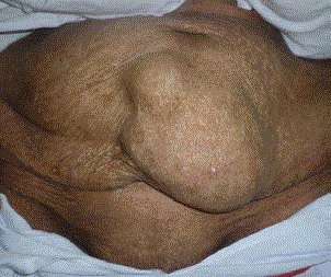
Figure 1
Case 1: Hypogastric hernias present as a midline lump in between the umbilicus and symphysis pubis.
Figure 2
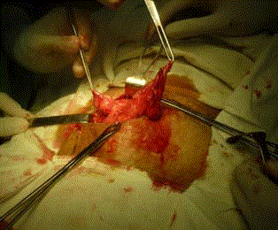
Figure 2
The defect and the hernial sac of the case 1 as seen during surgery.
Figure 3
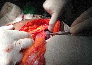
Figure 3
Case 2: The band of omentum (held between the arms of forceps)
could be seen herniating through the defect in the linea alba.
Figure 4
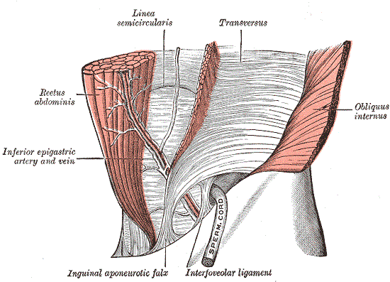
Figure 4
Posterior wall of the rectus sheath (Source: Henry Gray (1918)
Anatomy of the Human Body).
Figure 5
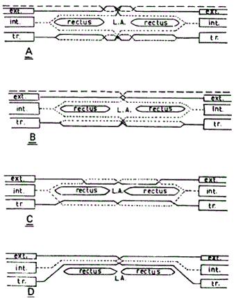
Figure 5
Arrangement of the aponeurotic fibres of the three anterior
abdominal wall muscles in the formation of the rectus sheath.
A: In 70% of cases between the costal margin and the level of umbilicus
B: In 30% of cases between the costal margin and the level of umbilicus
C: Between the level of umbilicus and iliac crest
D: Between the level of iliac crest and symphysis pubis
Figure 6
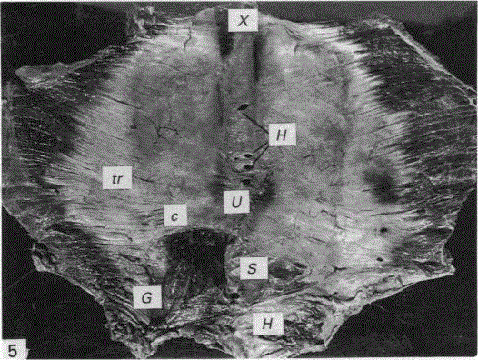
Figure 6
Arcuate line of Douglas (C). The specimen also shows many
hernial orifices (H) located above and below the umbilicus (U). tr-tTrans vs.
abdominis muscle, S- posterior wall of rectus sheath; G- inferior epigastric
artery.
Discussion
Midline abdominal hernias are common. These include epigastric (located in the epigastric area above the umbilicus), umbilical and para-umbilical (located in or close proximity to the umbilicus) and incisional (located anywhere in the abdomen through a previous laparotomy incision) hernias. A midline hernia below the umbilicus, unrelated to a surgical incision is a rare variety of hernias. The term ‘hypogastric hernia’ is more appropriate because it seems to mirror the epigastric hernia on either side of the umbilicus. The defect in this kind of hernia lies in the linea alba. This is different from Spigelian hernia where the hernia protrudes through a defect in the transversalis muscle lateral to the linea semilunaris. Hypogastric hernias differ significantly from epigastric hernias with respect to the prevalence. Epigastric hernia is the third commonest abdominal wall hernia after inguinal and umbilical hernias [1]. Hypogastric hernias are rare as evident by seeing only three hernias over a period of five years.
Pathogenesis
Epigastric hernias are thought to be due to the differential pull of the diaphragmatic fibres over the fibres of linea alba composed of external oblique, internal oblique and transversus abdominis [2,3]. A similar explanation can be given to hypogastric hernia noticing that it occurs at the level of the Arcuate Line of Douglas (AL), also known as linea semicircularis. The anatomy of the AL has been extensively studied by N.N Rizk [4,5]. According to Rizk, the arrangement of the fibres of the rectus sheath in each of the anterior and posterior layers is quite complex, each consisting of three layers contributed by a layer of each of bilaminar external oblique, internal oblique and transversus abdominis aponeurosis. The linea alba represents the line of decussation of those fibres. The transition of Figure 5 is usually not abrupt but gradual thinning out of the posterior layer due to the gradual transposition of the fibres of the posterior wall of the rectus sheath over the rectus abdominis muscle. As a result, the AL is not always clearly discernible [6,7]. Rizk had observed abrupt transition from Figure 5, forming a well demarcated AL, only in 1 out of 40 cases. Probably this is an uncommon pathological variation as noted by Rizk by the presence of multiple hernial orifices and fibrosis along the linea alba on that specimen. The pathogenesis of hypogastric hernia could be due to the pulling effect of the tendinous fibres of the internal oblique and transversus abdominis muscles through the posterior layer of the rectus sheath over the combined tendinous intersections of the fibres of the linea alba. Superior to the arcuate line, the direction of the pull of the fibres of the anterior and posterior layer of the rectus sheath in two directions angulated slightly anteriorly and posteriorly, whereas, below the arcuate line all the fibres of all three layers are in the one plane and the pull is stronger. As a result, the tendinous fibres of the linea alba at the level of the arcuate line is at risk of separation due to the differential pulling effect. The fact that the hypogastric hernias are not so common is also worth exploring. The separation of the fibres of the linea alba can occur only when the arcuate line is well demarcated as seen on the three cases described above. In the usual case of gradual thinning of the posterior wall of the rectus sheath the linea alba will not be subjected to such abrupt directional changes of pull. The well demarcated AL is not a common phenomenon. The factors like obesity, old age and pregnancy tend to make the linea alba broader and weaker making it more vulnerable to develop larger defects and hernia.
Conclusion
As with other hernias, hypogastric hernias can go into incarceration and strangulation. Therefore, surgery has to be recommended. As with other types of repairs of midline hernias both tissue repair and mesh repair techniques are viable options.
References
- Dabbas N, Adams K, Pearson K, Royle G. Frequency of abdominal wall hernias: is classical teaching out of date? JRSM Short Rep. 2011;2(1):5.
- Ponten JE, Somers KY, Nienhuijs SW. Pathogenesis of the epigastric hernia. Hernia. 2012;16(6):627-33.
- Moschcowitz AV. Epigastric hernia without palpable swelling. Ann Surg. 1917;66(3):300-7.
- Rizk NN. A new description of the anterior abdominal wall in man and mammals. J Anat. 1980;131(3):373-85.
- Rizk NN. The arcuate line of the rectus sheath--does it exist? J Anat. 1991;175:1-6.
- Loukas M, Myers C, Shah R, Tubbs RS, Wartmann C, Apaydin N, et al. Arcuate line of the rectus sheath: clinical approach. Anat Sci Int. 2008;83(3):140-4.
- Mwachaka PM, Saidi HS, Odula PO, Awori KO, Kaisha WO. Locating the arcuate line of Douglas: is it of surgical relevance? Clin Anat. 2010;23(1):84-6.