Clinical Video
Thoracic Pleomorphic Liposarcoma: Resection and Reconstruction>
Aaron M Kime*, Robbin G Cohen and P Michael McFadden
Department of Surgery, University of Southern California, USA
*Corresponding author: Aaron M Kime, Department of Surgery, University of Southern California, 1520 San Pablo St., Suite 4300, Los Angeles, CA 90033, USA
Published: 03 Aug, 2017
Cite this article as: Kime AM, Cohen RG, McFadden PM.
Thoracic Pleomorphic Liposarcoma:
Resection and Reconstruction. Clin
Surg. 2017; 2: 1783.
Clinical Images
A 21-year-old woman presented with abdominal pain and weight loss and was found to have a left thoracic mass with displacement of the heart, lung, and esophagus. Biopsy demonstrated pleomorphic liposarcoma and neoadjuvant chemotherapy was initiated, however, due to refractory supra ventricular arrhythmias and respiratory failure she was taken for resection (Figure 1). Through a left thoracoabdominal incision, en bloc tumor resection, including adherent Lung (L), esophagus, diaphragm, and pericardium, was accomplished. The left phrenic nerve was preserved on an uninvolved Pedicle (PNP). Diaphragm was reconstructed with Gore-Tex (G) and the pericardium replaced with bovine pericardium (BP) (Figure 2 and Video 1). An R0 tumor resection was accomplished. Pathology demonstrated a 16 cm × 13 cm × 11 cm pleomorphic liposarcoma. The patient was slow to wean from ventilator support and required tracheostomy. Her chemotherapy was reinstituted postoperatively. Pleomorphic liposarcomas are highly aggressive, lethal tumors, which are best treated with multimodal therapy that includes complete surgical resection and chemotherapy [1].
Figure 1
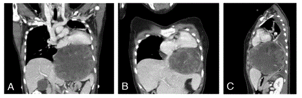
Figure 1
CT images of thoracic pleomorphic liposarcoma
Figure 2
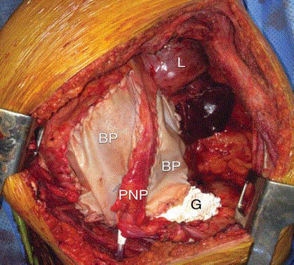
Figure 2
Peroneus brevis tendon tear prior to repair.