Clinical Image
Endolymphatic Hydrops Detected by HYDROPS Image of MRI
Kazuo Ishikawa*
Department of Otolaryngology Head and Neck Surgery, Akita University, Graduate School of Medicine, Japan
*Corresponding author: Kazuo Ishikawa, Department of Otolaryngology Head and Neck Surgery, Akita University, Graduate School of Medicine, 1-1-1, Hondo, Akita, 010-8543, Japan
Published: 30 Nov, 2017
Cite this article as: Ishikawa K. Endolymphatic Hydrops
Detected by HYDROPS Image of MRI.
Clin Surg. 2017; 2: 1782.
Clinical Image
Newly developed radiological image technique could enhance our understanding of various disease entity. Now, it has become possible to detect endolyphatic hydrops clearly by hybrid of reversed image of positive endolymph signal and negative image of positive perilymph signal (HYDROPS) of MRI. Until recently, audio-vestibular examinations such as ECoG, Glycerol Test and Lasix test should be performed to obtain indirect evidence of endolymphatic hydrops, and there had been no direct method to ascertain the endolymphatic hydrops except for histological procedure. Employing this imaging technique, those suspected causative factors of endolyphatic hydrops such as psychological stress, autoimmune reaction, allergic reaction, autonomic imbalance, viral infections, and dietary deficiencies and so on can be verified. In addition, the significance of those audio-vestibular examinations should also be confirmed in the future (Figure 1 and 2). Also, some relation between endolyphatic hydrops and inner ear diseases will also be elucidated.
Figure 1
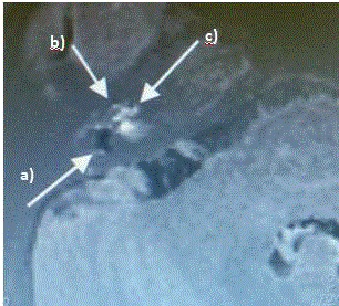
Figure 1
Dark portion indicates hydrops: balloon sacculus (a), and cochlear duct (b,c).
Figure 2
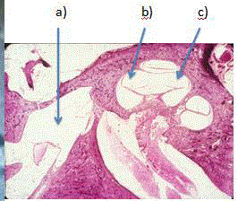
Figure 2
Histological findings of temporal bone in a case with Meniere’s disease.