Clinical Image
Bifid Ureter Obstructed with Single Ureteric Stone
Zor M*, Coguplugil AE and Bedir S
Department of Urology, Gulhane Research and Training Hospital, Turkey
*Corresponding author: Murat Zor, Department of Urology, Gulhane Research and Training Hospital, Ankara, Turkey
Published: 29 Nov, 2017
Cite this article as: Zor M, Coguplugil AE, Bedir S. Bifid
Ureter Obstructed with Single Ureteric
Stone. Clin Surg. 2017; 2: 1766.
Keywords
Bifid ureter; Ureteral stone; Hydronephrosis
Clinical Image
A 33 year-old female patient admitted to our emergency department with left flank pain. Physical examination showed costovertebral angle tenderness. Her blood analysis was normal but urinalysis revealed hematuria and leukocyturia. Ultrasonography pointed out left ureterohydronephrosis and suspicious urine extravasation. In the light of this information abdominal tomography was performed. Non-contrast series revealed 12 mm left ureteric stone (Figure 1A) and left hydronephrosis. But on contrast enhanced coronal images it was clear that the patient had bifid ureter and they were joining together distally to form one ureteric branch at the level of iliac crossing (Figure 1B). The ureteric stone was located immediately distal to the orifice of the bifid ureter so obstructed both ureteric units (Figure 1B). Ureteroscopy and laser lithotripsy was performed under general anesthesia. After stone removal initially double guide wires were placed to each ureter (Figure 2A) and double J stents were placed in both ureteric units (Figure 2B). Plain abdominal graphy was performed to confirm the correct placement of the stents (Figure 3). The patient was discharged on postoperative first day without any complication. The ureteric stents were removed on the second week of operation. Although renal collecting system duplication is one of the most common congenital abnormality of the collecting system [1], our case is a unique example of a ureteric stone obstructing both ureteric units. It also demonstrates the usefulness of contrastenhanced coronal imaging in diagnostic work-up when necessary.
Figure 1
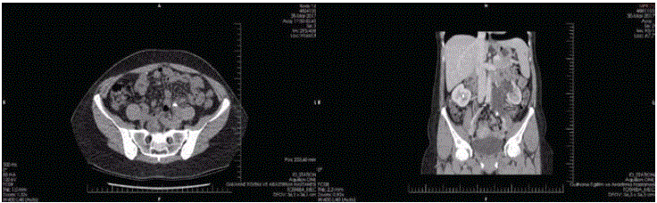
Figure 1
A: A 12 mm ureteric stone (black arrow) on non-contrast axial images of computed tomography; B:
Bifid ureter joining together (left and right black arrows) to form one ureteric branch at the level of iliac crossing
and a 12 mm stone obstructing two ureteric segments.
Figure 2
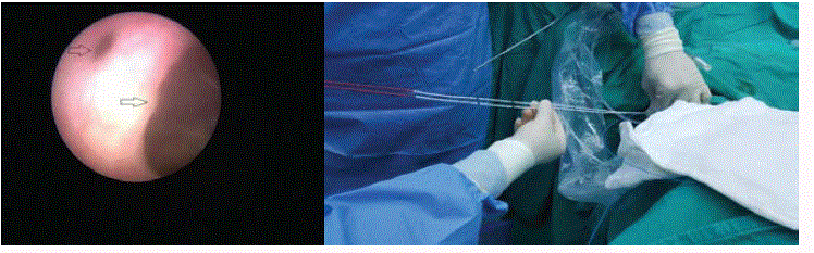
Figure 2
A: Ureteroscopic vision of both ureteric segments (left and right black arrows); B: Double J stents were
introduced with the guidance of previously placed guidewires.
Figure3
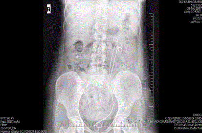
Figure 3
Plain abdominal graphy confirming the correct position of the stents.