Review Article
Plantar Fasciitis and Current Treatment Approaches
Ayse Abit Kocaman, Sulenur Yildiz and Nilgün Bek*
Department of Physiotherapy and Rehabilitation, Hacettepe University, Turkey
*Corresponding author: Nilgün Bek, Department of Physiotherapy and Rehabilitation, Hacettepe University, 06100 Samanpazari, Ankara, Turkey
Published: 22 Nov 2017
Cite this article as: Kocaman AA, Yildiz S, Bek N. Plantar
Fasciitis and Current Treatment
Approaches. Clin Surg. 2017; 2: 1752.
Abstract
Plantar fasciitis is the most common pathology of the musculoskeletal system, especially affecting
the plantar fascia. Invasive and non-invasive treatment approaches have been shown to be effective
when treatment mechanisms are considered biomechanically. In addition, non-invasive treatment
applications, such as applications for restoration of muscle, bony and articular structures, correct
alignment, enhancement of vascularization, and improvement of proprioceptive parameters, have
been recorded in the literature. In this review study, plantar fasciitis formation mechanisms, clinical
symptoms and evaluation, invasive and non-invasive treatment options, and physiotherapy and
rehabilitation applications in conservative treatment options are given together with the results of
the literature. In addition, the treatment protocol established with our clinical experiences is shared
with the readers.
Keywords: Foot; Plantar fasciitis; Treatment modalities; Rehabilitation
Foot and Plantar Fascia
Plantar aponeurosis is a connective tissue starting from the calcaneus, and composed of medial, central, and lateral segments. The medial and lateral segments surround the abductor digitiquinti and abductor hallucis muscles, and the central portion of the calcaneus, starting from the medial tubercle, is named as plantar fascia. Plantar fascia extends from the phalanges of the small fingers through the longitudinal septum and from the sesamoid bone to the thumb, and through the vertical fibers in the form of five bands passing along the arch of the foot [1]. Plantar fascia contributes to foot functions by providing static and dynamic shock absorption for the longitudinal arch [2]. The Windlass Mechanism is described by Hicks in 1954 as follows: plantar fascia pulls the distal end of the metatarsal head as it applies a continuous traction at the same time; as a result, the longitudinal arch ascends, the hind foot rotates, and the leg makes an external rotation as the hind foot makes inversion. This mechanism is completely dependent on bone and joint stability [3]. In summary, the elastic tension force stored during the stance phase provides the necessary force for the push phase with the contribution of the passive elastic structure of the plantar aponeurosis [4].
Plantar Fasciitis
Plantar Fasciitis (PF) is an inflammatory and painful condition occurring at the site where the medial part of the plantar fascia attaches to the calcaneus. This is one of the most common causes of plantar heel pain and is responsible for most of the foot pain, which is due to the biomechanical imbalance that occurs as a result of excessive strain in the fascia. Generally, the case may be accompanied by calcaneal epin, which may also be clinically asymptomatic. Plantar fasciitis-related heel pain occurs in a slowly increasing pattern, most notably in the morning when standing up or after long periods of inactivity [5].
Risk Factors
PF usually develops due to the coexistence of many etiologic factors. Identifying the risk factors
playing a role in the occurrence of plantar fasciitis is crucial for both the identification of etiology
and the successful management of preventable risk factors.
The most common risk factors:
• Restricted ankle dorsiflexion
• Increased foot pronation
• Body mass index over 30 kg/m²
• Carrying out activities such as weight lifting
• Long distance runners
• Patients with an arch problem (pes planus, pes cavus)
Among them, the most important risk factor has been reported
to be restricted ankle dorsiflexion. The PF risk rate has been cited
to be increase by the reduction of ankle flexion angle. It has been
shown that the probability of PF increases by 2.1 times in people
with < 10° angle flexion angle [6]. If there is a limitation in the ankle
dorsiflexion, advanced level pronation develops in the subtalar joint
as a compensation. This leads to an increase in the tension load in
the plantar fascia [7]. Another common cause is the biomechanical
dysfunction of the foot, as well as there may be infectious, neoplastic,
rheumatic, neurological, traumatic, and other systemic causes. The
tension occurring in gastrocnemius and soleus muscles and Achilles
tendon is among the important risk factors for PF because it affects
the foot biomechanics negatively. Aging and heel fat pad atrophy are
among other degenerative risk factors [7]. All of these risk factors lead
to recurrent intense-stress on the plantar fascia, and they sometimes
result in pain along with degenerative changes. It is also considered
that intrinsic muscle weakness may be inadequate to dynamically
support the medial longitudinal arch, increasing the tension on the
fascia, and thus, causing the formation of PF [8].
Figure 1
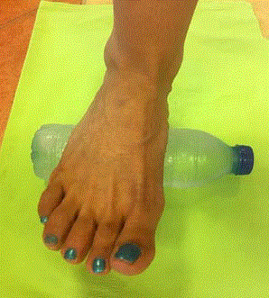
Figure 1
Iced bottle rolling exercise weight bearing position.
Table 1

Table 1
Hacettepe Protocol in the treatment of plantar fasciitis.
Clinical Symptoms
The main complaint is increasing pain that is felt throughout the heel. Pain in acute injury and avulsion-type lesions begins suddenly and intensely. Apart from the onset characteristics of pain, the clinical picture is similar in acute and chronic cases [9]. Pain is often felt at the origin of the plantar aponeurosis and distally about a centimeter of this area, and typically there exists severe pain during the first step when standing up in the morning, or at the first load after an extended rest. Pain continues to increase with increased load and continues to exacerbate progressively with daily activities [10]. Abnormal stretching of the plantar fascia caused by biomechanical factors does not constitute as a clinical symptom. However, risk factors such as increased standing pronation, high longitudinal arch (pes cavus), height inequality between extremes, body mass index over 30 kg/m², occupations requiring long standing and running (such as military personnel), sedentary life, the tension of the Achilles tendon and the intrinsic foot muscles, activity on the hard floor cause recurrent micro traumas on plantar fascia and other structures, leading to the appearance of clinical symptoms [5,9]. The stretching of the plantar fascia occurs during the gait terminal stance phase, when the heel is raised and the body weight is transferred to the front of the foot. The touch of the heel on the ground increases the traction force by 20%, and the force is even higher when running. Addition of the forces of the intrinsic muscles adhering to the medial tuberculosis increases the effect of traction on the medial calcaneal tuberculosis. Changes in activity (downhill and uphill walking, running) cause longer durations than normal walking. The tearing of the implant results in micro-tears and, consequently, inflammatory reaction and pain.
Evaluation
Diagnosis is usually clinically based on the characteristics of patients' pain complaints. Pain localization was defined in the middle of the heel (52%), in the medial tubercle region (42%) and in the lateral tubercle region (37%) [11]. Increased plantar heel pain score with weight-bearing, pain in the palpation of the proximal insertion of plantar fascia, positivity in the windlass test, negativity in the tarsal tunnel test, antalgic walking, limitation in active and/or passive talocrural joint dorsiflexion joint motion, and abnormal Foot Posture Index (FPI) score are important in diagnosis. In addition, diagnostic ultrasound is an evaluation method that has recently been used as effective as MRI and scintigraphy in the diagnosis of plantar fasciitis [7]. In the clinical evaluation of patients, pain intensity, as determined with the Visual Analogue Scale (VAS-FA) and the postural features of the foot, as determined by FPI, as well as the patient's narrative and imaging modalities can be used. Functional status is also assessed by customized valid and reliable scales such as the American Orthopedic Surgeons Foot and Foot Ankle Scale (AOFAS), the Foot and Foot Ankle Ability Measure (FAAM), the Foot Health Status Questionnaire (FHSQ), the Foot Function Index (FFI), and the Lower Extremity Functional Scale [12-14]|. PF has also been defined in accordance with the International Function Classification (ICF) system, which has been defined and used for various pathologies by health professionals in recent years. According to the ICF disorderhealth- disability-based category, heel pain is associated with bodily function-related codes b28015 Pain in lower limb, b2804 Radiating pain as a segment or region; s75023 Ligaments and fasciae of ankle and foot, s75028 Structures of ankle and foot, neural as body structure; d4500 Walking short distances, d4501 Walking long distances, and d4154 Maintaining a standing position in codes related to activity and participation [15].
Figure 2
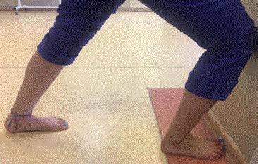
Figure 2
Gastrocnemius muscle stretching exercise.
Treatment Approaches
PF has a wide range of treatment modalities extending from
conservative approaches to surgery. While 90 percent of patients
are treated with conservative approaches, surgical approaches are
sometimes needed in chronic and persistent cases [5].
Conservative applications
Conservative treatment of plantar fasciitis involves non-steroidal
anti-inflammatory analgesics, local steroid injection, botulinum toxin
injection, and physiotherapy and rehabilitation (orthotic approaches,
stretching and strengthening exercises, electrophysical modalities,
taping, manual therapy and dry needling). Although it is not clear
which of the treatment options is more effective, these treatments
are often applied in combinations [5]. Local corticosteroid injection
is one of the first invasive treatment modalities where non-invasive
conservative treatment modalities are not effective. It has been
shown that this practice, which is often administered before surgical
approaches in patients with persistent pain, is faster and more
effective than other treatment options [16,17]. However, it is known
that corticosteroid injection has more side effects than Extracorporeal
Shock Wave Therapy, and it has been discussed whether the lowcost
corticosteroid injection or non-invasive ESWT should be used
first. In a previous study on the subject, it was emphasized that if the
patient has perifascial edema, first corticosteroid injection should be
used first, if not, ESWT should be used first [18]. Platelet Rich Plasma
(PRP) injection, which is considered to be among the minimally
invasive methods, have been reported to be effective in reducing
plantar fascia pain, but it does not have a positive effect on the
biomechanical function of the foot [19]. In another study comparing
PRP with Low Dose Radiation (LDR), after 6 months, pain (as
evaluated by VAS), AOFAS, and plantar fascia decreased significantly
and PRP was found to be at least as effective as LDR [20]. In another
study comparing the effect of PRP injection with corticosteroids in
chronic cases, it has been shown that PRP is as effective as steroids
in reducing pain at months 3 and 6, but PRP is more effective on
pain and function at month 12 [21]. Considering the biomechanical
basis of the development of pathology, the effects of restoration of
muscle, bony and articular structures as part of physiotherapy and
rehabilitation approaches, ensuring correct alignment, increasing
vascularization, and improving proprioceptive parameters are
absolute.
Karimzadeh et al. [22] compared the results of autologous
whole blood injection with local corticosteroid administration, and
it was found better than conservative treatment and it has nearly
similar outcomes with corticosteroid injection rates. Because of
improvements observed in the control group, researchers argued that
the first option should be non-invasive and conservative treatments
[22].
In a randomized controlled trial evaluating the efficacy of
botulinum toxin, another treatment agent that has been in use
recently, 50 patients were divided into two groups and one group
received botulinum toxin and the other group received saline
injection. Patients treated with botulinum toxin have been reported
to have better results in pain (as measured by VAS) and foot function
(as measured by FAAM), suggesting that this agent may be an
alternative to non-surgical methods [23]. Another conservative
practice is acupuncture. In a systematic review evaluating the effect
of acupuncture on PF-induced pain, few studies have found evidence
of short-term pain relief, while evidence of long-term responses is
inadequate. More work on the subject is needed [24].
Physiotherapy applications
Physiotherapy applications constitute the key part of conservativ treatments in PF. In particular, acute symptomatic patients experience
a symptomatic relief by these non-surgical methods in a short time.
Although there are different and numerous treatment modalities
administered, evidence is lacking which treatment method is most
effective and there is a need for studies in this regard.
Stretching exercises: It is the most important part of the treatment
program in the early period. Progressive stretching exercises of plantar
fascia and gastro-soleus muscle groups were shown to reduce pain
[25]. In an 8-week study, Healey et al. [26] found 52% improvement
in the group exercised only plantar fascia stretch, while there was
22% improvement in the group exercised only gastro-soleus muscle
stretching. Another study showed the importance of stretching
plantar fascia, and also that gastro-soleus muscle group and plantar
fascia stretching improved clinical symptoms, besides plantar fascia
stretching was more beneficial than Achilles tendon stretching [27].
There is no consensus on the optimal stretching method, although
studies have indicated that the effect of stretching on pain relief is
between 2 weeks and 4 months [28].
Strengthening exercises: The local stabilizing intrinsic foot
muscles, which contribute to the control of the foot arches, and the
extrinsic foot muscles responsible for the global motion, support the
foot arches as parts of the foot core structure and actively contribute
against the shocks during walking.
The core structure of the foot resembles the lumbopelvic core
system, and as a consequence of the weakness of these muscles along
with an injury in any of the foot core components, the stress on
the other structures increases and lower extremity injuries become
inevitable [29]. Studies investigating the possible effects of intrinsic
muscles on plantar fasciitis pathology are increasing. In a study
comparing runners with plantar fasciitis to healthy runners, it was
reported that symptoms in PF cases were associated with intrinsic
muscle atrophy [30]. In addition to weakening in plantar and finger
flexors and abductor hallucis resulting from PF, decreases in forefoot
muscle volume have been reported [31]. In the literature, for plantar
intrinsic muscle strengthening, the patients are often given towel curls
exercises or they are asked to pick up small objects like marbles with
their toes. However, these exercises do not provide isolated intrinsic
muscle training by activating the muscles of the flexor hallucislongus
and the flexor digitorum longus in addition to the intrinsic muscles.
After teaching the foot subtalar neutral position, the most effective
exercise in intrinsic muscle strengthening has been shown to be short
foot exercises that allow the isolated foot intrinsic muscles to contract
by continuing in passive modeling active modeling and active
modeling [32]. With the training of intrinsic foot muscles and their
successful integration to the foot core system, it is possible to prevent
plantar fasciitis due to increased foot control. Studies focusing on
extrinsic muscles have reported that high intensity strengthening
training leads to a faster reduction in pain and a faster development
in function [33]. The improvement of pain in all groups in patients
administered stretching and stretching plus strengthening exercises
showed the importance of stretching in the program. In conclusion,
strengthening intrinsic foot muscles is important in PF rehabilitation
[29,32,34,35].
Manual treatment methods: Manual therapy applications are a
common method used by physiotherapists. In a systematic review by
Martin et al. [15] it was reported that manual techniques involving
joint and soft tissue mobilization are effective in patients with plantar
fasciitis by increasing lower extremity joint mobility, improved
flexibility of the calf muscles, and in reducing pain.
Deep calf massage administered with neural mobilization
exercises, and ankle and midfoot mobilization techniques, all of
which are among manual techniques, are cited in the literature
[36,37]. In a previous study, a group of patients conducting
stretching exercises on their own was compared with a group
receiving therapeutic ultrasound application of flat-leg neural
mobilizations with deep myofascial massage and inflexible tape, and
positive results were reported in terms of function in the second
group [36]. When a treatment program is developed with many of
the physiotherapy and rehabilitation programs, tibial, subtalar, and
midtarsal joint mobilizations are performed along with stretching
exercises or electrophysiological agents. Stretching exercises and
physiotherapist-administered mobility of antero/posterior ankle
in weighted and weightless positions, subtalar mobilization in the
direction of inversion/eversion, and mobility of the midtarsal joint
mobilization in the direction of pronation/supination were shown to
yield similar results with therapeutic Ultrasonography (Usg), which
is a conventional application in functional scales and stretching
techniques [38].
In a study conducted by Celik et al. [39] a patient group that
received only steroid injection was compared with group that
received a program that included stretching exercises in addition to
mobilization techniques, and they observed brief relief from pain and
short-term improvement in functioning parameters in the injection
group, and long-term favorable developments were observed in
symptoms in the mobilization and stretching group. In a previous
study, transdermal Usg (3 Mhz, 1.5 W/cm², 100-Hz, 20 cycles/5 min)
administered with dexamethasone iontophoresis was compared
with manual physiotherapy techniques and exercise in plantar heel
pain. In this 4-week therapy and 2-week follow-up study, the manual
treatment group showed better results at both weeks 4 and 6 in terms of LEFS and FAAM scores [37].
Electrophysical modalities: Electrophysiological agents are
frequently used as treatment options in physiotherapy rehabilitation,
and Therapeutic Usg (TUsg) is one of these methods commonly used
in PF and plantar heel pain. However, in recent years, the results of
the evidence-based practice published in the literature in this regard
contradict with this condition. It was reported previously that no
positive effects of TUsg (0.5 w/cm2 3 MHz) application for eight
sessions per week were observed [40]. Shanks et al. [41] also reported
in a review that TUsg use in this patient group did not produce the
expected positive results. Based on these results, TUsg applications
were found to be ineffective in the treatment of patients with plantar
fasciitis. Electric stimulation is used with the assumption that it
alleviates pain and accelerates healing in plantar fasciitis patients.
In a study in which the effect of monophasic intermittent current
was compared with stimulation plus stretch exercises, improvement
was observed in both groups in terms of pain and function, but no
significant difference was observed between groups. Because authors
use monophasic intermittent current less frequently than stretching,
they recommend it as an alternative [42].
Stratton et al. [43] conducted a randomized controlled trial and
observed that low frequency electrical stimulation (10 Hz, 20 min)
added to plantar fascia stretching exercises and prefabricated orthosis
treatment has no effect after a 4-week and 3-month follow-up after
treatment. In a previous study, all plantar facial patients received
whirlpool bath, orthopedic shoe use, and exercise, and after that one
group received phonophoresis and ketoprofen gel and the other group
received 1 Hz, 1 w/cm2 TUsg. In this study, treatment continued for
6 min to 8 min 5 days for 3 weeks, and the measured pain intensity,
range of joint motion, and muscle strength improved in both groups,
but the results in the phonophoresis group were better [44]. The aim
of laser application in PF therapy is to reduce the pain by affecting
the cellular metabolism, protein synthesis and wound healing, but
the desired results have not been reported in the literature. Basford
et al. [45] did not observe a significant difference compared to the
control group after 12 sessions of laser application. Extracorporeal
Shock Wave Therapy (ESWT) is another non-invasive treatment
used in the treatment of plantar fasciitis. The working principle
of the treatment is to provide healing by neovascularization of the
degenerative tissue [5]. Pain, ecchymosis, distress and stiffness can
be seen after treatment, which is recommended for cases that did not
benefit from conservative treatment for six months. A meta-analysis
of 897 patients treated with ESWT showed improvement in the first
step taken in the morning [46]. It was stated that ESWT may be the
preferred treatment method in patients with plantar fascia before
opting for surgical intervention due to this marked improvement
in the pain intensity parameter [2]. Another study reported a 60%
improvement in VAS pain parameters after treatment with ESWT in
plantar fascia patients [47]. In a study of 363 PF feet of 284 patients
treated with ESWT, a single session was sufficient for most cases, but
it was noted that having longer breaks between sessions in patients
requiring more than one session was more effective on pain [48].
ESWT is one of the electrophysiological treatments, on which there
is no established consensus regarding its long-term efficacy as well
as the number of treatments, and it is recommended for patients
receiving conservative treatment for at least six months with no effect
[18].
Taping techniques: Therapeutic taping is a commonly used
method in the conservative treatment of musculoskeletal problems
in recent years for purposes such as reducing pain, supporting joints,
and increasing proprioception [49]. The purpose of taping with
elastic or non-elastic tapes using different techniques is to reduce
the load on the plantar fascia and to control the pronation of the
foot. Taping practice is effective in reducing calcaneal eversion,
preservation of arch height, lateral transfer of plantar pressure to the
midfoot, reduction of tibialis posterior and tibialis anterior muscle
activity, and limitation of leg abduction and medial deviation of talus.
Anti-pronation tape up to three weeks was reported to be effective
in reducing pain and increasing function. It has also been noted that
taping of the gastro-soleus muscle group and plantar fascia for one
week with a flexible tape is more effective in reducing the pain and the
thickness of plantar fascia compared to only physiotherapy treatment
[50].
The Low-Dye taping technique has recently been used for plantar
fasciitis to restrict the increase in pronation and to raise medial
longitudinal arch. Using this taping method, it is aimed to bring
the origo and insertion of the plantar fascia close to each other, and
also to decrease the increased pronation of the foot and lower the
fascial tension by fixing the subtalar joint, and thereby to relieve the
symptoms [26,51]. Goff et al. [5] used this technique and observed
that it is suitable for patients with mild-to-moderate symptoms, but it
is not very effective in chronic patients. Podolsky et al. [49] reported
that taping is an effective method to reduce pain in short term. In
another study comparing the efficacy of US and the Low-Dye taping
technique, it was observed that first step pain significantly reduced in
the taping group [52].
Saxelby et al. [53] examined, via pedobarography, the pressure
changes on the soles of the feet created by taping and observed that
peak pressure under the heel and at the heads of the second and
third metatarsals reduced, and it increased at the heads of the fourth
and fifth metatarsals. These results were noted as an indication of a
decrease in foot pronation [53]|. In a study investigating the longterm
effect of taping, it was noted that medial longitudinal arch height
increased by 0.16 cm on average after 12-day taping [54]. Patients
who benefit from tape are suitable for orthosis applications. Arch
supports and night splints used to reduce foot pronation are among
orthosis approaches.
Pedorthotics: For the correction of the deformed biomechanics
of the foot, in addition to the elimination of pathologic risk factors,
orthosis applications, such as orthotic insoles, arch supports, heel cups,
and night splints and footwear modifications are used. In a systematic review of the effects of orthoses on the kinematic parameters of the
foot and ankle, shock absorption parameters, and neuromotor control
data, it was determined that the orthoses reduced hind foot eversion
and tibial internal rotation and, as neuromotor control mechanism,
tibialis anterior and fibularislongus muscle activity increased [55].
The most effective means of benefiting from the positive effects
of the extended stretching is night splints. These devices keep the
foot in neutral position or in the plantar flexor during the night,
preventing the foot’s plantar movement throughout the night [51].
Using a custom-made orthosis, the subtalar joint is held in the neutral
position and the midtarsal joint is supported by maximum pronation.
The disadvantage of night splints is that they cannot be used for a long
time because of discomfort during night use. It has been reported that
night splints are better tolerated than posterior night splints [56]. In
a study involving plantar fasciopathy patients in which the control
group was compared with a therapeutic group of patients who
received an ankle dorsiflexion dynamic splint in addition to NSAIDs,
orthosis, and corticosteroid injections, significant differences in pain
and functionality were recorded in patients using ankle dorsiflexion
dynamic splint [57]. In a randomized controlled study, custom made
orthosis, anterior night splint, and custom-made orthosis+anterior
night splint were compared, and it was shown that both anterior night
splint and foot orthosis provided short-term pain relief and improved
function [58]. Findings suggest that custom-made or prefabricated
orthoses improve pain and function within 1 to 3 months, but it is
not clear which type of orthosis is better in improving pain [59]. In
patients with plantar heel pain, the purpose of shoe modifications
that can be used with orthosis or alone is to reduce the tension of
the plantar fascia during thrust phase of gait and to ensure that the
resulting load is transferred to the shoe. Mizel observed improved
symptoms in more than half of the patients with the anterior rockerbottom
and steel support shoes, and Fong reported that the combined
use of rocker-bottom shoe and foot orthoses reduced medial heel
pain more than single use. The combined use of the rocker bottom
shoes with foot orthoses is an add-on that reduces the weight on the
plantar fascia and facilitates the thrust phase, especially in plantar
fascia patients eperiencing prolonged walking and standing [60-62].
0
Dry needling: Dry needling is one of the alternative methods
that can be used to treat plantar heel pain. There are studies showing
that dry needling application, which is one of the minimally invasive
methods applied by therapists to myofascial trigger points in recent
years, can be effectively used for reducing pain and functional return
by applying to the trigger points in the plantar extrinsic foot muscle
structure and plantar proximal muscles that play an important role
in plantar heel pain. On the other hand, in a systematic review of the
subject, it was shown that there is insufficient evidence of the efficacy
of applying dry needling and/or injection. Future work is needed
because of inadequacy of results, lack of sufficient number of studies,
and methodological problems [62,63].
Figure3
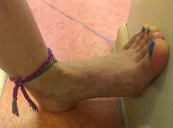
Figure 3
Plantar fascia stretching exercise.
Our Own Treatment Protocol (Hacettepe Protocol)
Patients diagnosed with plantar heel pain due to plantar fascia
are frequently referred to our unit, and a standardized treatment
protocol based on home program is implemented in accordance
with our cumulated experience with this condition. Our treatment
protocol, which we call the Hacettepe protocol and whose results
have not yet been published, show some differences based on patients
having acute onset, chronic, and accompanying heel stiffness. The
basic parameters of the treatment program we developed are shown
in Table 1. The treatment protocol summarized under headings in
Table 1 is taught to patients with plantar fascia who are referred to
our unit for the first time, and they are instructed to exercise this
protocol for 6 weeks at home. At week 6, symptoms, complaints,
and foot biomechanical properties of the patient are evaluated in
accordance with the initial status of the patient. After an evaluation
of the results, the decision to administer an intensive physiotherapy
program or ESWT is made, or the patient is referred back to his/her
doctor. The patients are advised to apply before the check-up date
if they experience increased symptoms during home treatment. Our
clinical experience shows that NSAID use and non-elastic taping are
effective in rapidly reducing symptoms in patients presenting with
acute complaints. In addition, it has been observed that ESWT, which
is applied repeatedly in chronic patients, affects the increase of pain in
some cases. Our clinical experience shows that NSAID use and nonelastic
taping are effective in rapidly reducing symptoms in patients
presenting with acute complaints. It is considered that ESWT, which
is applied repeatedly in chronic patients, has increased pain in some
cases.
Surgical applications
Surgical applications should be considered only when all other
treatments fail in PF cases. Most cases are treated with stretching,
taping, orthotic approach, and inflammation-treatment medicine,
but only a small portion requires surgery. The main purpose in
this application is to loosen the plantar fascia. The most commonly
used method is partial open or closed plantar fasciotomy. Plantar
fasciotomy is a popular approach where up to half of the fascia is
removed in some cases. The open method requires a 3 cm to 6 cm
plantar medial incision to release the fascia. Resection of nerve
decompression and/or calcaneal dissection is also performed during
this procedure. Endoscopy is used to release fascia in the closed
method. In this method, the resection of the calcaneal protrusion
is not performed. According to the results of a previous study, both
types of treatment were reported to be of equal benefit [64]. The
success rate in surgical methods is around 70% to 90%. The duration
of healing after this surgery may range from a few weeks to several
months [65]. Today, increasingly popular endoscopic plantar
fasciotomy affects the plantar fasciitis at minimal extent and this
method results in minimally invasive visualization of fascial band.
As a result of this application, the healing process also decreases [66].
Figure 4

Figure 4
Short Foot Exercise.
Figure 5
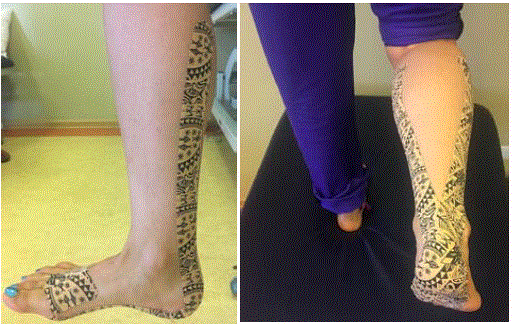
Figure 5
Plantar fasciataping technique with dynamic tape.
Figure 6
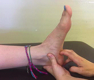
Figure 6
Transverse friction massage.
Results
Plantar fasciitis is a soft tissue pathology that is most often associated with pathomechanical problems in the foot, is a painful and inflammatory condition that can cause secondary problems if not treated, and responds to physical therapy and rehabilitation. Although the choice of treatment modalities to be applied varies according to the characteristics of the patient and the pathology, there is a consensus that stretching, strengthening methods, manual techniques, and orthosis applications are effective. The use of new and not yet fully proven methods, along with other methods, will also have an impact on increasing effectiveness. PF is foot pathology with a wide range of treatment options with many risk factors.
References
- Toomey EP. Plantar heel pain. Foot Ankle Clin. 2009;14(2):229-45.
- Othman AM, Ragab EM. Endoscopic plantar fasciotomy versus extracorporeal shock wave therapy for treatment of chronic plantar fasciitis. Acta Orthop Trauma Surg. 2010;130(11):1343-7.
- Orchard J. Plantar fasciitis. BMJ. 2012;345:e6603.
- Bolla LA, Malone TR. Plantar fasciitis and the windlass mechanism: a biomechanical link to clinical practice. J Athl Train. 2004;39(1):77-82.
- Goff JD, Crawford R. Diagnosis and treatment of plantar fasciitis. Am Fam Physician. 2011;84(6):676-82.
- Riddle DL, Pulisic M, Pidcoe P, Johnson RE. Risk factors for plantar fasciitis: a matched case-control study. J Bone Joint Surg Am. 2003;85-A(5):872-7.
- Roxas M. Plantar Fasciitis: Diagnosis and Therapeutic Considerations Altern Med Rev. 2005;10(2):83-93.
- Warren BL, Jones CJ. Predicting plantar fasciitis in runners. Med Sci Sports Exerc. 1987;19(1):71-3.
- Schepsis AA, Leach RE, Gorzyca J. Plantar fasciitis. Etiology, treatment, surgical results, and review of the literature. Clin Orthop Relat Res. 1991;(266):185-96.
- Johnson RE, Haas K, Lindow K, Shields R. Plantar fasciitis: what is the diagnosis and treatment? Orthop Nurs. 2014;33(4):198-204.
- Agyekum EK, Ma K. Heel pain: A systematic review. Chin J Traumatol. 2015;18(3):164-9.
- Eechaute C, Vaes P, Van Aerschot L, Asman S, Duquet W. The clinimetric qualities of patient-assessed instruments for measuring chronic ankle instability: a systematic review. BMC Musculoskelet Disord. 2007;8:6.
- SooHoo NF, Samimi DB, Vyas RM, Botzler T. Evaluation of the validity of the Foot Function Index in measuring outcomes in patients with foot and ankle disorders. Foot Ankle Int. 2006;27(1):38-42.
- Martinelli N, Scotto GM, Sartorelli E, Bonifacini C, Bianchi A, Malerba F. Reliability, validity and responsiveness of the Italian version of the Foot Function Index in patients with foot and ankle diseases. Qual Life Res. 2014;23(1):277-84.
- Martin RL, Davenport TE, Reischl SF, McPoil TG, Matheson JW, Wukich DK, et al. Heel pain-plantar fasciitis: revision 2014. J Orthop Sports Phys Ther. 2014;44(11):A1-33.
- Mardani-Kivi M, Karimi Mobarakeh M, Hassanzadeh Z, Mirbolook A, Asadi K, Ettehad H, et al. Treatment Outcomes of Corticosteroid Injection and Extracorporeal Shock Wave Therapy as Two Primary Therapeutic Methods for Acute Plantar Fasciitis: A Prospective Randomized Clinical Trial. J Foot Ankle Surg. 2015;54(6):1047-52.
- Yucel I, Ozturan KE, Demiraran Y, Degirmenci E, Kaynak G. Comparison of high-dose extra- corporeal shockwave therapy and intralesional corticosteroid injection in the treatment of plantar fasciitis. J Am Podiatr Med Assoc. 2010;100(2):105-10.
- Sorrentino F, Iovane A, Vetro A, Vaccari A, Mantia R, Midiri M. Role of high-resolution ultrasound in guiding treatment of idiopathic plantar fasciitis with minimally invasive techniques. Radiol Med. 2008;113(4):486-95.
- Ragab EM, Othman AM. Platelets rich plasma for treatment of chronic plantar fasciitis. Arch Orthop Trauma Surg. 2012;132(8):1065-70.
- Gogna P, Gaba S, Mukhopadhyay R, Gupta R, Rohilla R, Yadav L. Plantar fasciitis: A randomized comparative study of platelet rich plasma and low dose radiation in sportspersons. Foot (Edinb). 2016;28:16-19.
- Jain K, Murphy PN, Clough TM. Platelet rich plasma versus corticosteroid injection for plantar fasciitis: A comparative study. Foot (Edinb). 2015;25(4):235-7.
- Karimzadeh A, Raeissadat SA, ErfaniFam S, Sedighipour L, Babaei-Ghazani A. Autologous whole blood versus corticosteroid local injection in treatment of plantar fasciitis: A randomized, controlled multicenter clinical trial. Clin Rheumatol. 2017;36(3):661-9.
- Ahmad J, Ahmad SH, Jones K. Treatment of Plantar Fasciitis With Botulinum Toxin. Foot Ankle Int. 2017;38(1):1-7.
- Thiagarajah AG. How effective is acupuncture for reducing pain due to plantar fasciitis? Singapore Med J. 2017;58(2):92-97.
- Hyland MR, Weber-Gaffney A, Cohen L, Licht- man PT. Randomized controlled trial of cal- caneal taping, sham taping and plantar fascia streching for the short term management of plantar heel pain. J Ortop Sports Phys Ther. 2006;36(6):364-71.
- Healey K, Chen K. Plantar fasciitis: current diagnostic modalities and treatments. Clin Podiatr Med Surg. 2010;27(3):369-80.
- Landorf KB, Menz HB. Plantar heel pain and fasciitis. BMJ Clin Evid. 2008;2008:1111.
- Sweeting D, Parish B, Hooper L, Chester R. The effectiveness of manual stretching in the treatment of plantar heel pain: a systematic review. J Foot Ankle Res. 2011;4:19.
- McKeon PO, Hertel J, Bramble D, Davis I. The foot core system: a new paradigm for understanding intrinsic foot muscle function. Br J Sports Med. 2015;49(5):290.
- Cheung RT, Sze LK, Mok NW, Ng GY. Intrinsic foot muscle volume in experienced runners with and without chronic plantar fasciitis. J Sci Med Sport. 2016;19(9):713-5.
- Santos BD, Corrêa LA, Teixeira Santos L, Filho NA, Lemos T, Nogueira LA. Combination of Hip Strengthening and Manipulative Therapy for the Treatment of Plantar Fasciitis: A Case Report. J Chiropr Med. 2016;15(4):310-3.
- McKeon PO, Fourchet F. Freeing the foot: integrating the foot core system into rehabilitation for lower extremity injuries. Clin Sports Med. 2015;34(2):347-61.
- Rathleff MS, Molgaard CM, Fredberg U, Kaalund S, Andersen KB, Jensen TT, et al. High-load strength training improves outcome in patients with plantar fasciitis: A randomized controlled trial with 12-month follow-up. Scand J Med Sci Sports. 2015;25(3):292-300.
- Kamonseki DH, Gonçalves GA, Yi LC, Júnior IL. Effect of stretching with and without muscle strengthening exercises for the foot and hip in patients with plantar fasciitis: A randomized controlled single-blind clinical trial. Man Ther. 2016;23:76-82.
- Moon DC, Kim K, Lee SK. Immediate Effect of Short-foot Exercise on Dynamic Balance of Subjects with Excessively Pronated Feet. J Phys Ther Sci. 2014;26(1):117-9.
- Saban B, Deutscher D, Ziv T. Deep massage to posterior calf muscle in combination with neural mobilization exercises as a treatment for heel pain: A pilot rondomized clinical trial. Man Ther. 2014;19(2):102-8.
- Cleland JA, Abbott JH, Kidd MO, Stockwell S, Cheney S, Gerrard DF, et al. Manual physical therapy and exercise versus electrophysical agents and exercise in the management of plantar heel pain: A multicenter randomized clinical trial. J Orthop Sports Phys Ther. 2009;39(8):573-85.
- Shashua A, Flechter S, Avidan L, Ofir D, Melayev A, Kalichman L. The effect of additional ankle and midfoot mobilizations on plantar fasciitis: A randomized controlled trial. J Ortop Sports Phys Ther. 2015; 45(4):265-72.
- Celik D, Kus G, Sirma SO. Joint Mobilization and Stretching Exercise vs Steroid Injection in the Treatment of Plantar Fasciitis: A Randomized Controlled Study. Foot Ankle Int. 2016;37(2):150-6.
- Crawford F, Thomson C. Interventions for treating plantar heel pain. Cochrane Database Syst Rev. 2003;(3):CD000416.
- Shanks P, Curran M, Fletcher P, Thompson R. The effectiveness of therapeutic ultrasound for musculoskeletal conditions of lower limb: A literature review. Foot (Edinb) 2010;20(4):133-9.
- Alotaibi AK, Petrofsky JS, Daher NS, Lohman E, Laymon M, Syed HM. Effect of Monophasic Pulsed Current on Heel Pain and Functional Activities caused by Plantar Fasciitis. Med Sci Monit. 2015;21:833-9.
- Stratton M, McPoil TG, Cornwall MW, Patrick K. Use of low-frequency electrical stimulation for the treatment of plantar fasciitis. J Am Podiatr Med Assoc. 2009;99(6):481-8.
- Jasiak-Tyrkalska B, Jaworek J, Franczuk B. Efficacy of two different physiotherapeutic procedures in comprehensive therapy of plantar calcaneal spur. FizioterPolska. 2007;7:145-54.
- Basford JR, Malanga GA, Krause DA, Harmsen WS. A randomized controlled evaluation of low-intensity laser therapy: plantar fasciitis. Arch Phys Med Rehabil. 1998;79(3):249-54.
- Thomson CE, Crawford F, Murray GD. The effectiveness of extra corporeal shock wave therapy for plantar heel pain: a systematic review and meta-analysis. BMC Musculoskelet Disord. 2005;6:19.
- Kudo P, Dainty K, Clarfield M, Coughlin L, Lavoie P, Lebrun C. Randomized, placebo-controlled, double-blind clinical trial evaluating the treatment of plantar fasciitis with an extracoporeal shockwave therapy (ESWT) device: a North American confirmatory study. J Orthop Res. 2006;24(2):115-23.
- Scheuer R, Friedrich M, Hahne J, Holzapfel J, Machacek P, Ogon M, et al. Approaches to optimize focused extracorporeal shockwave therapy (ESWT) based on an observational study of 363 feet with recalcitrant plantar fasciitis. Int J Surg. 2016;27:1-7.
- Podolsky R, Kalichman L. Taping for plantar fasciitis. J Back Musculoskelet Rehabil. 2015;28(1):1-6.
- Morris D, Jones D, Ryan H, Ryan CG. The clinical effects of Kinesio® Tex taping: A systematic review. Physiother Theory Pract. 2013;29(4):259-70.
- Johnson RE, Haas K, Lindow K, Shields R. Plantar fasciitis: what is the diagnosis and treatment? Orthop Nurs. 2014;33(4):198-204.
- Radford JA, Landorf KB, Buchbinder R, Cook C. Effectiveness of low-Dye taping for the short-term treatment of plantar heel pain: a randomised trial. BMC Musculoskelet Disord. 2006;7:64.
- Saxelby J, Bygrave C. ‘Low-Dye’ taping on the foot in the management of plantar- fasciitis. The Foot. 1997;7(4):205-9.
- Franettovich M, Chapman A, Blanch P, Vicenzino B. Continual use of augmented low-Dye taping increases arch height in standing but does not influence neuromotor control of gait. Gait Posture. 2010;31(2):247-50.
- Mills K, Blanch P, Chapman AR, McPoil TG, Vicenzino B. Foot orthoses and gait: a systematic review and meta-analysis of literature pertaining to potential mechanisms. Br J Sports Med. 2010;44(14):1035-46.
- Roos E, Engström M, Söderberg B. Foot orthoses for the treatment of plantar fasciitis. Foot Ankle Int. 2006;27(8):606-11.
- Sheridan L, Lopez A, Perez A, John MM, Willis FB, Shanmugam R. Plantar fasciopathy treated with dynamic splinting: a randomized controlled trial. J Am Podiatr Med Assoc. 2010;100(3):161-5.
- Uden H, Boesch E, Kumar S. Plantar fasciitis - to jab or to support? A systematic review of the current best evidence. J Multidiscip Healthc. 2011;4:155-64.
- Lewis RD, Wright P, McCarthy LH. Orthotics Compared to Conventional Therapy and Other Nonsurgical Treatments for Plantar Fasciitis. J Okla State Med Assoc. 2015;108(12):596-8.
- Mizel MS, Marymont JV, Trepman E. Treatment of plantar fasciitis with a night splint and shoe modification consisting of a steel shank and anterior rocker bottom. Foot Ankle Int. 1996;17(12):732-5.
- Fong DT, Pang KY, Chung MM, Hung AS, Chan KM. Evaluation of combined prescription of rocker sole shoes and custom-made foot orthoses for the treatment of plantar fasciitis. ClinBiomech (Bristol, Avon). 2012;27(10):1072-7.
- Cotchett MP, Landorf KB, Munteanu SE. Effectiveness of dry needling and injections of myofascial trigger points associated with plantar heel pain: a systematic review. J Foot Ankle Res. 2010;3:18.
- Cotchett MP, Munteanu SE, Landorf KB. Effectiveness of trigger point dry needling for plantar heel pain: a randomized controlled trial. Phys Ther. 2014;94(8):1083-94.
- Cornwall MW, McPoil TG. Plantar fasciitis: etiology and treatment. J Orthop Sports Phys Ther. 1999;29(12):756-60.
- Young CC, Rutherford DS, Niedfeldt MW. Treatment of plantar fasciitis. Am Fam Physician. 2001;63(3):467-74,477-8.
- Hake DH. Endoscopic plantar fasciotomy: a minimally traumatic procedure for chronic plantar fasciitis. Ochsner J. 2000;2(3):175-8.