Clinical Image
Fungal Infection of the Brain: A Diagnostic Dilemma Resolved
Seema Rohilla1*, Ishwar Singh2 and Dhara B Dhaulakhandi3
1Department of Radiodiagnosis & Imaging, Post Graduate Institute of Medical Sciences, Pt. B.D. Sharma
University of Health Sciences, Rohtak 124 001 (Haryana), India
2Department of Neurosurgery, Post Graduate Institute of Medical Sciences, Pt. B.D. Sharma University of Health
Sciences, Rohtak 124 001 (Haryana), India
3Department of Biotechnology & Molecular Medicine Regional Cancer Centre, Post Graduate Institute of Medical
Sciences, Pt. B.D. Sharma University of Health Sciences, Rohtak 124 001 (Haryana), India
*Corresponding author: Seema Rohilla, Department of Radiodiagnosis & Imaging, Post Graduate Institute of Medical Sciences, Pt. B.D. Sharma University of Health Sciences, Rohtak 124 001 (Haryana), India
Published: 03 Aug, 2017
Cite this article as: Rohilla S, Singh I, Dhaulakhandi
DB. Fungal Infection of the Brain: A
Diagnostic Dilemma Resolved. Clin
Surg. 2017; 2: 1741.
Clinical Image
A 35 year old male presented with burning and tingling on left side of face (in the distribution of trigeminal nerve) and headache off and on. He was suspected to have trigeminal neuralgia and underwent contrast enhanced CT scan (image not available) on which an enhancing mass was seen in left temporal area adjacent to cavernous sinus and was reported to have meningioma. Contrast enhanced MRI of his brain was then done which showed a mass lesion in the same area which was isointense on T1W images (Figure A), very hypointense on T2W images (Figure B) and showed enhancement on contrast images (Figure C). There appeared to be some extension into the orbit. In addition to meningioma, fungal involvement of trigeminal nerve was kept as a possibility. It turned out to be fungal infection on surgery and patient responded well to antifungal treatment.
Figure 1(A-C)
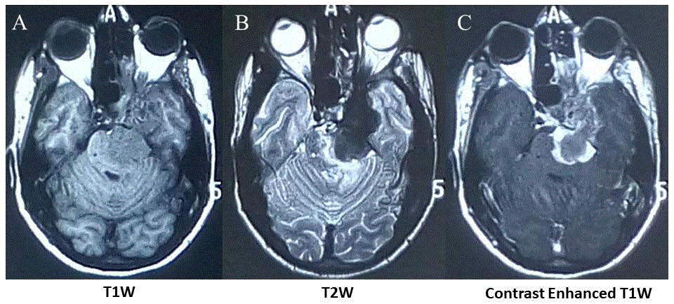
Figure 1(A-C)
Fungal Infection of the Brain.