Clinical Image
Isolated Cervical Rib Fracture
Serkan Özbay1, Asli Gül Temel2* and Salih Topçu3
1Department of Thoracic Surgery, Giresun University Faculty of Medicine, Turkey
2Department of Thoracic Surgery, University of Health Sciences Faculty of Medicine, Turkey
3Department of Thoracic Surgery, Kocaeli University Faculty of Medicine, Turkey
*Corresponding author: Asli Gül TEMEL, Department of Thoracic Surgery, University of Health Sciences Faculty of Medicine, Şişli Etfal Training and Research Hospital, 8th Floor, Istanbul, Turkey
Published: 27 Oct, 2017
Cite this article as: Özbay S, Temel AG, Topçu S. Isolated
Cervical Rib Fracture. Clin Surg. 2017;
2: 1697.
Keywords
Thorax; Trauma; Imaging
Clinical Image
We have had many experiences with thoracic outlet syndrome. Cervical ribs on chest roentgenograms are detected incidentally or due to the symptoms of the patients. We have seen an isolated cervical rib fracture in our clinic for the first time. Bilateral cervical ribs with fracture at right sided were detected on the chest X-ray (Figure 1A) of a 3 year-old male patient who admitted to our clinic because of the trauma of falling from a 1.5 meters height and it is also well defined at spiral computed tomography with three dimensional reconstruction (Figure 1B).
Figure 1
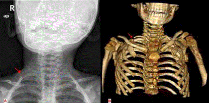
Figure 1
Cervical rib fracture at chest X-ray and 3D spiral tomography.