Clinical Image
Left Atrial Free Wall Myxoma
Hüseyin Ali Tünel* and Orhan Saim Demirtürk
Department of Cardiovascular Surgery, Baskent University Adana Dr. Turgut Noyan Teaching and Medical
Research Center, Turkey
*Corresponding author: Hüseyin Ali Tünel, Department of Cardiovascular Surgery, Baskent University, Adana Teaching and Medical Research Center, Dadaloglu mah. Serinevler 2591. Sok. No:4/A 01250, Yüregir, Adana, Turkey
Published: 11 Oct, 2017
Cite this article as: Tünel HA, Demirtürk OS. Left Atrial
Free Wall Myxoma. Clin Surg. 2017; 2:
1665.
Clinical Image
A 64 year-old woman suffered a cerebrovascular infarction affecting the left cerebrum. She was hospitalized by the neurology department for right hemiplegia. One week after the admission she had undergone transthoracic echocardiography revealing a pulsatile, mobile mass or thrombus moving in a to-and-fro fashion through the mitral valve. Upon Transesophaeal Echocardiogram (TEE) left atrial tumor of 6x3 cm of pedunculated morphology which entered and exited the left ventricle in each cardiac cycle was detected (Figure 1). Excision of the tumor after complete neurological recovery and stabilization was decided. Four weeks after the acute stroke a cardiac operation under Cardiopulmonary Bypass (CPB) was conducted. The left atrium was opened and a 3x5 cm gelatinous tumor which was attached to the free wall of the left atrium was observed. The mass was removed together with the free left atrial wall requiring a patch repair of the free left atrial wall using fresh autolous pericardium (Figure 2). The cross-clamp and total CPB times were 30 and 45 minutes respectively. The operation and the postoperative period were eventless. The patient stayed for one day in the intensive care unit. She was discharged on postoperative day 5. She is cardiovascularly well two months after the operation. Her neurological condition is stable but her right hemiplegia persists. Cardiac myxomas frequently present with neurological symptoms and the anterior circulation, namely the middle cerebral artery, is frequently affected as has been seen in our patient [1]. Stroke due to cardiac myxoma is rare but may cause devastating clinical conditions [2]. Immediate operative treatment is indicated but if stroke occurs before surgery can be performed, the surgical intervention requires waiting for neurological stabilization and avoidance of possible exacerbation of cerebral hemorrhage after heparinization for CPB. This period usually requires one to four weeks after the initial stroke [2,3]. Full thickness resection of the tumor with clear margins is necessary to prevent recurrences [3].
Figure 1
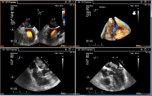
Figure 1
Clockwise, the myxoma in color doppler echocardiogram; 3-D echocardiographic image of the tumor;
tumor in the left atrium shown by arrow; tumor which moved into the left ventricle through the mitral valve shown
by arrow.
Figure 2
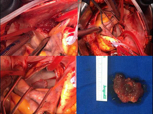
Figure 2
Clockwise, atrial myxoma as seen after atriotomy; the atrium after excision of the tumor showing the
defect in the free left atrial wall; the left atrium after pericardial patch repair; the myxoma (excisional specimen).
References
- Andreu JP, Parrilla G, Arribas JM, Garcia- Villalba B, Lucas JJ, Navarro MG, et al. Neurological manifestations of cardiac myxoma: experience in a referral hospital. Neurologia. 2013;28(9):529-34.
- Yuan S, Humuruola G. Stroke of a cardiac myxoma origin. Braz J Cardiovasc Surg. 2015;30(2):225-34.
- Mishra A, Shah M, Sharma P, Kothari J, Malhotra A. Operative management of intracardiac myxomas: a single center experience. Med J Armed Forces India. 2014;70(1):5-9.