Case Report
Delayed Jejunal Perforation due to Taekwondo Kick: A Rare Case
Aydin Yavuz1, Alp Yildız1, Hüseyin Göbüt1, Kürşat Dikmen2, Hasan Bostancı2, Aybala Ağaç1 and
Hande Köksal2*
1Department of General Surgery, Yenimahalle Education and Research Hospital, General Surgery Clinic, Ankara,
Turkey
2Department of General Surgery, Gazi University School of Medicine, Ankara, Turkey
*Corresponding author: Hande Köksal, Department of General Surgery, Gazi University School of Medicin, Ankara, Turkey
Published: 11 Oct, 2017
Cite this article as: Yavuz A, Yildız A, Göbüt H, Dikmen K,
Bostancı H, Ağaç A, Köksal H. Delayed
Jejunal Perforation due to Taekwondo
Kick: A Rare Case. Clin Surg. 2017; 2:
1664.
Abstract
Background: One of the most frequently injured hollow organs following blunt abdominal trauma
is the jejunum; however, isolated jejunal perforation is rare.
Case Report: A 19 year-old male patient received a blow during taekwondo competition and
admitted to emergency department with complaint of abdominal pain. Laboratory work-up
and chest X-ray examinations did not reveal any pathology and the patient was discharged with
recommendation of analgesics. Two days later, the patient presented to our emergency department
with intense abdominal pain, nausea and vomiting with presence of abdominal tenderness, muscular
defense, and rebound signs, whereas chest X-ray was completely normal. The patient was examined
with computed abdominal tomography (CT), which revealed jejunal perforation. Following proper
debridement, primary closure was performed. The patient was discharged on the postoperative 6th
day.
Discussion: Although isolated intestinal injury after blunt abdominal trauma is extremely
rare, it should certainly be considered particularly in cases with persistent abdominal pain. As
it is particularly demonstrated in our case, failure to recognize hollow organ damage after blunt
abdominal trauma by relying on the initially normal examination findings may lead to delay in the
diagnosis for hours and even days as in our case. Considering that delayed diagnosis is an important
cause of increased morbidity and mortality, we would like to remind that close follow-up of patients
with repeated physical examinations and prolonging the hospital stay time have great benefits even
if the initial examinations are normal.
Keywords: Abdominal trauma; Blunt; Jejunal perforation
Introduction
With blunt abdominal trauma, the frequency of injury to the solid organs is higher than the frequency of injury to hollow organs [1]. One of the most frequently injured hollow organs following blunt abdominal trauma is the jejunum; however, isolated jejunal perforation is rare (1%) [2]. The blunt trauma causes compression of the intestinal segment against abdominal wall and the vertebra, and sudden rise in intraluminal pressure results in a bursting type of tear in the intestinal wall [3]. In literature, there are very few reports of intestinal perforations that occurred as a result of blunt traumas due to various causes (traffic accidents, falling, being kicked, etc.) [1,4]. Since the appearance of clinical manifestations may take some time following injury to intestines after blunt trauma, the initial physical examinations may be normal. This leads to delays in diagnosis and treatment, and causes significant increases in morbidity and mortality [1,3,5].
Patients and Methods
A 19 year-old male patient received a kick during taekwondo competition, and despite wearing protective gear, he could not complete the match, and presented to emergency department of a local hospital with complaint of abdominal pain. Laboratory tests and abdominal and chest X-ray examinations did not reveal any pathology and the patient was discharged with recommendation of analgesics. Two days later, the patient presented to emergency department of university hospital with intense abdominal pain, nausea and vomiting. His vital signs were within normal limits. In physical examination, he had acute abdominal pain with presence of abdominal tenderness, muscular defense, and rebound signs. His hemoglobin (Hb) level was 12.9 g/dL and white blood cell (WBC) count was 12.300/uL, whereas chest and abdominal X-ray were completely normal (Figure 1). The patient was examined with contrast abdominal CT, which revealed pneumoperitoneum (Figure 2). The patient underwent surgery with initial diagnosis of hollow organ perforation. Diagnostic laparoscopy was performed. Since the site of perforation could not be detected, the operation was converted to open surgery, and during exploration, a jejunal perforation site was detected 60 cm away from the ligament of Treitz. The perforation site was limited with the omentum and was surrounded by fibrin clots (Figure 3). Following proper debridement, primary suturing was performed. The patient was discharged upon recovery on the postoperative 6th day.
Figure 1
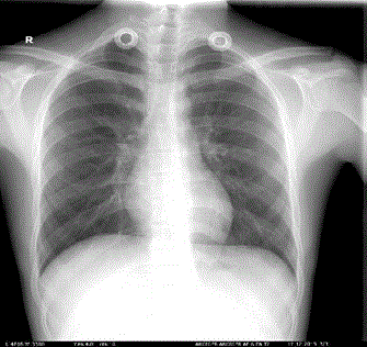
Figure 1
Abdominal X-ray.
Figure 2
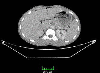
Figure 2
Revealed pneumoperitoneum.
Figure 3
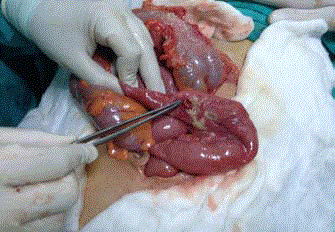
Figure 3
The perforation site was limited with the omentum and was
surrounded by fibrin clots.
Discussion
Solid organ injuries are more common in blunt abdominal
trauma, with liver and spleen being the most frequently injured
organs. The incidence of isolated intestinal injury following blunt
trauma is about 1%, and perforation occurs only in the rate of 0.3%
[1,6]. More than 75% of blunt traumas are caused by motor vehicle
accidents, followed by pounding, occupational accidents, fall from
height and being crushed [3]. Intestinal injury during blunt trauma is
often caused by compression of the intestinal segment against anterior
abdominal wall and the vertebra, which results in a sudden rise of
intraluminal pressure. Sometimes the sudden forward movement
of the intestines results in injury to the immobile structures such
as ligament of Treitz, the ileocecal valve and mesentery root. Blunt
injury to the intestines can manifest as intramural hematoma,
contusion, laceration or rupture [7,8]. Hollow organ injury can
present with acute abdomen during examination, although it may
not manifest any signs in the early phase. Allen et al. [2] reported
that diagnostic reliability of abdominal physical examination alone
after blunt abdominal trauma is 30%. Following trauma, the clinical
picture may be initially obscure, or perforations may occur at the
late phase due to ischemia. In our case, the clinical manifestations
were severe since the beginning; however, because of the normal
laboratory results, and due to the fact that hollow organ injury after
blunt trauma is extremely rare, the diagnosis was delayed. Therefore,
in such cases, the patient should be observed closely with repeated
physical examinations and imaging studies, since early diagnosis of
intestinal perforation is essential to reduce morbidity and mortality.
In one study by Watts et al. [9] mortality rate in case of unnoticed
perforation and more than 24 hour delay in the operation was 16%,
which is nearly 5 times the rate when intervention is made within the
first few hours of perforation. Similarly, morbidities such as sepsis,
intra-abdominal abscess and wound dehiscence are at least two times
more frequent when the operation is delayed [10].
Even if the initial examination does not reveal any pathology
(patient's consciousness state and administration of analgesics may be
misleading), repeated examinations should be carried out. In laboratory
tests, leukocytosis and elevated amylase level may be of significance.
Focused abdominal sonography for trauma (FAST), abdominal CT,
diagnostic peritoneal lavage (DPL), and chest-abdominal X-rays may
be of guidance for detecting intestinal perforation. In our case, chest
and erect plain abdominal X-rays obtained at the time of presentation
and two days later were both normal, yielding false negative results
which were surpassingly misleading. Our case represents a good
example demonstrating the importance of appraising the clinical
situation and continuing examinations with further imaging
studies. On the other hand, ultrasonography does not have much
of a diagnostic value particularly in the early phase of the intestinal
injuries. Plain X-rays are also unreliable much of the time, as
demonstrated by our case. Majority of cases do not manifest with
intraperitoneal free air. CT, as the most beneficial diagnostic method
in these cases has sensitivity and specificity values as 92% and 94%,
respectively. CT signs that are suggestive of intestinal perforation are
contrast material extravasation and presence of extraluminal free air.
Other non-diagnostic but supportive signs include presence of free
fluid in the absence of solid organ damage, thickening in intestinal
wall and intestinal dilatation [11,12].
In treatment, explorative laparotomy and drainage of septic
peritoneal fluid along with lavage are important. Additionally,
prophylactic antibiotic is necessary. The type of the repair of
perforation depends on the localization of the injury and the defect
size. Primary repair of small defects, and resection of large defects
and ischemic segments with primary anastomosis may be successfully
performed even in the delayed cases [13].
Although isolated intestinal injury after blunt abdominal trauma
is extremely rare, it should certainly be considered particularly in cases
with persistent abdominal pain. As it is particularly demonstrated
in our case, failure to recognize hollow organ damage after blunt
abdominal trauma by relying on the initially normal examination
findings may lead to delay in the diagnosis for hours and even days as
in our case. Considering that delayed diagnosis is an important cause
of increased morbidity and mortality, we would like to remind that
close follow-up of patients with repeated physical examinations and
prolonging the hospital stay time have great benefits even if the initial
examinations are normal.
References
- Coskun AK, Yarici M, Ulke E, Mentes O, Kozak O, Tufan T. Perforation of isolated jejunum after a blunt trauma: case report and review of the literature. Am J Emerg Med. 2007;25(7):862.
- Allen GS, Moore FA, Cox CS Jr, Wilson JT, Cohn JM, Duke JH. Hollow visceral injury and blunt trauma. J Trauma. 1998;45(1):69-75.
- Munshi IA, DiRocco JD, Khachi G. Isolated jejuna perforation after blunt thoracoabdominal trauma. J Emerg Med. 2006;30(4):393-5.
- Houshian S. Traumaticduodenalrupture in a soccerplayer. Br J Sports Med. 2000;34:218-9.
- Analay H, Gokgoz MŞ, Yıldırır C, Turan M, Aker B. İnce BağırsakYaralanmaları. Ulusal Travma Dergisi. 1997;3:288-90.
- Fraga GP, Silva FH, Almeida NA, Curi JC, Mantovani M. Blunt abdominal trauma with small bowel injury: are isolated lesions riskier than associated lesions? Acta Cir Bras. 2008;23(2):192-7.
- Dongo AE, Kesieme EB, Irabor DO, Ladipo JK. A review of post traumatic bowel injuries in ibadan. ISRN Surg. 2011;2011:478042.
- Watts DD, Fakhry SM; EAST Multi-Institutional Hollow Viscus Injury Research Group. Incidence of hollow viscus injury in blunt trauma: an analysis from 275.557 trauma admissions from the East multi-institutional trial. J Trauma. 2003;54(2):289-94.
- Fakhry SM, Watts DD, Luchette FA; EAST Multi-Institutiona l Hollow Viscus Injury Research Group. Current diagnostic approaches lack sensitivity in the diagnosis of perforated blunt small bowel injury: analysis from 275.557 trauma admissions from the EAST multi-institutional HVI trial. J Trauma. 2003;54(2):295-306.
- Saku M, Yoshimitsu K, Murakami J, Nakamura Y, Oguri S, Noguchi T, et al. Small bowel perforation resulting from blunt abdominal trauma: intervalchange of radiological characteristics. Radiat Med. 2006;24(5):358-64.
- Kostantinidis C, Pitsinis V, FragulidisG. Isolated jejuna perforation following blunt abdominal trauma. Ulus Travma Acil Cerrahi Derg. 2010;16(1):87-9.
- Sozuer EM, Bedirli A, İkizceli İ, Yeşilkaya Y. Kunt travmaya bağlı izole incebağırsak yaralanmalarında cerrahi tedavi. Ulusal Travma Dergisi. 1997;3:298-302.
- Mukhopadhyay M. Intestinal Injury from Blunt Abdominal Trauma: A Study of 47 Cases. Oman Med J. 2009;24(4):256-9.