Case Report
Medical Necessity and Evidence Basis for Extra-osseous Talotarsal Joint Stabilization
Michael E Graham*
Department of Foot & Ankle Surgery, GraMedica & Graham International Implant Institute, USA
*Corresponding author: Michael E Graham, Department of Foot & Ankle Surgery, GraMedica & Graham International Implant Institute, USA
Published: 11 Oct, 2017
Cite this article as: Graham ME. Medical Necessity and
Evidence Basis for Extra-osseous
Talotarsal Joint Stabilization. Clin Surg.
2017; 2: 1663.
Abstract
Extra-Osseous Talotarsal Joint Stabilization (EOTTS) is a minimally invasive surgical procedure that involves the insertion of an internal fixation device, stent, into the sinus tarsi. The goal of this stent is to realign and stabilize the “foundation joint” of the body, while still allowing a normal joint range of motion. There are more than 100 peer-reviewed, published articles that prove the medical necessity for and the effectiveness of EOTTS.
Background
EOTTS implants are cleared for use in more than 70 countries. Obtaining the clearance from the Food and Drug Administration (FDA) and CE mark for European countries is a major hurdle. This means that the government has validated the evidence that the device contains sufficient valid scientific evidence that provides reasonable assurance that the device is safe and effective for its intended use or uses. These sinus tarsi stents are advocated for and inserted by leading orthopedic and podiatric foot surgeons around the world. The end-users, foot surgeons, recommend and use these sinus tarsi stents for a reason. It works and patients get better. The only reason why more patients are not offered this solution is simply due to limited insurance coverage.
EOTTS Advantages
Of importance is the fact that EOTTS is a surgical procedure that does not involve cutting and/
or shifting of any bones. It does not have a negative impact on the very important joints of the
hindfoot. In fact, the EOTTS procedure should reverse or delay arthritic damage caused to joints
due to the partial dislocation of the TTJ. There are many advantages of EOTTS over other forms
of “treatment” of talotarsal joint dislocation. EOTTS can be used as a “stand-alone procedure”,
meaning there are situations where the only form of treatment required is EOTTS. It can also be
used in conjunction with both external measures, or along with other surgical procedures to correct
co-foot/ankle deformities. EOTTS can be performed on both children and adults. Patients can
walk, run, and perform any activity of their choice, once the tissues have recovered from the initial
procedure. The muscles, ligaments, and tendons will work more efficiently. Patients claim to “run
faster” and “jump higher” due to their realigned talotarsal joint.
One of the many other advantages of this procedure is that it is reversible. This is very important
when compared to other surgical procedures where tendons are sacrificed, bones are cut and shifted,
or joints are fused to eliminate motion. EOTTS is the only reversible surgical procedure that can
realign and stabilize the talotarsal joint.
Primary Indication of EOTTS
There is only one primary indication/diagnosis that a surgeon would recommend EOTTS to
a patient and that is talotarsal joint dislocation. There are clear, validated clinical and radiologic
measurements that are used to diagnose this complex orthopedic deformity. The majority of cases
where EOTTS is performed are for Recurrent Talotarsal Joint Dislocation (RTTJD), as opposed to
the minority of cases where there is a fixed deformity such as a tarsal coalition. RTTJD is a pathologic
anomaly where the talus repeatedly dislocates on the calcaneus and/or the navicular bone. This
deformity wills never auto-repair. It won’t get better; it will only get worse and has been proven to
be the underlying etiology to many chronic lower extremity pathologies [1-28].
“Recurrent” means something that happens repeatedly. With RTTJD, the talus repeatedly
dislocates on the tarsal mechanism (calcaneus and/or navicular bones). There are two main phases
during the walking, or gait cycle. The swing phase is when the foot is in the air not touching the weightbearing surface. During this phase, the talus will be aligned on
the tarsal mechanism. The second phase is stance. This is when the
foot makes contact or touches the weightbearing surface. The talus
becomes dislocated or malpositioned during this portion of the gait
cycle.
Dislocation is a generic term meaning the articular facets are no
longer anatomically aligned. “Talotarsal Joint Dislocation” means
that the joint surfaces of the talus are abnormally aligned on the
heel and/or navicular bones. The articular surfaces, joints, of the
body must remain in constant congruent contact to be considered
stable and aligned. There are many additional descriptors that are
added to dislocation because there are many forms of dislocation. A
total dislocation where the articular facets have completely lost any
contact. Partial dislocation means that a portion of the joint remains
in contact. Both total and partial are considered pathology and
neither is considered “normal”. Dislocation can occur from trauma
and would be considered “acute”, where others could be congenital
where the joint was always misaligned from birth.
Dislocation is a pathologic condition that is addressed with
manipulation. There are situations where the dislocated parts can
“pop” back into normal alignment, but there are also situations where
surgery is required to maintain the alignment and stability. This is
the situation with RTTJD. The only way to permanently maintain the
alignment and stability of the talus on the tarsal mechanism is with
surgical correction. External measures are not capable of realigning
and stabilizing the talotarsal joint.
Clinical signs of RTTJD are found with non-weightbearing and
weightbearing examination. The hindfoot is put through a range-ofmotion
while the patient is in the exam chair. The motions of the
talotarsal joint are called supination and pronation. There should only
be a minimum amount of pronation, up to 5 or 6 degrees maximum.
The weightbearing exam occurs when the patient stands and walks.
Typical findings include bulging of the head of the talus on the inner
ankle, which gives the appearance of two inner ankle bones. There is
also the “too-many-toes” sign which is being able to see more than the
5th toe when looking at the back of the heel forward. Arch height is
not a very reliable finding. Many patients can have a lowering of their
arch, but this is not a consistent finding. Also, another possible finding
is looking at the back of the heel bone. Many times, the heel can turn
outward, but this observation is not always present (Figure 1 and 2).
Weightbearing radiographic imaging (x-rays) provides confirmation
of RTTJD. There are validated radiographic measurements and
findings that document it. The issue with clinical examination is that
the findings are inconsistent from examiner to examiner, whereas
radiographic findings are consistent measurements that provide
specific measurements. The first indication of RTTJD is that there
is partial to full obliteration of the sinus tarsi, as seen on the lateral
radiograph. Partial closure of that space only occurs when the ankle
bone is partially dislocated on the heel bone.
RTTJD can occur in one or more geometric planes: transverse,
sagittal, and/or frontal. A diagnosis of RTTJD can be made if there
is one plane of deformity. It does not require all three cardinal
planes. There are many radiographic measurements to determine the
alignment of the talotarsal joint. Two commonly used measurements
are the talar second metatarsal angle on dorsoplantar view and the
talar declination angle on the lateral x-ray. There can be many other
possible radiographic findings that are also present Figure 3 and 4.
Figure 1
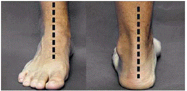
Figure 1
Aligned and stabile talotarsal joint.
Figure 2
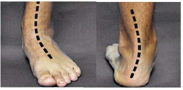
Figure 2
Misaligned and unstable talotarsal joint.
Figure 3
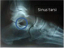
Figure 3
Aligned and stabile talotarsal joint with an “open” sinus tarsi.
Figure 4
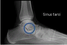
Figure 4
Misaligned and unstable talotarsal joint with a collapsed/obliterated
sinus tarsi.
The Importance of an Aligned and Stable Talotarsal Joint (TTJ)
Walking is one of the most common conscious functions of the
body, next to breathing. The average person takes approximately
6,000 to 8,000 steps a day. A healthy goal is to take at least 10,000
steps at least day. The average person will take more than 72 million
steps by the time they reach 40 years of age and over 100 million steps
by the time they are 60. This is why the alignment and stability of the
foot and ankle is so important.
The entire weight of the body rests on the talotarsal or “foundation
joint.” This complex joint is composed of 4 separate articulations that balance the forces from the body above and the weight bearing
surface below. These forces are subdivided so that slightly more
than half of the force should act posteriorly to the back of the heel
and the remaining forces pass through the front of the foot and out
through the toes Figure 5 and 6. Just as important is the complex
range of motion of the talotarsal joint, supination and pronation.
Supination locks the bones of the foot to make them stable during
the walking cycle. Pronation is the opposite of supination where
there is an unlocking of the foot bones to allow the foot to adapt to
an uneven weightbearing surface. The TTJ should only exhibit a very
slight amount of pronation motion during the beginning portion
of the stance phase of the walking cycle Figure 7. RTTJD creates an
imbalance of joint forces. There is a shift of force from the back of the
foot to the inner-front. Increased forces will now act on the front of
the foot. This means the foot bones will be unlocked and weakened
more than they should. The combination of excessive forces acting
on a weakening joint structure will lead to increased strain to the
ligaments. This signals a nerve reflex mechanism that causes certain
muscle-tendon structures to contract in order to compensate for
the excessive hindfoot motion. Because these excessive forces act on
abnormally while standing, walking, or running, it is only a matter of
time until a critical threshold is reached. At that point, the tissues can
no longer withstand the repeated forces and they become damaged.Eventually, these damaged tissues will cause pain. If the TTJ is not
internally realigned and stabilized, those tissues will continue to
become further damaged until they partially or fully rupture. Once
that occurs, other muscles and tendons will now have to make up for
the failure of those tissues to stabilize the foot Figure 8.
Figure 5
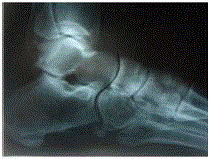
Figure 5
Balanced joint forces
Figure 6
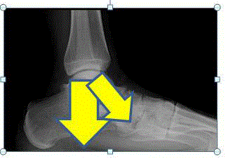
Figure 6
RTTJD shifts the joint forces anteriorly.
Figure 7
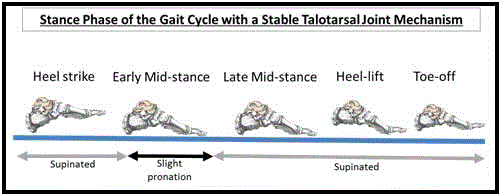
Figure 7
An aligned and stabile talotarsal joint allows a normal amount of
pronation and supination
Figure 8
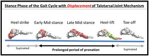
Figure 8
A misaligned and unstable talotarsal joint leads to an excessive
duration of pronation. The supporting tissues have increased strain, and
eventually over-use injuries develop.
Figure 9
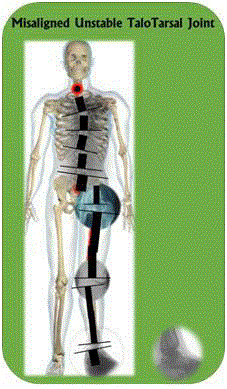
Figure 9
Human amniotic membrane allograft wrapped around the repaired
peroneus brevis tendon.
Figure 10
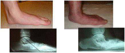
Figure 10
The possible long-term progression of the RTTJD deformity.
Figure 11
Figure 11
Human amniotic membrane allograft wrapped around the repaired
peroneus brevis tendon.
Figure 12
Figure 12
Example of typical hindfoot realignment surgery.
Secondary Symptoms of RTTJD
A symptom is something that is experienced, physically or
mentally, by someone. It is an indication that something is wrong.
For instance, it shouldn’t hurt when you breathe. If it hurts when you
breathe, that means there is a problem. Pain is a warning signal. Pain
is the symptom, but there is an underlying cause to the pain. There
is an underlying etiology to every symptom. However, pain is not
the only “symptom” that would warrant treatment. There are many
diseases where “pain” is not found, yet those diseases are still treated.
There is wisdom in the adage “a chain is as strong as its weakest
link.” This translates to - the weakest tissue of the lower extremity
will become the first to become symptomatic due to RTTJD. For
some individuals, these symptoms show up in childhood, whereas
there are others who don’t develop symptoms until old age. There
are many factors that contribute to the development of symptoms.
But the bottom line is it’s not “if,” it’s “when.” Childhood symptoms
of RTTJ begin with growing pains, shin splints, children not wanting
to participate in sports, wanting to be carried more than usual, to suffering after a period of walking or running Figure 9. Later in
life these RTTJD patients will develop heel pain, heel spurs, plantar
fasciitis, posterior tibial tendon dysfunction, bunions, big toe joint
arthritis, hammertoes, plantar neuropathy, ankle joint problems, knee
joint instability and arthritis, hip pain, sciatica, sacroiliac instability,
and many issues to their spine. Even worse are the long-term health
issues that are related to inactivity. If it hurts when you walk, you
stop walking or exercising. This leads to decreased metabolism and
weight gain. Eventually, the person becomes obese. Obesity is a link
to many other metabolic diseases such as diabetes, hypertension,
heart disease, artery disease, and even certain forms of cancer. The
leading recommendation to these diseases is simply to go out and
walk, to increase their metabolic rate. But the patient is not able to
perform this most simple form of exercise because they suffer, as a
result. Therefore, those diseases slowly get worse and the patients are
never “cured.”
Treatment Option of RTTJD
RTTJD is an internal orthopedic deformity that requires
internal correction. Medical literature has confirmed that external
measures are not able to realign or stabilize the TTJ. The use of
external measures should be considered subtherapeutic and therefore
below the standard-of-care in the treatment of RTTJD. The goal of
“treatment” is that the talus is realigned and stabilized on/with the
calcaneus and navicular bones.
The role of “observation” by the physician should be considered
“medical neglect.” There is no evidence of “auto-correction” of RTTJD.
There are no radiographic studies that show pediatric patients with
RTTJD get better. Rather, the long-term evidence suggests that not
only will it continue to get worse, but that other secondary tissues will
become damaged and destroyed. This is the worst form of “treatment”
that could ever be recommended to a patient with RTTJD Figure 10.
The use of arch supports or foot orthotics/orthosis is a multi-billiondollar
industry, globally. These unregulated devices claim that they
can realign the foot bones, however, the medical literature continues
to disprove these “claims.” [29-44]. There is no published evidence
that an arch support can realign and/or stabilize the talus on the tarsal
bones. Therefore, the claim that these “foot orthotics” can realign
the foot is false advertising. There are studies that showed these foot
supports failed to realign the foot structure; they made it even more
misaligned Figure 11.
Other forms of treatment to realign and stabilize the talus include
aggressive, irreversible surgical procedures. Many surgeons have
advocated cutting and shifting the back portion of the heel bone. This
requires a long period of non-weightbearing and most of the patients
must be taken back to the operating room to have painful screws
removed. The published literature shows that this procedure still
has a limited affect to realign and stabilize the talus [45-53]. Another
option is to cut the outer front of the calcaneus and insert a bone
graft. This again is a long surgery that has shown to cause arthritis
at the adjacent joint and that is not able to realign the TTJ Figure 12.
Finally, the last option is to fuse the posterior subtalar joint. This
is a very destructive, devastating option. The joint-articular surfaces
are resected; the bones are pushed together and one or more screws
are inserted from one bone into the other. The cutting out of the
joints leads to a further loss in height to that limb and will lead to
more secondary pathology to the lower extremity and spine. There
are many patients whose bones don’t fuse that will require more
surgery. Most patients will require the removal of painful screws.
There is a long list of complications including mal-union, nonunion,
and arthritic changes to adjacent joints [54-76]. EOTTS is the
superior treatment of choice for many reasons. Unlike observation,
foot orthotics, and many other surgical procedures, EOTTS is proven
to realign and stabilize the TTJ while still allowing a normal range of
motion [77-92].
Evidence Basis of EOTTS
The primary reason why many in the insurance industry have
denied the reimbursement of the EOTTS procedure is due to “limited
published data” on the effectiveness of EOTTS.
What is the minimum evidence required to show that EOTTS is
an effective treatment option?
1. Is there evidence that the EOTTS procedure maintains the
stability and alignment of the TTJ clinically and radiographically?
YES!
2. Is there evidence that the EOTTS procedure normalizes the range of motion of the TTJ? YES!
3. Is there evidence that the EOTTS procedure normalizes the
joint forces acting on the tarsal mechanism? YES!
4. Is there evidence that the EOTTS procedure decreases
strain acting on supporting soft tissue structures? YES!
5. What are the reported “worst case” complications? Limited
and no long lasting “complications”, simply the stent was removed
without negative long-term issues.
Armed with that information, the insurance carriers should
decide that the EOTTS procedure should be reimbursed.
There have been numerous scientific papers looking at the
short and long-term effects of the EOTTS procedure [77-92]. There
are multiple papers that have proven both clinical and radiologic
normalization of TTJ alignment and stability. Research shows that
following the EOTTS procedure, there are decreased forces acting
on the front of the foot and increased forces acting on the heel [93].
Likewise, there are studies that have shown a significant decrease in
tissue strain and elongation [94-98]. Furthermore, long-term studies
have shown limited post-EOTTS complications. There have only
been a few reported cases of “worst case” complications [99-103].
There estimates that as many as 250,000 EOTTS procedures have
been performed globally. Yet, only a few “complications” have been
reported? The worst of these involved sinus tarsi implant that shifted
180 degrees from where it was initially placed and it became partially
embedded into the ankle bone. The device was removed and there
was no long-term negative effect. The article made it appear as if the
ankle bone was fractured. There are many surgeons who partially
insert screws into either the ankle bone or heel bone in a similar
fashion to block excessive ankle bone motion [104-113]. That is not
a true “complication.” Another “complication” case-report occurred
when the surgeon failed to remove a dislocated stent and allowed the
patient to continue to walk on their foot, knowing full well the stent
was not placed in the desired alignment. Later that patient developed
arthritic changes. The issue is that it occurred “spontaneously” after
more than 2 years. It should have been a malpractice case against the
surgeon, rather than a knock against sinus tarsi implants. This was a
complication that could have been prevented if the surgeon would
have intervened when the initial displacement was visualized on the
x-ray. What has not been reported is that not a single patient ended
up with a serious complication. Typically, the worst thing that can
occur is prolonged pain and the EOTTS implant must be removed.
There is not a single reported case where a patient required more
extensive surgical procedures because of undergoing an EOTTS
procedure. Most times, patients would be told they need more
aggressive surgery and the EOTTS procedure is performed as a more
conservative option.
Benefit-Risk Analysis of EOTTS
A few of the benefits include a minimally invasive procedure that internally realigns and stabilizes the TTJ while still allowing a normal range of motion. This procedure is reversible and can be used in conjunction with both external measures and other surgical procedures. It is commonly performed in both children and adults. Patients, once recovered, can participate in sporting activities of their choosing. Besides the very important benefits listed above, is the fact that not only is the TTJ realigned, but also the normalization of forces acting within the foot structures. Instantly, there are decreased forces acting on the bones, joints, tendons, ligaments, and neurovascular structures of the foot. The ankle joint is realigned, and there are positive effects to the knee, hips, pelvis, and spine. Patients who have had the EOTTS procedure have been able to increase their activity levels, increase their metabolism, decrease their weight, and improve their blood sugar levels and blood pressure. Simply put, they get their lives back. They don’t have to think about whether an activity is worth the pain they would experience due to walking or standing. They just get back to living. What are the potential risks of EOTTS? First off, they are nowhere near the potential risks of traditional corrective surgery. There are no chances of non-union or implant breakage (orthopedic screws have been known to break from timeto- time). The worst possible issue is simply that there is prolonged pain due to soft tissue adaptation. The EOTTS stent can be removed or downsized. Occasionally the stent can displace. This is because it is only anchored by the soft tissues in the sinus tarsi space and many times those tissues no longer exist due to the duration of the RTTJD deformity. Other times, there could be a need to down or up-size the stent. The size of the EOTTS implant is determined while the patient is lying on a procedure table. The best way to determine size would be to insert the stent and have the patient walk. However, this cannot be performed during the procedure, so the surgeon must use their best judgment.
Summary
EOTTS is a time-tested, evidence-based solution that makes sense. The fact that this procedure has been approved for use by the health officials in 70 countries and it is performed by leading orthopedic and podiatric surgeons globally, is very telling. The EOTTS procedure makes sense, fixes the underlying etiology of many chronic lower extremity deformities, and the benefits far outweigh any potential risks. There is a crisis in the treatment of musculoskeletal disorders. The governments around the world are trying to figure out how they can lower healthcare expenditures to chronic diseases. The answer is prevention, yet it is all talk and no action. When one considers the existing healthcare frame of mind, it is focused on symptomrelief rather than fixing the underlying etiology. If you address the symptoms without fixing what is broken, it is only a matter of time until the symptoms reoccur. The underlying problem is still present and will continue to cause the same condition that made that symptom occur in the first place.
References
- Zhang TJ, Wang Y, Lin SJ, Ma X. Correlation between hindfoot joint three-dimensional kinematics and the changes of the medial arch angle in stage II posterior tibial tendon dysfunction flatfoot. Clin Biomech (Bristol, Avon). 2015;30(2):153-8.
- Wearing SC, Smeathers JE, Urry SR, Hennig EM, Hills AP. The pathomechanics of plantar fasciitis. Sports Med. 2006;36(7):585-611.
- Wong DW, Zhang M, Yu J, Leung AK. Biomechanics of first ray hypermobility: an investigation on joint force during walking using finite element analysis. Med Eng Phys. 2014;36(11):1388-93.
- Rao S, Song J, Kraszewski A, Backus S, Ellis SJ, Deland JT, et al. The effect of foot structure on the 1sy metatarsophalangeal joint flexibility and hallucal loading. Gait Posture. 2011;34(1):131-7.
- Gibbs RC, Boxer MC. Abnormal biomechanics of feet and their cause of hyperkeratoses. J Am Acad Dermatol. 1982;6(6):1061-9.
- Barker AR, Rosson GD, Dellon AL. Pressures changes in the medial and lateral plantar and tarsal tunnels related to ankle position: a cadaver study. Foot Ankle Int. 2007;28(2):250-4.
- Nubé VL, Molyneaux L, Yue DK. Biomechanical risk factors associated with neuropathic ulceration of the hallux in people with diabetes mellitus. J Am Podiatr Med Assoc. 2006;96(3):189-97.
- Bonnel F, Toullec E, Mabit C, Tourne Y, Sofcot. Chronic ankle instability: biomechanics and pathomechanics of ligaments injury and associated lesions. Orthop Traumatol Surg Res. 2010;96(4):424-32.
- Friedman MA, Draganich LF, Toolan B, Brage ME. The effects of adult acquired flatfoot deformity on tibiotalar joint contact characteristics. Foot Ankle Int. 2001;22(3):241-6.
- Valderrabano V, Horisberger M, Russell I, Dougall H, Hintermann B. Etiology of ankle osteoarthritis. Clin Orthop Relat Res. 2009;467(7):1800-6.
- Winkelmann ZK, Anderson D, Games KE, Eberman LE. Risk Factors for Medial Tibial Stress Syndrome in Active Individuals: An Evidence-Based Review. J Athl Train. 2016;51(12):1049-1052.
- Delacerda FG. A study of anatomical factors involved in shinsplints. J Orthop Sports Phys Ther. 1980;2(2):55-9.
- Nigg BM1. The role of impact forces and foot pronation: a new paradigm. Clin J Sport Med. 2001;11(1):2-9.
- Van Gheluwe B, Kirby KA, Hagman F. Effects of simulated geu valgum and genu varum on ground reaction forces and subtalar joint function during gait. J Am Podiatr Med Assoc. 2005;95(5):531-41.
- Rodriques P, Change R, TenBroek T, van Emmerik R, Hamill J. Evaluating the coupling between foot pronation and tibial internal rotation continuously using vector coding. J Appl Biomech. 2015;31(2):88-94.
- Levinger P, Menz HB, Fotoohabadi MR, Feller JA, Bartlett RJ, Bergman NR. Foot posture in people with medial compartment knee osteoarthritis. J Foot Ankle Res. 2010;3:29.
- Hertel J, Dorman JH, Braham RA. Lower extremity malalignments and anterior cruciate ligament injury history. J Sports Sci Med. 2004;3(4):220-5.
- Rothbart BA. Relationship of functional leg-length discrepancy to abnormal pronation. J Am Podiatr Med Assoc. 2006;96(6):499-504.
- Duval K, Lam T, Sanderson D. The Mechanical relationship between the rearfoot, pelvis and low-back. Gait Posture. 2010;32(4):637-40.
- Khamis S, Dar G, Peretz C, Yizhar Z. The relationship between foot and pelvic alignment while standing. J Hum Kinet. 2015;46:85-97.
- Duval K, Lam T, Sanderson D. The mechanical relationship between the rearfoot, pelvis and low-back. Gait Posture. 2010;32(4):637-40.
- Cibulka MT. Low back pain and its relation to the hip and foot. J Orthop Sports Phys Ther. 1999;29(10):595-601.
- Farahpour N, Jafanezhad A, Damavandi M, Bakhtiari A, Allard P. Gait ground reaction force characteristics of low back pain patients with pronated foot and able-bodied individuals with and without foot pronation. J Biomech. 2016;49(9):1705-10.
- Khamis S, Dar G, Peretz C, Yizhar Z. The Relationship Between Foot and Pelvic Alignment While Standing. J Hum Kinet. 2015;46:85-97.
- Farokhmanesh K, Shirzadian T, Mahboubi M, Shahri MN. Effect of foot hyperpronation on lumbar lordosis and thoracic kyphosis in standing position using 3-dimensional untrasound-based motion analysis system. Glob J Health Sci. 2014;6(5):254-60.
- Khamis S, Yizhar Z. Effect of feet hyperpronation on pelvic alignment in a standing position. Gait Posture. 2007;25(1):127-34.
- Menz HB, Dufour AB, Riskowski JL, Hillstrom HJ, Hannan MT. Foot posture, foot function and low back pain: the Framingham Foot Study. Rheumatology (Oxford). 2013;52(12):2275-82.
- Barwick A, Smith J, Chuter V. The relationship between foot motion and lumbopelvic-hip function: a review of the literature. Foot (Edinb). 2012;22(3):224-31.
- Steber S, Kolodziej L. Analysis of Radiographic Outcomes Comparing Foot Orthosis to Extra-Osseous Talotarsal Stabilization in the Treatment of Recurrent TaloTarsal Joint Dislocation. J Min Inv Orthop. 2015;2(1):8.
- Banwell HA, Mackintosh S, Thewlis D. Foot orthoses for adults with flexible pes planus: a systematic review. J Foot Ankle Res. 2014;7(1):23.
- Reina M, Lafuente G, Munuera PV. Effect of custom-made foot orthoses in female hallux valgus after one-year follow up. Prosthet Orthot Int. 2013;37(2):113-9.
- Bok SK, Kim BO, Lim JH, Ahn SY. Effects of custom-made rigid foot orthosis on pes planus in children over 6 years old. Ann Rehabil Med. 2014;38(3):369-75.
- Andreasen J, Molgaard CM, Christensen M, Kaalund S, Lundbye-Christensen S, Simonsen O, et al. Exercise therapy and custom-made insoles are effective in patients with excessive pronation and chronic foot pain- a randomized controlled trial. Foot (Edinb). 2013;23(1):22-8.
- Eslami M, Ferber R. Can orthoses and navicular drop affect foot motion patterns during running? J Sci Med Sport. 2013;16(4):377-81.
- Cheung RT, Chung RC, Ng GY. Efficacies of different external controls for excessive foot pronation: a meta-analysis. Br J Sports Med. 2011;45(9):743-51.
- Hirano T, McCullough MB, Kitaoka HB, Ikoma K, Kaufman KR. Effects of foot orthoses on the work of friction of the posterior tibial tendon. Clin Biomech (Bristol, Avon). 2009;24(9):776-80.
- Whitford D, Esterman A. A randomized controlled trial of two types of in-shoe orthoses in children with flexible excess pronation of the feet. Foot Ankle Int. 2007;28(6):715-23.
- Christopher RC, Drouin JM, Houglum PA. The influence of a foot orthotic on lower extremity transverse plane kinematics in collegiate females athletes with pes planus. J Sports Sci Med. 2006;5(4):646-55.
- Rome K, Brown CL. Randomized clinical trial into the impact of rigid foot orthoses on balance parameters in excessively pronated feet. Clin Rehabil. 2004;18(6):624-30.
- Tochigi Y. Effect of arch supports on ankle-subtalar complex instability: a biomechanical experimental study. Foot Ankle Int. 2013;24(8):634-9.
- Rose HM, Shultz SJ, Arnold BL, Gansneder BM, Perrin Dh. Acute orthotic intervention does not affect muscular response times and activation patterns at the knee. J Athl Train. 2002;37(2):133-40.
- Kitaoka HB, Luo ZP, Kura H, An KN. Effect of foot orthoses on 3-dimensional kinematics of flatfoot: a cadaveric study. Arch Phys Med Rehabil. 2002;83(6):876-9.
- Nester CJ, Hutchins S, Bowker P. Effect of foot orthoses on rearfoot complex kinematics during walking gait. Foot Ankle Int. 2001;22(2):133-8.
- Kilmartin TE, Barrington RL, Wallace WA. A controlled prospective trial of a foot orthosis for juvenile hallux valgus. J Bone Joint Surg Br. 1994;76(2):210-4.
- Jastifer JR, Coughlin MJ. Hindfoot deformity and calcaneal tuberosity osteotomies. Foot Ankle Spec. 2015;8(1):50-8.
- Cao HH, Tang KL, Lu Wz, Wu JZ. Medial displacement calcaneal osteotomy with posterior tibial tendon reconstruction for the flexible flatfoot with symptomatic accessory navicular. J Foot Ankle Surg. 2014;53(5):539-43.
- Klaue K, Kaliyaperumal K, Swanson SA, Low WC. Central calcaneal osteotomy for correction of flexible pes planovalgus deformity. Foot Ankle Int. 2013;34(8):1079-89.
- Feuerstein CA, Weil L Jr, Weil LS Sr, Klein EE, Agerakis NG, Akram U. The calcaneal scarf osteotomy: surgical correction of the adult acquired flatfoot deformity and radiographic results. Foot Ankle Spec. 2013;6(5):367-71.
- Iossi M, Johnson JE, McCormick JJ, Klein SE. Short-term radiographic analysis of operative correction of adult acquired flatfoot deformity. Foot Ankle Int. 2013;34(6):781-91.
- Scott RT, Berlet GC. Calcaneal Z osteotomy for extra-articular correction of hindfoot valgus. J Foot Ankle Surg. 2013;52(3):406-8.
- Chan JY, Williams BR, Nair BR, Nair P, Young E, Sofka C, et al. The contribution of the medializing calcaneal osteotomy on hindfoot alignment in the reconstruction of the stage II adult acquired flatfoot deformity. Foot Ankle Int. 2013;34(2):159-66.
- Guha AR, Perera AM. Calcaneal osteotomy in the treatment of adult acquired flatfoot deformity. Foot Ankle Clin. 2012;17(2):247-58.
- Lee MS. Posterior calcaneal displacement osteotomy for the adult acquired flatfoot. Clin Podiatr Med Surg. 2005;22(2):277-89, vii.
- Yang C, Xu X, Zhu Y, Liu J, Wei B. A Long-Term Study of the Effect of Subtalar Arthrodesis on the Ankle and Hindfoot Joints. J Am Podiatr Med Assoc. 2016;106(1):47-53.
- Hutchinson ID, Baxter JR, Gilbert S, Hogan MV, Ling J, Saunders SM, et al. How Do Hindfoot Fusions Affect Ankle biomechanics: A Cadaver Model. Clin Orthop Relat Res. 2016;474(4):1008-16.
- Hentges MJ, Gesheff MG, Lamm BM. Realignment Subtalar Joint Arthrodesis. J Foot Ankle Surg. 2016;55(1):16-21.
- Xu X, yang C, Zhu Y, Liu J, Wei B. A retrospective clinical study on 37 subtalar arthrodesis patients of nine years follow up. J Am Podiatr Med Assoc. 2015.
- Röhm J, Zwicky L, Horn Lang T, Salentiny Y, Hintermann B, Knupp M. Mid- to long-term outcome of 96 corrective hindfoot fusions in 84 patients with rigid flatfoot deformity. Bone Joint J. 2015;97-B(5):668-74.
- Lin JS, Myerson MS. The management of complications following the treatment of flatfoot deformity. Instr Course Lect. 2011;60:321-34.
- Hermus JP. Osteonecrosis of the talus after talonavicular arthrodesis: a case report and review of the literature. J Foot Ankle Surg. 2011;50(3):343-6.
- Iaguinto JM, Wayne JS. Effects of surgical correction for the treatment of adult acquired flatfoot deformity: a computational investigation. J Orthop Res. 2011;29(7):1047-54.
- Jordan TH, Rush SM, Hamilton GA, Ford LA. Radiographic outcomes of adult acquired flatfoot corrected by medial column arthrodesis with or without a medializing calcaneal osteotomy. J Foot Ankle Surg. 2011;50(2):176-81.
- Basioni Y, El-Ganainy AR, El-Hawary A. Double calcaneal osteotomy and percutaneous tenoplasty for adequate arch restoration in adult flexible flatfoot. Int Orthop. 2011;35(1):47-51.
- Frost NL, Grassbaugh JA, baird G, Caskey P. Triple arthrodesis with lateral column lengthening for the treatment of planovalgus deformity. J Pediatr Orthop. 2011;31(7):773-82.
- Camasta CA, Menke CR, Hall PB. A review of 51 talonavicular joint arthrodeses for flexible pes valgus deformity. J Foot Ankle Surg. 2010;49(2):113-8.
- Haeseker GA, Mureau MA, Faber FW. Lateral column lengthening for acquired adult flatfoot deformity caused by posterior tibial tendon dysfunction stage II: a retrospective comparison of calcaneus osteotomy with calcaneocuboid distraction arthrodesis. J Foot Ankle Surg. 2010;49(4):380-4.
- Joveniaux P, Harisboure A, Ohl X, Dehoux E. Long-term results of in situ subtalar arthrodesis. Int Orthop. 2010;34(8):1199-205.
- Tuijthof GJ, Beimers L, Kerkhoffs GM, Dankelman J, Dijk CN. Overview of subtalar arthrodesis techniques: options, pitfalls and solutions. Foot Ankle Surg. 2010;16(3):107-16.
- de Groot IB, Reijman M, Luning HA, Verhaar JA. Long-term results after a triple arthrodesis of the hindfoot: function and satisfaction in 36 patients. Int Orthop. 2008;32(2):237-41.
- Bolt PM, Coy S, Toolan BC. A comparison of lateral column lengthening and medial translational osteotomy of the calcaneus for the reconstruction of the adult acquired flatfoot. Foot Ankle Int. 2007;28(11):1115-23.
- Francisco R, Chiodo CP, Wilson MG. Management of the rigid adult acquired flatfoot deformity. Foot Ankle Clin. 2007;12(2):317-27, vii.
- Van der Krans A, Louwerens JW, Anderson P. Adult acquired flexible flatfoot, treated by calcaneocuboid distraction arthrodesis, posterior tibial tendon augmentation, and percutaneous Achilles tendon lengthening: a prospective outcome study of 20 patients. Acta orthop. 2006;77(1):156-63.
- Kadakia AR, Haddad SL. Hindfoot arthrodesis for the adult acquired flat foot. Foot Ankle Clin. 2003;8(3):569-94, x.
- Maenpaa H, Lehto MU, Belt EA. What went wrong in triple arthrodesis? An analysis of failures in 21 patients. Clin Orthop Relat Res. 2001;391:218-23.
- Pell RF 4th, Myerson MS, Schon LC. Clinical outcome after primary triple arthrodesis. J Bone Joint Surg Am. 2000;82(1):47-57.
- Cooper PS, Nowak MD, Shaer J. Calcaneocuboid joint pressures with lateral column lengthening (Evans) procedure. Foot Ankle Int. 1997;18(4):199-205.
- Steber S, Kolodziej L. Analysis of Radiographic Outcomes Comparing Foot Orthosis to Extra-Osseous Talotarsal Stabilization in the Treatment of Recurrent TaloTarsal Joint Dislocation. J Min Inv Orthop. 2015;1-11.
- Graham ME, Jawrani NT, Chikka A, Rogers RJ. Surgical Treatment of Hyperpronation Using an Extra-Osseous TaloTarsal Stabilization Device: Radiographic Outcomes in Adult Patients. J Foot Ankle Surg. 2012;51(5):548-55.
- Graham ME, Jawrani NT, Chikka A. Radiographic Evaluation of Navicular Position in the Sagittal Plane – Correction Following an Extra-Osseous TaloTarsal Stabilization Procedure. J Foot Ankle Surg. 2011;50(5):551-7.
- Chong DY, Macwilliams BA, Hennessey TA, Teske N, Stevens PM. Prospective comparison of subtalar arthroereisis with lateral column lengthening for painful flatfeet. J Pediatr Orthop B. 2015;24(4):345-53.
- Jay RM, Din N. Correcting pediatric flatfoot with subtalar arthroereisis and gastrocnemius recession: a retrospective study. Foot Ankle Spec. 2013;6(2):101-7.
- Baker JR, Klein EE, Weil L JR, Weil LS Sr, Knight JM. Retrospective analysis of the survivability of absorbable versus nonabsorbable subtalar joint arthroereisis implants. Foot Ankle Spec. 2013;6(1):36-44.
- Fernandez de Retana P, Alvarez F, Bacca G. Is there a role for subtalar arthroereisis in the management of adult acquired flatfoot. Foot Ankle Clin. 2012;17(2):271-81.
- Hazany S, Ly N, Hazany D, Bader S, Ostuka N. Outcomes of subtalar arthroereisis for the planovalgus foot. J Surg Orthop Adv. 2012;21(3):147-50.
- Brancheau SP, Walker KM, Northcutt DR. An analysis of outcomes after use of the Maxwell-Brancheau Arthroereisis implant. J Foot Ankle Surg. 2012;51(1):3-8.
- Metcalfe SA, Bowling FL, Reeves ND. Subtalar joint arthroereisis in the management of pediatric flexible flatfoot: a critical review of the literature. Foot Ankle Int. 2011;32(12):1127-39.
- Fernández de Retana P, Alvarez F, Viladot R. Subtalar arthroereisis in pediatric flatfoot reconstruction. Foot Ankle Clin. 2010;15(2):323-35.
- Scharer BM, Black BE, Sockrider N. Treatment of painful pediatric flatfoot with Maxwell-Brancheau subtalar arthroereisis implant a retrospective radiographic review. Foot Ankle Spec. 2010;3(2):67-72.
- Koning PM, Heesterbeek PJ, de Visser E. Subtalar arthroereisis for pediatric flexible pes planovalgus: fifteen years’ experience with the cone-shaped implant. J Am Podiatr Med Assoc. 2009;99(5):447-53.
- Adelman VR, Szczepanski JA, Adelman RP. Radiographic evaluation of endoscopic gastrocnemius recession, subtalar joint arthroereisis, and flexor tendon transfer for surgical correction of stage II posterior tibial tendon dysfunction: a pilot study. J Foot Ankle Surg. 2008;47(5):400-8.
- Cicchinelli LD, Pascual Huerta J, Carcia Carmona FJ, Fernandez Morato D. Analysis of gastrocnemius recession and medial column procedures as adjuncts in arthroereisis for the correction of pediatric pes planovalgus: a radiographic retrospective study. J Foot Ankle Surg. 2008;47(5):385-91.
- Soomekh DJ, Baravaraian B. Pediatric and adult flatfoot reconstruction: subtalar arthroereisis versus realignment osteotomy surgical options. Clin Podiatr Med Surg. 2006;23(4):695-708.
- Graham ME, Parikh R, Goel V, Mhatre D, Matyas A. Stabilization of joint forces of the subtalar complex via HyProCure sinus tarsi stent. J Am Podiatr Med Assoc. 2011;101(5):390-9.
- Graham ME, Jawrani, NT, Goel VK. Effect of Extra-Osseous TaloTarsal Stabilization on Posterior Tibial Tendon Strain in Hyperpronating Feet. J Foot Ankle Surg. 2011;50(6):676-81.
- Graham ME, Jawrani NT, Goel VK. Evaluating plantar fascia strain in hyperpronating cadaveric feet following an extra-osseous talotarsal stabilization procedure. J Foot Ankle Surg. 2011;50(6):682-6.
- Graham ME. Jawrani NT, Goel VK. Effect of extra-osseous talotarsal stabilization on posterior tibial nerve strain in hyperpronating feet: a cadaveric evaluation. J Foot Ankle Surg. 2011;50(6):672-5.
- Graham ME, Jawrani NT, Goel VK. The effect of HyProCure(®) sinus tarsi stent on tarsal tunnel compartment pressures in hyperpronating feet. J Foot Ankle Surg. 2011;50(1):44-9.
- Corpuz M, Shofler D, Labovitz J, Hodor L, Yu K. Fracture of the talus as a complication of subtalar arthroereisis. J Foot Ankle Surg. 2012;51(1):91-4.
- Van Ooij B, Vos CJ, Saouti R. Arthroereisis of the subtalar joint: an uncommon complication and literature review. J Foot Ankle Surg. 2012;51(1):114-7.
- Cook EA, Cook JJ, Basile P. Identifying risk factors in subtalar arthroereisis explantation: a propensity-matched analysis. J Foot Ankle Surg. 2011;50(4):395-401.
- Lui TH. Spontaneous subtalar fusion: an irreversible complication of subtalar arthroereisis. J Foot Ankle Surg. 2014;53(5):652-6.
- Kumar V, Clough TM. Talar neck fracture -a rare by important complication following subtalar arthroereisis. Foot (Edinb). 2014;24(4):169-71.
- Samaila E, Bonetti I, Bruno C, Argentini E, Magnan B. Navicular tenosuspension with anterior tibialis tendon (Young procedure) associated to calcaneo-stop for the treatment of paediatric flexible flatfoot clinical and ultrasound study. Acta Biomed. 2016;87(1S):69-74.
- Calvo Calvo S, Marti Niruelos R, Rasero Ponferrada M, Gonzaled de Orbe G, Vina Fernandez R. More than 10 years of follow of the stop screw technique. Rev Esp Cir Ortop Traumatol. 2016;60(1):75-80.
- Abbara-Czardybon M, Frank D, Arbab D. The talus stop screw arthroereisis for flexible juvenile pes planovalgus. Oper Orthop Traumtol. 2014;26(6):625-31.
- Pavone V, Costarella L, Testa G, Conte G, Riccioli M, Sessa G. Calcaneo-stop procedure in the treatment of the juvenile symptomatic flatfoot. J Foot Ankle Surg. 2013;52(4):444-7.
- Richter M, Zech S. Arthrorisis with calcaneostop screw in children corrects Talo-1st Metatarsal-Index (TMT-Index). Foot Ankle Surg. 2013;19(2):91-5.
- Usuelli FG, Montrasio UA. The calcaneo-stop procedure. Foot Ankle Clin. 2012;17(2):183-94.
- Kellermann P, Roth S, Gion K, Boda K, Tóth K. Calcaneo-stop procedure for paediatric flexible flatfoot. Arch Orthop Trauma Surg. 2011;131(10):1363-7.
- Jerosch J, Schunck J, Abdel-Aziz H. The stop screw technique - a simple and reliable method in treating flexible flatfoot in children. Foot Ankle Surg. 2009;15(4):174-8.
- Roth S, Sestan B, Tudor A, Ostojic Z, Sasso A, Durbesic A. Minimally invasive calcaneo-stop method for idiopathic, flexible pes planovalgus in children. Foot Ankle Int. 2007;28(9):991-5.
- De Pellegrin M1. [Subtalar screw-arthroereisis for correction of flat foot in children]. Orthopade. 2005;34(9):941-53, quiz 954.