Case Report
Destination Therapy with the Heartware HVAD Ventricular Assist Device for Systemic Ventricular Failure with Transposition of the Great Arteries Corrected by the Mustard Procedure
Babar B Chaudhri*
Department of Cardiac Surgery, KG Mittal Hospital, Mumbai 400004, India
*Corresponding author: Babar B. Chaudhri, Department of Cardiac Surgery, KG Mittal Hospital, Marine Drive, Mumbai 400004, India
Published: 21 Sep, 2017
Cite this article as: Chaudhri BB. Destination Therapy with
the Heartware HVAD Ventricular Assist
Device for Systemic Ventricular Failure
with Transposition of the Great Arteries
Corrected by the Mustard Procedure.
Clin Surg. 2017; 2: 1631.
Abstract
Systemic ventricular failure is a recognised late consequence of the atrial switch procedure for transposition of the great arteries. There is a significant population status post atrial switch. We report one such patient with a failing Mustard procedure who was treated by surgical implantation of a Heartware HVAD rotary blood pump into the systemic ventricle.
Introduction
Following atrial correction for transposition of the great arteries late systemic ventricular failure may occur [1,2]. The pumping chamber in the systemic position is the morphological right ventricle and is prone to failure in this situation. In contrast the morphologically left ventricle is well placed in the pulmonary circulation. These are an attractive subset of patients in terms of candidacy for destination therapy for ventricular assist device placement. This is especially reinforced by the paucity of donor hearts available for transplantation. We report the successful implantation of the Heartware HVAD ventricular assist device (Heartware Inc, Framingham, MA) into the failing systemic ventricle as a strategy for destination therapy.
Patients and Methods
The patient was a 37 year old male with transposition of the great arteries and left SVC. He was palliated with a right classical Blalock Taussig (BT) shunt. AT 18 months of age he underwent atrial switch with a Mustard procedure and take down of the BT shunt. In the interval leading to this presentation he sustained a perioperative right hemiplegia with full resolution, and severe failure of his systemic ventricle (morphologically right ventricle) with severe tricuspid regurgitation. A biventricular pacemaker and automated implantantble cardioverter defibrillator were implanted. There was occlusion of the SVC baffle. There was evidence of end organ dysfunction. His renal function was impaired (creatinine 375 μM/L). Echocardiography showed impaired biventricular function and severe tricuspid regurgitation. Cardiac catheterisation was undertaken. This showed that the systemic Sao2 of 95% and mixed venous of 50% with no shunts (Figure 1). The systemic venous pressure was 15 mm Hg with the pulmonary/morphologically left ventricular pressure of 80/14. The mean PA was 46 mmHg. The systemic/morphologically right ventricular end diastolic pressure was 14-18mmHg. The calculated PVR was in excess of 10 Wood units. The cardiac index of 2l/min/m2. The decision was taken to implant a VAD and support the failing systemic ventricle. Full cardiopulmonary bypass was instituted via femoral venous and arterial cannulation. Cooling was commenced to 32oC. Repeat sternotomy was safely performed. The inferior wall of the morphological RV was chosen. The sewing ring was implanted in this position. The heart was fibrillated, the aorta was not cross-clamped. A considerable amount of trabeculated myocardium was excised in order to allow unobstructed inflow into the VAD. The inflow was carefully positioned to face the atrioventricular valve. The outflow graft was anastomosed to the ascending aorta. The position of the inflow cannula was confirmed by intraoperative transoesophageal echocardiography (TOE). The VAD was started once weaned from cardiopulmonary bypass. TOE confirmed adequate decompression of the systemic ventricle and a balanced position of the interventricular septum. The total cardiopulmonary bypass time was 52 minutes. The patient was managed in the intensive care unit. His circulation was managed with an infusion of milrinone which was discontinued. Anticoagulation was commenced with warfarin and anti platelet therapy. He was safely discharged home and remains well and active.
Figure 1
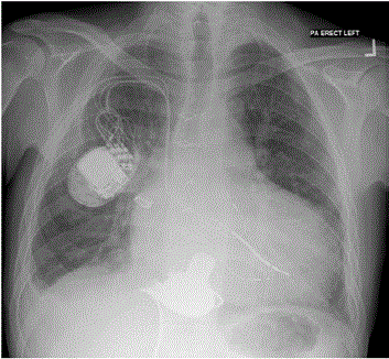
Figure 1
Chest radiograph showing the implanted Heartware HVAD
posteriorly positioned in the systemic ventricle.
Discussion
An atrial switch operation for transposition of the great arteries has been superseded by the arterial switch operation3. There is a significant population of individual’s status post atrial switch, who survive beyond the second decade of life. 30 year survival is 79.3% [1,2]. The incidence of sudden death is 7% [2]. Right ventricular failure has been observed in long-term cohorts ranging from 18% to 44% [1,2]. Baffle obstruction, occurred much more frequently in patients with a Mustard procedure. Usage of VADs in the setting of congenital heart disease is extremely rare [4-6]. LVAD flow is dependent upon RV function to facilitate device inflow. For all indications for LVADs, a major limiting factor is the unpredictable nature of the RV which may fail and thus necessitate biventricular support [7]. Their use in patients with a failing systemic ventricle post atrial switch is attractive particularly as the durable morphological LV is in the pulmonary position, thus making it less likely to fail. The Heartware HVAD is a third generation rotary blood pump with magnetically levitated rotors. It has the advantage of being small with a short inflow cannula. This allows for complete intrapericardial placement, eliminating the need for a pump pocket and ease of surgical implantation. In addition it is easy to orientate the inflow cannula to face the atrioventricular valve, thereby optimizing ventricular unloading. Initial clinical experience with this device has been reported as extremely favorable [8] and longer term data is emerging regarding its efficacy. In particular this device has been implanted in the right ventricle as well as the left in one patient, and has been reported as providing biventricular support. In the technique described by Strueber et al. [9] the outflow graft of the RVAD was constricted in order to provide sufficient after load to allow the pump to generate requisite physiological flows. In the situation of a patient following atrial switch, the morphological RV is already working against systemic vascular resistance and thus this modification is not required. The growing incidence of heart failure worldwide is coupled to a lack of suitable heart donors for transplantation. This has generated demand for durable mechanical blood pumps. We have demonstrated the utility of VAD implantation using the Heartware HVAD in a patient with a failing Mustard procedure.
References
- Moons P, Gewillig M, Sluysmans T, Verhaaren H, Viart P, Massin M, et al. Long term outcome up to 30 years after the Mustard or Senning operation: a nationwide multicentre study in Belgium. Heart. 2004;90(3):307-13.
- Wilson NJ, Clarkson PM, Barratt-Boyes BG, Calder AL, Whitlock RM, Easthope RN, et al. Long-term outcome after the mustard repair for simple transposition of the great arteries. 28-year follow-up. J Am Coll Cardiol. 1998;32(3):758-65.
- Yacoub MH, Kakihara R, Arensman FW, Radley-Smith R. Current status of arterial switch operation for transposition of the great arteries. Nippon Kyobu Geka Gakkai Zasshi. 1983;31(5):623-33.
- Wiklund L, Svensson S, Berggren H. Implantation of a left ventricular assist device, back-to-front, in an adolescent with a failing mustard procedure. J Thorac Cardiovasc Surg. 1999;118(4):755-6.
- Mohapatra B, Vick GW 3rd, Fraser CD Jr, Clunie SK, Towbin JA, Sinagra G, et al. Short-term mechanical unloading and reverse remodeling of failing hearts in children. J Heart Lung Transplant. 2010;29(1):98-104.
- Kirklin JK, Naftel DC, Kormos RL, Stevenson LW, Pagani FD, Miller MA, et al. Second INTERMACS annual report: more than 1,000 primary left ventricular assist device implants. J Heart Lung Transplant. 2010;29(1):1-10.
- Slaughter MS, Pagani FD, Rogers JG, Miller LW, Sun B, Russell SD, et al. Clinical management of continuous-flow left ventricular assist devices in advanced heart failure. J Heart Lung Transplant. 2010;29(4):S1-39.
- Wieselthaler GM, Driscoll O, Jansz P, Khaghani A, Strueber M. Initial clinical experience with a novel left ventricular assist device with a magnetically levitated rotor in a multi-institutional trial. J Heart Lung Transplant. 2010;29(11):1218-25.
- Strueber M, Meyer AL, Malehsa D, Haverich A. Successful use of the Heart Ware HVAD rotary blood pump for biventricular support. J Thorac Cardiovasc Surg. 2010;140(4):936-7.