Case Report
Charcot Cysts: A Rarely Reported Finding
Jaminelli Banks, Annie Jong and Robert Frykberg*
Department of Podiatry, University of Arizona College of Medicine-Phoenix, Phoenix VA Health Care System, USA
*Corresponding author: Robert Frykberg, Department of Podiatry, University of Arizona College of Medicine-Phoenix, Phoenix VA Health Care System, 650 East Indian School Road, Phoenix, Arizona 85012, USA
Published: 21 Sep, 2017
Cite this article as: Banks J, Jong A, Frykberg R. Charcot
Cysts: A Rarely Reported Finding. Clin
Surg. 2017; 2: 1630.
Abstract
Aims/Goals: Charcot arthropathy is a debilitating condition affecting the lower extremity of persons
with long established peripheral neuropathy. Deformities of the midfoot can place the foot at risk
of ulceration and subsequent amputation if infection ensues. Although surgical management has
become much more common for this disorder, there has been little, if any, mention of a peculiar
cystic proliferation noted in the deep soft tissues upon surgical dissection. We herein report our
observations on several patients who were found to have what we have termed “Charcot Cysts.”
Methods: Three type 2 diabetic patients are presented who reported to our High Risk Foot Clinic
with Charcot arthropathy of the midfoot represented by a collapse of the midfoot and loss of
calcaneal inclination. These patients were treated per our customary treatment of wound care,
offloading boots, and instructions for non weight bearing as indicated. Although all patients were
given instructions and supplies for wound care and provided with therapeutic footwear appropriate
for their condition, two patients persisted with midfoot ulceration. The other went on to develop
a plantar soft tissue mass. Despite further casting, the ulcers and mass remained recalcitrant to
conservative care. All patients were taken to surgery for a simple plantar exostectomy to reduce the
plantar bony prominence underlying the chronic ulceration or mass.
Results: Upon deep exploration a translucent, fluctuant, multi-loculated cystic mass was encountered
that extended throughout a considerable portion of the dissection. Upon resection, the masses were
found to be spongy and contained what appeared to be synovial fluid. Pathology confirmed that
these were simple benign cysts. Importantly, however, we found a delay in wound healing caused by
persistent synovial fluid leakage.
Conclusions: This rarely reported complication of the diabetic Charcot foot has been noted by the
senior author (RGF) in many such operative patients over the last several decades. Since most patients
with Charcot foot are treated conservatively, these cysts are not usually recognized surgically. Once
the foot becomes ulcerated or is treated surgically, however, the Charcot cysts can be shown to have
impeded normal wound healing. While the pathogenesis of Charcot neuroarthropathy has been
previously described, the formation of such related cysts has yet to be explained. We, therefore,
encourage clinicians and surgeons to corroborate our findings with further study of this interesting
pathology.
Keywords: Charcot; Charcot foot; Charcot arthropathy; Charcot neuroarthropathy; Cyst;
Synovial cyst
Introduction
Charcot Neuropathic osteoarthropathy (CN), commonly referred to as the Charcot foot is a
serious and potentially limb-threatening lower-extremity complication of diabetes. First described in
1883, by a French neurologist, Jean-Martin Charcot (1825–1893), this condition continues to remain
a challenge even for the most experienced practitioners [1,2]. Now considered an inflammatory
syndrome, the diabetic Charcot foot is characterized by varying degrees of destruction of osseous
and articular structures of the foot and ankle joint secondary to underlying neuropathy, trauma,
and perturbations of bone metabolism [3,4]. It has been suggested that the onset of the condition is
triggered by a preceding event such as minor trauma, previous ulcer, infection or foot surgery with
the common factor to all of these events is local inflammation [5].
Charcot arthropathy is a debilitating condition affecting the lower extremity of persons with
long established peripheral neuropathy caused by many complicated yet interconnected etiologies;
however, diabetic neuropathy has become the most common etiology [3,6-8]. The interaction of
certain key factors (diabetes, sensory-motor neuropathy, autonomic neuropathy, trauma, and
metabolic abnormalities of bone) results in an acute localized inflammatory condition that may lead to varying degrees and patterns of bone destruction, subluxation, dislocation, and deformity. The hallmark deformity associated with
this condition is midfoot collapse, described as a “rocker-bottom”
foot [3,9]. Deformities of the midfoot can place the foot at risk of
ulceration and subsequent amputation if infection ensues. Annually,
$25 billion are expended for the treatment of chronic wounds, with
the number growing due to the aging population and increased
incidence of diabetes and obesity [10]. Therefore a timely, orderly and
effective wound management and treatment are crucial.
In poorly controlled diabetics, peripheral sensory neuropathy
allows for repetitive micro or macro trauma to the foot and ankle
[11]. This causes an increased release of inflammatory cytokines IL-
6, IL-1, and TNF-a, which, in turn, promotes osteoclast recruitment,
differentiation, and proliferation [12]. Receptor activator of nuclear
factor-kappaB (RANK) ligand (RANKL) has been identified as an
essential mediator of osteoclast formation and activation. RANKL
mediates the process of osteoclastogenesis by binding to its RANK,
which is expressed on mononuclear osteoclast precursors [13,14].
The effects of RANKL–RANK interaction are physiologically
counterbalanced by osteoprotegerin (OPG), which acts as a soluble
receptor decoy for RANKL and blocks the interaction of RANKL with
RANK [15-17]. The ratio of RANKL to OPG has been suggested to
regulate the extent of osteoclast formation and resorption. Therefore,
any alteration in the RANKL/OPG ratio could be critical in the
pathogenesis of osteolytic bone disorders [14-18]. The likelihood
that this pathway is involved is increased by the fact that the same
signaling system, RANKL/OPG, is also intimately involved in the
process of calcification of the media of arterial cell walls and such
calcification is a feature of diabetic neuropathy and especially of CN
[19]. The association between neuropathy and increased osteoclastic
activity can also be attributed to Calcitonin Gene Related Peptide
(CGRP), a neuropeptide that functions within the circulatory and
digestive systems and aids in the maintenance and growth of stem
cells [16-20]. Decreased levels of CGRP have been demonstrated
in study specimens with Charcot neuroarthropathy and diabetic
neuroarthropathy [21]. Because CGRP also acts as a RANKL
inhibitor, antagonizing its functions in osteocalstogenesis and bone
resorption, a decrease in CGRP allows for increased RANKL and
receptor binding, leading to unrestrained bony resorption [16-21].
Furthermore, decreases in nitric oxide synthase (eNOS) have also
been liked to diabetic sensory neuropathy [22]. Nitric Oxide (NO),
a free radical mediated by eNOS, functions at low levels to promote
osteoclastic bone resorption [23]. A decrease in NO levels, thus,
potentiates unrestricted osteoclast proliferation and bony resorption.
Poor glycemic control also produces an increase in advanced
glycation end products (AGE) [24], glycated compounds that form
due to glucose exposure [25]. These products within the intra and
extra cellular environment contribute too many of the vascular and
nephrologic complications of diabetes [25]. Along with RANKL,
however, AGE has also been demonstrated to play a role in inhibiting
osteoblastic proliferation and differentiation [26] and enhancing
induced osteoclastogenesis [27]. This is yet another inflammatory
pathway associated with Charcot arthropathy that leads to increased
bony resorption. The diagnosis of Charcot neuroarthropathy is based
upon the clinical examination of the patient. During the acute phase,
a patient will present with gross warmth (calor), redness (erythema),
and edema to their foot and/or ankle, which is often clinically
indistinguishable from infection [2,28,29]. These patients will present
with insensitivity to a Semmes-Weinstein 5.07 monofilament, which
is indicative of peripheral neuropathy; however, they will have a
palpable, often bounding, pulse. The patient will often complain of
instability in the foot and a feeling of “crunching” with ambulation
[2,3,30]. Plain X-ray may or may not document evidence of fracture
and/or dislocation at presentation. In those in whom the X-ray is
normal, an isotope bone scan or magnetic resonance imaging (MRI)
will provide evidence of inflammation involving the bone, as well as
adjacent soft tissues, although differentiation from osteomyelitis may
be difficult in those who have an overlying ulcer. Loss of protective
sensation will increase the likelihood of trauma to the foot, while
motor neuropathy could result in altered structure of the foot (with
exaggeration of the plantar arch and clawing) and changed gait with
resultant abnormal loading.3 With the full collapse and destruction
of the normal foot architecture, the patient's residual deformity will
assume a characteristic rocker-bottom appearance caused by plantar
flexion and lateral deviation of the talus in conjunction with collapse
of the midfoot joints. This plantar prominence leads to increased
plantar pressures and shear stress during ambulation causing a
breakdown of the skin and ulceration to form. It is imperative to
treat and prevent ulcerations from increasing in depth and severity
to prevent erosion of soft tissue down to the level of bone. A wound
that probes to bone leaves the boney structures susceptible to
osteomyelitis complicating the wound healing process and making
healing more challenging. Patients with such presentation or those
patients who have had wounds overlying bony deformity for some
arbitrary period of time are likely to have osteomyelitis. It is unlikely
that these patients will achieve resolution of the osteomyelitis without
resection of the infected bone.
The medical treatment of CN is aimed at offloading the foot
to alleviate pressures, treating bone disease with antibiotic therapy
(based on wound pathogen cultures), and preventing further foot
fractures [4,31]. In addition, employing a thorough patient and
wound assessment, optimizing glycemic control, and weekly sharp
debridement until healthy, granular, bleeding tissue is obtained is
of importance [2,4,32-35]. Treatment of chronic wounds should be
essentially directed against the main etiologic factors responsible for
the wound [36-41]. Because of the various etiologies of increased
local bone resorption and/or secondary osteoporosis in patients with
CN and limited randomized placebo-controlled trials in this area,
treatment guidelines are largely based on professional opinion rather
than the highest level of clinical evidence. The main stay of acute
phase nonsurgical treatment is immobilization. This is often done
using a total contact cast, which is changed every 1 to 2 weeks. During
the course of this active phase of the disease, the bones remain fragile
and there is evidence of increased bone turnover and associated
osteolysis. If the inflamed foot is splinted in a non-removable cast,
the inflammation usually settles rapidly – even though the underlying
process remains active. Splinting often needs to be maintained for up
to 3-6 months or until the physician can accurately discern that the
patient has now transitioned into stage III of the Charcot cascade by
comparing both radiographic and clinical exam evidence. At this time
the patient is evaluated for long-term management of their condition.
The surgical treatment of Charcot foot arthropathy has historically
been limited to debridement of infected wounds, correction of
deformity where accommodative bracing has been unsuccessful
and amputation when the foot was deemed non-reconstructable. A
recent review that aimed to provide an update on the current surgical
procedures routinely performed included amputation, arthrodesis,
debridement of ulcers, drainage of infections, and exostectomy. The use of internal or external fixation and the need for posterior muscle
group lengthening was also recorded. Though there remains to be a
lack of randomized, prospective, multicenter trials, Schneekloth et
al. [42] also found that published data now exist comparing fixation
techniques, reconstruction and amputation, and cost evaluations
of limb salvage. The group reported that arthrodesis, especially
tibiotalocalcaneal (TTC) arthrodesis seems to be gaining popularity
as a surgical treatment option for CN [42]. The goal of treatment,
whether nonoperative or operative, remains the same: to achieve a
plantigrade, stable foot that remains ulcer free. Although surgical
management has become relatively more common for this disorder,
there has been little, if any, mention of a peculiar cystic proliferation
noted in the deep soft tissues upon surgical dissection. We herein
report our observations on three patients who reported to our High
Risk Foot Clinic with Charcot arthropathy of the midfoot. All three
patients were type two diabetic patients that presented with a collapse
of the midfoot and loss of calcaneal inclination that were found to
have what we have termed “Charcot Cysts.”
Figure 1
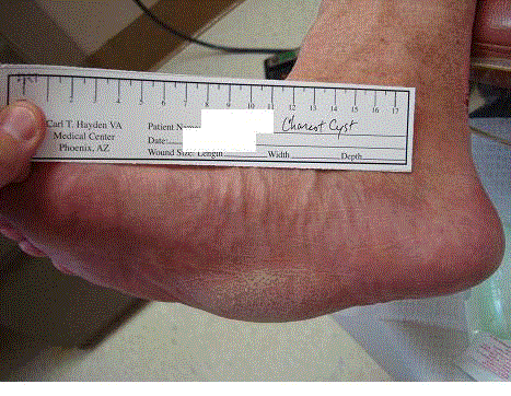
Figure 1
Physical exam reveals a solid, spongy/boggy, mobile, compressible
mass located near the plantar lateral fourth and fifth metatarsal bases.
Figure 2
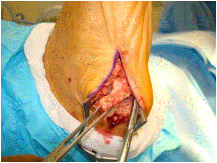
Figure 2
Surgical dissection Charcot cyst.
Methods and Results
Case 1
A sixty-one year old male with past medical history of type 2
diabetes, peripheral neuropathy, chronic inflammatory demyelinating
polyneuropathy for which he takes methadone and tiagabin, posttraumatic
stress disorder, hypertension, hypercholesterolemia,
history of abdominal aortic aneurysm, benign prostatic hyperplasia,
and a twenty-two-and-a-half pack year tobacco history. His chief
complaint was a one month history of right midfoot collapse with
associated swelling, change in shape, and a clicking noise with fourth postoperative appointment, where an incision site dehiscence
was noted centrally. Patient was started on a seven day course of
Augmentin. Appligraft was applied at the patient’s fifth postoperative
visit. On patient’s thirteenth postoperative visit, physical exam
revealed mild, localized bogginess on plantar lateral right foot with
calor. Serous sanguinous fluid was drained without need of incision
or anesthesia. Patient was instructed to finish another seven day
course of Augmentin. By patient’s nineteenth postoperative visit, the
incision site closed, and there were no open lesions noted. Patient
now continues to ambulate without issues in extra depth shoes.
Case 2
A fifty-five year old male with past medical history of type 2
diabetes, peripheral neuropathy, hyperlipidemia, and hypertension
presented to clinic with a chief complaint of a left foot plantar ulcer.
Patient reports about sixteen months ago he fell, sustained a midfoot
fracture, and subsequently developed the plantar ulcer. Patient relates
he was weight bearing on the left foot for about six weeks before he
was diagnosed with the Charcot foot collapse. At the time of patient’s
initial presentation, his blood glucose was measured at 422 mg/dL
and rechecked at 429 mg/dL. Focused physical exam of left foot
revealed palpable pedal pulses, diminished sensation to Semmes
Weinstein monofilament, a sub cuboid ulcer measuring two-and-ahalf
centimeters by three centimeters without acute signs of infection,
decreased ankle joint dorsiflexory range of motion, collapse of midfoot
consistent with Charcot, and plantar bony prominence corresponding
to ulcer location. Radiographs, negative for osteomyelitis, revealed
advanced deformity and stable appearing disorganization with lateral
subluxation of the midfoot tarsometatarsal joint complex relative to
tarsal navicular bones. Patient was treated with wound debridement,
betadine wet to dry dressings with supplies for daily changes, and
was instructed to remain in walking boot offloaded with padding.
Preoperatively, this patient was seen every three weeks and treated
with wound debridements and dressings to include Promogran,
betadine ointment, and covaderm, each time instructed to remain
in offloaded walking boot. Fourteen weeks after initial presentation,
patient underwent surgical intervention on the left foot. A four
centimeter incision was made directly over the ulceration site, and
the ulcer was excised (Figure 5), as was an underlying soft tissue mass
to be sent for pathological examination. Midfoot plantar planing
was performed to resect bony prominences via osteotome. A Tendo
Achilles lengthening was also performed via three stab incisions.
Patient was initially placed in a posterior splint. Per Pathology, the
“cyst left foot” consisted of two fragments of tan-pink soft tissue
with the larger mass measuring three-and-a-half centimeters by
one-and-a-half centimeters by one centimeter. This larger fragment
on cut section showed a one centimeter ill-defined, gelatinous and
tan-white central portion. Diagnosis was fibrotendinous tissue
with edema, myxoid and hyaline degeneration (Figure 6 and 7).
Postoperatively, patient was treated about every one to three weeks
based on postoperative healing and wound severity for about twoand-
a-half years. Wound care treatments included negative pressure
therapy, Dermagraft, Prisma, Oasis, Betadine, and Iodosorb. Patient
was also treated with offloading modalities, including posterior splint,
offloaded walking boots, total contact casts, and, ultimately, modified
custom shoes. Radiographs throughout this treatment period did
not demonstrate significant bony changes in patient’s Charcot foot
deformity. Upon final wound closure, the patient was graduated to
regularly scheduled preventative appointments.
Case 3
A fifty-eight year old male with a past medical history of type
2 diabetes, peripheral neuropathy, congestive heart failure, and
chronic obstructive pulmonary disease, history of cerebral vascular
accident, peripheral vascular disease, and hypertension presented to
clinic with a chief complaint of a one year history of right midfoot
collapse with new associated plantar midfoot wound. Patient denied
pain. Pertinent physical exam revealed nonpalpable pedal pulses,
absent sensation to Semmes Weinstein monofilament, and a rocker
bottom appearance with equinus deformity. The wound was also
noted to have serous drainage. Patient was treated with silvadene, dry
sterile dressings and custom diabetic shoes. Throughout the course of
patient’s preoperative care, he was followed every one to three weeks
with continued wound care and offloading shoe gear. Four months
after initial presentation, patient’s wound remained open with
persistent serous drainage and patient received an excisional biopsy
of wound tissue. Surgical pathology revealed soft tissue with necrosis,
acute and chronic inflammation and granulation tissue. Five months after initial presentation, patient’s wound still remained unhealed.
A wound culture was taken, revealing the presence of Methicillin
Sensitive Staph Aureus (MSSA). Radiographs revealed pes planus
with Charcot joint changes, negative for osteomyelitis. Patient was
started on Dicloxacillin 250 milligrams four times a day and received
continued wound care and offloading shoe gear. Six months after
initial presentation, patient presented with complaints of new onset
right lower leg redness and swelling, foot pain and purulent drainage
from wound. Patient was admitted to hospital for clinical diagnosis
of right leg cellulitis with associated Charcot foot wound and possible
osteomyelitis. Pertinent physical exam revealed erythema noted on
foot and leg, extending to knee level and a positive probe to bone test
in the foot wound. Patient was started on Vancomycin and Zosyn
and admission labs and imaging were ordered. MRI revealed findings
consistent with known neuropathic joint, surrounding cellulitis and
myositis with osteomyelitis of cuboid (Figure 8). Based on culture
and sensitivities (MSSA), patient’s antibiotics were also narrowed to
Nafcillin. On hospital day seven, patient received plantar exostectomy/
planning and a Tendo Achilles lengthening. Intraoperatively, cystic
material was noted upon deep wound debridement. The wound site
was primarily closed, and patient was placed in a posterior splint,
instructed to be non-weight bearing. Surgical pathology of wound
specimen revealed soft tissue with granulation, mild acute and
chronic inflammation and benign bone with attached soft tissue
showing fibrosis and mild chronic inflammation. On hospital day ten,
patient was discharged on Keflex with total contact cast placement.
Post operatively, patient was followed every week, and sutures were
removed at post op week five. Patient then transitioned into CAM
boot. Radiographs at this time were negative of osteomyelitis. By post
op week seven, the plantar ulcer was closed.
Figure 3
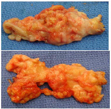
Figure 3
Excised Charcot cyst.
Figure 4
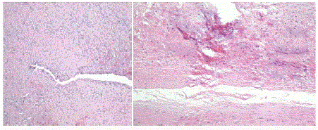
Figure 4
Pathology analysis revealing tan to pink portions of benign simple
cyst and cystic wall.
Figure 5
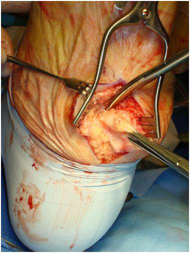
Figure 5
Surgical excision of Charcot cyst.
Figure 6
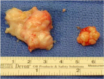
Figure 6
Charcot cyst..
Figure 7
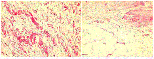
Figure 7
Pathology analysis revealing ill-defined, gelatinous and tan-white
central portion of mass. Diagnosis consistent with fibrotendinous tissue with
edema, myxoid and hyaline degeneration.
Figure 8
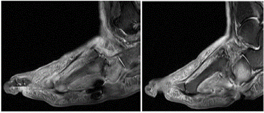
Figure 8
MRI revealing findings consistent with known neuropathic joint,
surrounding cellulitis, and myositis with osteomyelitis of cuboid.
Discussion
Although surgical management has become relatively more common for this disorder, there has been little, if any, mention of a peculiar cystic proliferation noted in the deep soft tissues upon surgical dissection. In our experience such cysts drain synovial fluid, impeding healing of ulcerations. Most times these patients have adequate blood flow to heal what appears to be the usual ulcers but remain resistant to traditional wound care and even advanced modalities. Brenner et al. [43] the only related study that we have found, reports a case of a sixty-two year old diabetic neuropathic male with a Charcot foot type with an obvious palpable mass five centimeters in diameter located superior to the left calcaneocuboid joint with consistency suggestive of cystic neoplasia. Fluid aspirations, vascular studies, and laboratory tests were negative and/or within normal limits. Intraoperatively, a large spherical, fluid-filled sac was revealed that extended deeply within the calcaneocuboid joint, confirmed to be a synovial cyst. Consistent with his findings, our patients also exhibited a translucent, fluctuant, multi-loculated cystic mass present throughout a considerable portion of our dissection. Upon resection, the masses were found to be spongy and contained what appeared to be synovial fluid. Pathology confirmed that these were simple benign cysts. Importantly, however, we found all patients exhibited a delay in wound healing caused by persistent synovial fluid leakage. Some differential diagnoses include dermoid cyst, teratoma, and epidermal inclusion cyst, and steatocystoma. Contents of these cysts may be keratin as in posttraumatic cysts, skin and its appendages as in dermoid cysts, and germ cell derivatives as in teratomas [44]. During wound healing, trapped squamous epithelium, undergoing keratinisation leads to cyst formation. Very few authors have reported such condition making it difficult to retrieve proper articles in the medical literature and derive a relevant message regarding incidence and guidelines for diagnosis and management. A long term follow-up after surgical removal is highly recommended.
Conclusions
Three (3) example cases illustrated what seems to be simple benign cysts all had similar characteristics of translucent, fluctuant, multi-loculated appearance that extended throughout a considerable portion of the dissection. Though observed in many of our diabetic Charcot patients, we have also observed such pathology in our nondiabetic population. Interestingly, we have found a delay in wound healing in these patients due to the persistent synovial fluid leakage. Since most patients with Charcot foot are treated conservatively, these cysts are not usually recognized surgically. While the pathogenesis of Charcot neuroarthropathy has been described, the formation of such related cysts has yet to be explained. We, therefore, encourage clinicians and surgeons to corroborate our findings with further study of this interesting pathology.
References
- PÅ‚aza M, Nowakowska-PÅ‚aza A, Walentowska-Janowicz M, Chojnowski M, SudoÅ‚-SzopiÅ,ska I. Charcot arthropathy in ultrasound examination - a case report. J Ultrason. 2016;16(65):210-5.
- Burson LK, Schank CH. Charcot Neuroarthropathy of the Foot and Ankle. Home Healthc Now. 2016;34(3):135-9.
- Rogers LC, Frykberg RG, Armstrong DG, Boulton AJM, Edmonds M, Van GH, et al. The Charcot foot in diabetes. J Am Podiatr Med Assoc. 2011;101(5):437-46.
- Frykberg RG, Sage RA, Wukich DK, Pinzur MS, Schuberth JM. Charcot arthropathy. Foot Ankle Spec. 2012;5(4):262-71.
- Jeffcoate WJ, Chipchase SY, Ince P, Game FL. Assessing the outcome of the management of diabetic foot ulcers using ulcer-related and person-related measures. Diabetes care. 2006;29(8):1784-7.
- Frykberg RG. Neuropathic arthropathy: the diabetic Charcot foot. Diabetes Educ. 1984;9(4):17-20.
- Frykberg RG, Belczyk R. Epidemiology of the Charcot foot. Clin Podiatr Med Surg. 2008;25(1):17-28.
- Pinzur MS. Surgical treatment of the Charcot foot. Diabetes Metab Res Rev. 2016;32 Suppl 1:287-91.
- Larson SA, Burns PR. The pathogenesis of Charcot neuroarthropathy: current concepts. Diabet Foot Ankle. 2012;3.
- Karapanagioti EG, Assimopoulou AN. Naturally occurring wound healing agents: An evidence-based review. Curr Med Chem. 2016;23(29):3285-321.
- Vasquez V, Henderson S. Charcot foot? Charcot arthropathy caused by lisfranc fracture-dislocation in a diabetic. West J Emerg Med. 2010;11(2):146-7.
- Schuerwegh AJ, Dombrecht EJ, Stevens WJ, Van Offel JF, Bridts CH, De Clerck LS. Influence of pro-inflammatory (IL-1 alpha, IL-6, TNF-alpha, IFN-gamma) and anti-inflammatory (IL-4) cytokines on chondrocyte function. Osteoarthritis Cartilage. 2003;11(9):681-7.
- Yasuda H, Shima N, Nakagawa N, Yamaguchi K, Kinosaki M, Mochizuki S, et al. Osteoclast differentiation factor is a ligand for osteoprotegerin/osteoclastogenesis-inhibitory factor and is identical to TRANCE/RANKL. Proc Natl Acad Sci U S A. 1998;95(7):3597-602.
- Mabilleau G, Petrova NL, Edmonds ME, Sabokbar A. Increased osteoclastic activity in acute Charcot's osteoarthropathy: the role of receptor activator of nuclear factor-kappaB ligand. Diabetologia. 2008;51(6):1035-40.
- Weitzmann MN. The Role of Inflammatory Cytokines, the RANKL/OPG Axis, and the Immunoskeletal Interface in Physiological Bone Turnover and Osteoporosis. Scientifica (Cairo). 2013;2013:125705.
- Wang L, Shi X, Zhao R, Halloran BP, Clark DJ, Jacobs CR, et al. Calcitonin-gene-related peptide stimulates stromal cell osteogenic differentiation and inhibits RANKL induced NF-kappaB activation, osteoclastogenesis and bone resorption. Bone. 2010;46(5):1369-79.
- Kauther MD, Xu J, Wedemeyer C. Alpha-calcitonin gene-related peptide can reverse the catabolic influence of UHMWPE particles on RANKL expression in primary human osteoblasts. Int J Biol Sci. 2010;6(6):525-36.
- Hofbauer LC, Schoppet M. Clinical implications of the osteoprotegerin/RANKL/RANK system for bone and vascular diseases. JAMA. 2004;292(4):490-5.
- Jeffcoate WJ. Charcot neuro-osteoarthropathy. Diabetes Metab Res Rev. 2008;24 Suppl 1:S62-5.
- Akopian A, Demulder A, Ouriaghli F, Corazza F, Fondu P, Bergmann P. Effects of CGRP on human osteoclast-like cell formation: a possible connection with the bone loss in neurological disorders? Peptides. 2000;21(4):559-64.
- La Fontaine J, Harkless LB, Sylvia VL, Carnes D, Heim-Hall J, Jude E. Levels of endothelial nitric oxide synthase and calcitonin gene-related peptide in the Charcot foot: a pilot study. J Foot Ankle Surg. 2008;47(5):424-9.
- Sasaki T, Yasuda H, Maeda K, Kikkawa R. Hyperalgesia and decreased neuronal nitric oxide synthase in diabetic rats. Neuroreport. 1998;9(2):243-7.
- van't Hof RJ, Ralston SH. Cytokine-induced nitric oxide inhibits bone resorption by inducing apoptosis of osteoclast progenitors and suppressing osteoclast activity. J Bone Miner Res. 1997;12(11):1797-804.
- Yan SF, Ramasamy R, Naka Y, Schmidt AM. Glycation, inflammation, and RAGE: a scaffold for the macrovascular complications of diabetes and beyond. Circ Res. 12 2003;93(12):1159-69.
- Goldin A, Beckman JA, Schmidt AM, Creager MA. Advanced glycation end products: sparking the development of diabetic vascular injury. Circulation. 2006;114(6):597-605.
- Sanguineti R, Puddu A, Mach F, Montecucco F, Viviani GL. Advanced glycation end products play adverse proinflammatory activities in osteoporosis. Mediators Inflamm. 2014;2014:975872.
- Franke S, Siggelkow H, Wolf G, Hein G. Advanced glycation endproducts influence the mRNA expression of RAGE, RANKL and various osteoblastic genes in human osteoblasts. Arch Physiol Biochem. 2007;113(3):154-61.
- Brown CW, Jones B, Donaldson DH, Akmakjian J, Brugman JL. Neuropathic (Charcot) arthropathy of the spine after traumatic spinal paraplegia. Spine (Phila Pa 1976). 1992;17(6):S103-8.
- Shem KL. Neuroarthropathy of the wrist in paraplegia: A case report. J Spinal Cord Med. 2006;29(4):436-9.
- Smith DG, Barnes BC, Sands AK, Boyko EJ, Ahroni JH. Prevalence of radiographic foot abnormalities in patients with diabetes. Foot Ankle Int. 1997;18(6):342-6.
- Rogers LC, Frykberg RG, Armstrong DG, Boulton AJ, Edmonds M, Van GH, et al. The Charcot foot in diabetes. Diabetes Care. 2011;34(9):2123-9.
- Frykberg RG. Diabetic foot ulcers: current concepts. J Foot Ankle Surg. 1998;37(5):440-6.
- Frykberg RG. Diabetic foot ulcers: pathogenesis and management. Am Fam Physician. 2002;66(9):1655-62.
- Frykberg RG, Mendeszoon E. Management of the diabetic Charcot foot. Diabetes Metab Res Rev. 2000;16 Suppl 1:S59-65.
- Frykberg RG, Zgonis T, Armstrong DG, Driver VR, Giurini JM, Kravitz SR, et al. Diabetic foot disorders. A clinical practice guideline (2006 revision). J Foot Ankle Surg. 2006;45(5 Suppl):S1-66.
- Frykberg RG, Banks J. Challenges in the Treatment of Chronic Wounds. Adv Wound Care (New Rochelle). 2015;4(9):560-582.
- Anichini R, Zecchini F, Cerretini I, et al. Improvement of diabetic foot care after the implementation of the International Consensus on the Diabetic Foot (ICDF): results of a 5-year prospective study. Diabetes Res Clin Pract. 2007;75(2):153-8.
- Ayuk SM, Abrahamse H, Houreld NN. The Role of Matrix Metalloproteinases in Diabetic Wound Healing in relation to Photobiomodulation. J Diabetes Res. 2016;2016:2897656.
- Boulton AJ. The diabetic foot: a global view. Diabetes Metab Res Rev. 2000;16(1):S2-5.
- Boulton AJ. Pressure and the diabetic foot: clinical science and offloading techniques. Am J Surg. 2004;187(5A):17S-24S.
- Boulton AJ, Vileikyte L, Ragnarson-Tennvall G, Apelqvist J. The global burden of diabetic foot disease. Lancet. 2005;366(9498):1719-24.
- Schneekloth BJ, Lowery NJ, Wukich DK. Charcot Neuroarthropathy in Patients with Diabetes: An Updated Systematic Review of Surgical Management. J Foot Ankle Surg. 2016;55(3):586-90.
- Brenner MA, Kalish SR, Lupo PJ, Rokhsar B. The diabetic foot with synovial cyst. Cutis. 1990;46(2):142-4.
- Amaranathan A, Sinhasan SP, Dasiah SD. Median raphe cysts of the prepucial skin, with triple histological linings: a case report and review of the literature. J Clin Diagn Res. 2013;7(7):1466-8.