Short Communication
Advances in Plastic Surgery: From Microsurgery to Tissue Engineering and Biofabrication?
Raymund E. Horch1*, Annika Weigand1, Harald Wajant2 and Andreas Arkudas1
1Department of Plastic and Hand Surgery and Laboratory for Tissue Engineering and Regenerative Medicine,
University Hospital Erlangen, Germany
2Department of Molecular Internal Medicine in the Medical Clinic and Polyclinic II, University Hospital Wuerzburg,
Germany
*Corresponding author: Raymund E. Horch, Department of Plastic and Hand Surgery and Laboratory for Tissue Engineering and Regenerative Medicine, University Hospital Erlangen, Friedrich-Alexander University Erlangen-Nuernberg FAU Krankenhausstrasse 12, 91054 Erlangen, Germany
Published: 20 Sep, 2017
Cite this article as: Horch RE, Weigand A, Wajant H,
Arkudas A. Advances in Plastic
Surgery: From Microsurgery to Tissue
Engineering and Biofabrication?. Clin
Surg. 2017; 2: 1622.
Short Communication
Although Plastic Surgery as a mono-speciality has not been widely academically implemented
until recently it has always been an integral part of surgery. Oftentimes groundbreaking
developments of new methods have driven relevant innovations and have influenced the whole field
of surgical specialities. Detailed and still valuable operation techniques, such as how to reconstruct
a nose for example, have been reported as early as 600 BC in the Indian Sushruta Ayurveda. For
centuries complex methods of reconstruction using delayed pedicled random pattern flaps in multi
stage approaches were known and were successfully performed before the advent of anaesthesia.
Tagliacozzi´s two volume book De Curtorum Chirurgia per Insitionem (1597) detailed the different
surgical steps with graphic illustrations that became a hallmark of surgical textbooks ever since. It
was the Plastic Surgeon John Murray who was awarded the Nobel Prize for his groundbreaking
achievements in kidney transplantation, just to name a few examples. The advances in microsurgery
with transplantation of vascularized tissue to almost any part of the body has influenced many other
specialities as well. It is the interdisciplinary character of modern reconstructive and oncological
concepts that underlines the value of Plastic Surgery as a problem solving discipline within the
concert of medical specialities and that contributes significantly to the improvement of patient´s
quality of life.
One meanwhile widely established example of interdisciplinary surgical concepts is the salvage
of hypovascularized wounds in the lower extremity by transferring well vascularized tissue utilizing
microsurgery. This concept is based on the surgically induced angiogenesis to treat chronic,
hypovascularized wounds, especially diabetic ulcers and ulcers following arteriosclerotic disease
[1-5]. When necessary autologous venous bypass grafts can be used as a prolongation or as arteriovenous
loops to allow for a distal free flap connection even in the absence of appropriate local vessels.
We have studied a larger cohort of such patients with bypasses and free flaps and have proposed an
algorithm based on our results and from literature data [6-11].
Whereas previously limb salvage was based mainly on myocutaneous or muscle flaps recently
perforator flaps have now gained widespread use and have been advocated to be another soft
tissue choice for all zones of the lower extremity, recognizing that function preservation is their
major asset as no muscle need be included (Figure 1 and 2). In patients without a compromise
of arterial inflow and microcirculation, however, it has been propagated that in certain instances
even peninsular, propeller, or advancement perforator flaps can be regarded as valuable local nonmicrosurgical
flap alternatives [12]. The differential indication for a local flap or a free tissue transfer
however depends on the localization and the size of the defect as well as on the vascular situation
of the recipient site [13]. In diabetic foot ulcers for instance the use of local flaps is very limited.
It also has to be taken into account that any local flap does not only cause a donor site defect but
also may further deteriorate the vascular supply of the distal extremity. Experimental studies have
highlighted a potential role of (neo-) angiogenesis at the non-ischemic/ ischemic interfaces, as they
appear after free vascularized flap transfers into an ischemic wound [14,15]. It is of interest that the
age of patients eligible for free flap transfers to the lower extremity does no longer seem to be an
obstacle against this technique, since a correlation between flap loss and increased risk factors and
age was not found so far [16-18].
Our results with more than 100 patients receiving a bypass/or av-loop along with a free flap
show that the donor site morbidity is comparatively low and acceptable. Strict patient selection and strategic planning can help to keep the rate of complications at an
acceptable level.
Since Plastic Surgeons always are engaged in replacing lost tissue
it is of no wonder that this specialty also has been involved into
techniques of Tissue Engineering (TE) and Regenerative Medicine
(RM). Despite the often times considerable results of generating
replacement tissue in the laboratory TE and RM have not easily found
their way into daily clinical practice yet. One hindrance is the lack of
vascularization especially in large constructs that suffer from sufficient
blood supply to nourish cells from the beginning of their inset. One
way is the prevascularization of scaffolds utilizing microsurgically
created arterio-venous (av-) loops to three dimensionally vascularize
large constructs before cells are inoculated and can then be
successfully transplanted [19-22]. Methods derived from these
approaches have been successfully implemented into the clinical
scenario with application of av-loops together with the patient´s
own bone marrow stem cells and have been proven to permanently
replace and restore human bone defects [23]. Latest advances include
the integration of 3D bioprinting of cells and proteins together with
biodegradable matrices, generally now perceived as the new field
of “biofabrication” [24]. It has been postulated by researchers that
bioprinting would now be on the cusp of entering the translational
phase where laboratory research practices can be scaled up into
manufacturing products specifically designed for individual patients
[25]. In addition to tissue replacement fighting systemic conditions
such as diabetes mellitus or malignant diseases are considered in
view of being treated with methods of biofabrication. Ravinc et al.
report on recent successful attempts to generate beta cells and how
this can be coupled with bioprinting technologies in order to fabricate
pancreas tissues, which holds great potential for type 1 diabetes [26].
They postulate the possibility of integrating vascularization and
encapsulation in bioprinted tissues which would lend other future
prospects, such as pancreas-on-a-chip [26]. Our own group is actively
investigating the value of bioprinting to generate 3D vascularized
containers that can then be loaded with protein producing cells that
can continuously express functional substances and address specific
functions in the recipient organism. This approach combines again
the expertise of plastic surgical microvascular techniques together
with bioengineering and biological methods and seems promising.
In conclusion consolidated findings from regenerative medicine and
tissue engineering merging into the new field of biofabrication will
enrich the daily practice of surgical reconstruction to the benefit of
our patients [27,28].
Figure 1
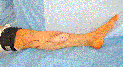
Figure 1
Figure 2
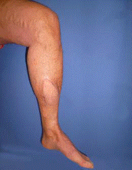
Figure 2
Figure3
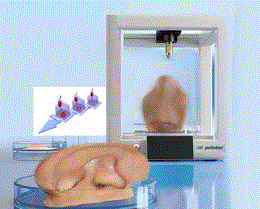
Figure 3
References
- Biemer E, Jaeger K. [Drug treatment, after care and secondary interventions following lower leg reconstruction by free tissue transfer]. Chirurg. 1986;57(3):140.
- Hay EL, Shealy GJ, Hanel DP, Stoddard LC. Lower extremity salvage with microvascular free flaps. J S C Med Assoc. 1985;81(6):311-6.
- Hallock GG. Severe lower-extremity injury. The rationale for microsurgical reconstruction. Orthop Rev. 1986;15(7):465-70.
- Walgenbach KJ, Voigt M, Andree C, Stark GB, Horch RE. Management of hypovascularized wounds not responding to conventional therapy by means of free muscle transplantation. Vasa. 2001;30(3):206-11.
- Ducic I, Rao SS, Attinger CE. Outcomes of microvascular reconstruction of single-vessel lower extremities: limb salvage versus amputation. J Reconstr Microsurg. 2009;25(8):475-8.
- Daigeler A, Kneser U, Fansa H, Riester T, Uder M, Horch RE. [Reconstruction of the vascular compromised lower extremity - report of the consensus workshop at the 35. Meeting of the DAM (Deutschsprachige Gemeinschaft fur Mikrochirurgie der peripheren Nerven und Gefasse) 2013 in Deidesheim]. Handchir Mikrochir Plast Chir. 2014; 46(4):248-55.
- Kneser U, Arkudas A, Beier JP, Dragu A, Stübinger A, Lang W, et al. [Extended skin and soft tissue defects after vascular wounds: plastic surgical concepts]. Zentralbl Chir. 2013;138(5):536-42.
- Horch RE, Horbach T, Lang W. The nutrient omentum free flap: revascularization with vein bypasses and greater omentum flap in severe arterial ulcers. J Vasc Surg. 2007;45(4):837-40.
- Horch RE, Lang W, Arkudas A, Taeger C, Kneser U, Schmitz M, et al. Nutrient free flaps with vascular bypasses for extremity salvage in patients with chronic limb ischemia. J Cardiovasc Surg (Torino). 2014;55(2 Suppl 1):265-72.
- Horch RE, Lang W, Meyer A, Schmitz M. Distal pedal bypasses combined with free microsurgical flaps in chronic limb ischaemia for problematic wounds. Int Wound J. 2016;13(3):425-6.
- Horch RE, Walgenbach KJ, Voigt M, Stark GB. [The free "emergency" rectus abdominis flap transfer for coverage of complex hand injuries]. Langenbecks Arch Chir Suppl Kongressbd. 1998;115:1194-6.
- Hallock GG. A paradigm shift in flap selection protocols for zones of the lower extremity using perforator flaps. J Reconstr Microsurg. 2013;29(4):233-40.
- Colen LB. Limb salvage in the patient with severe peripheral vascular disease: the role of microsurgical free-tissue transfer. Plast Reconstr Surg. 1987;79(3):389-95.
- Walgenbach KJ, Horch R, Voigt M, Andree C, Tánczos E, Stark GB. [Free microsurgical flap-plasty in reconstructive therapy of diabetic foot ulcer]. Zentralbl Chir. 1999;124:40-4.
- Walgenbach KJ, Voigt M, Horch R, Stark GB. [Surgically-induced angiogenesis as basic principle in treatment ov hypovascularized wounds--the nutritive flap]. Langenbecks Arch Chir Suppl Kongressbd 1998;115:1186-8.
- Goldberg JA, Alpert BS, Lineaweaver WC, Buncke HJ. Microvascular reconstruction of the lower extremity in the elderly. Clin Plast Surg. 1991;18(3):459-65.
- Lorenzetti F, Giordano S, Tukiainen E. Intraoperative hemodynamic evaluation of the latissimus dorsi muscle flap: a prospective study. J Reconstr Microsurg 2012;28(4):273-8.
- Ludolph I, Lehnhardt M, Arkudas A. Plastic reconstructive microsurgery in the elderly patient. Consensus statement of the German Speaking Working Group for Microsurgery of the Peripheral Nerves and Vessels. Handchir Mikrochir Plast Chir. 2017;49:1-8.
- Weigand A, Beier JP, Arkudas A, Al-Abboodi M, Polykandriotis E, Horch RE, et al. The Arteriovenous (AV) Loop in a Small Animal Model to Study Angiogenesis and Vascularized Tissue Engineering. J Vis Exp. 2016(117).
- Weigand A, Beier JP, Hess A, Gerber T, Arkudas A, Horch RE, et al. Acceleration of vascularized bone tissue-engineered constructs in a large animal model combining intrinsic and extrinsic vascularization. Tissue Eng Part A. 2015;21(9-10):1680-94.
- Arkudas A, Beier JP, Heidner K, Tjiawi J, Polykandriotis E, Srour S, et al. Axial prevascularization of porous matrices using an arteriovenous loop promotes survival and differentiation of transplanted autologous osteoblasts. Tissue Eng. 2007;13(7):1549-60.
- Arkudas A, Tjiawi J, Saumweber A, Beier JP, Polykandriotis E, Bleiziffer O, et al. Evaluation of blood vessel ingrowth in fibrin gel subject to type and concentration of growth factors. J Cell Mol Med. 2009;13(9A):2864-74.
- Horch RE, Beier JP, Kneser U, Arkudas A. Successful human long-term application of in situ bone tissue engineering. J Cell Mol Med 2014;18(7):1478-85.
- Groll J, Boland T, Blunk T, Jason A Burdick, Dong-Woo Cho, Paul D Dalton, et al. Biofabrication: reappraising the definition of an evolving field. Biofabrication 2016;8(1):013001.
- Wu C, Wang B, Zhang C, Wysk RA, Chen YW. Bioprinting: an assessment based on manufacturing readiness levels. Crit Rev Biotechnol. 2017;37(3):333-54.
- Ravnic DJ, Leberfinger AN, Ozbolat IT. Bioprinting and Cellular Therapies for Type 1 Diabetes. Trends Biotechnol. 2017.
- Horch RE. Future perspectives in tissue engineering. J Cell Mol Med. 2006;10(1):4-6.
- Horch RE, Weigand A, Beier JP, Arkudas A, Boos AM. The Potential Role of Telocytes for Tissue Engineering and Regenerative Medicine. Adv Exp Med Biol. 2016;913:139-147.