Research Article
The Value of Medical Needling in Burn Scars
Matthias Aust1 and Desmond Fernandes2*
1Department of Plastic and Aesthetic Surgery, Hannover Medical School, Germany
2Department of Plastic and Reconstructive Surgery, Shirnel Clinic - Cape Town, South Africa
*Corresponding author: Desmond Fernandes, Department of Plastic and Reconstructive Surgery, Shirnel Clinic, 822 Fountain Medical Centre Heerengracht Cape Town 8001, South Africa
Published: 18 Sep, 2017
Cite this article as: Aust M, Fernandes D. The Value of
Medical Needling in Burn Scars. Clin
Surg. 2017; 2: 1612.
Abstract
We aim to show that deeper needling of skin (1 mm to 3 mm depth) is a valuable tool for treating people with burn scars. Currently numerous ablative cutaneous treatments and surgical procedures are the mainstay of treating burn scars. Needling takes us to a new paradigm of instigating a regenerative phase to remove scar collagen and replace it with normal lattice patterned collagen and thereby reducing the appearance of nodular hypertrophic scars and contractions. This occurs because skin needling causes perforation of blood vessels and an automatic cascade of platelet derived growth factors etc. The dominant growth factor is TGF-β-3 which surges after needling in a way not seen in normal adult tissues but which the usual pattern is found in foetal regenerative wound repair. By repeating needling treatments one can flatten hypertrophic/ burn keloid scars and softens contractures and get closer to the ideal treatment of scars. The authors believe skin needling should be done reasonably soon after the initial injury.
Introduction
Patients with burn scar deformities frequently request help to improve both the aesthetic and
functional complications of their scars. There are numerous methods available for surgeons to treat
burn scars but nowadays, there is a demand for less invasive and more cost-effective procedures to
give the desired benefits.
Non-invasive treatments such as silicone patches, pressure garments etc. are important ways
to control scars. Minimally invasive techniques such as cortisone injections also have a place.
Surgeons have generally depended on surgical interventions such as scar excision, W- or Z-pasties,
and pedicled or free skin flaps to treat contractures or severe irregular scars. The quest for better
results has also led to the application of many different topical therapies such as laser resurfacing,
dermabrasion and deep chemical peels.
The last-mentioned methods all follow the same principal: they are ablative; they change the
scar by destroying the epidermis partially or completely and scarring the dermis. The death of tissue
leads to an inflammatory response. In the process of trying to treat dermal scarring the epidermis
may be completely destroyed and replaced by a thinner epidermis with flatter rete ridges covering
parallel-orientated scar collagen which is distinctive for scarred skin [1-3]. Furthermore, the skin
becomes more vulnerable to infections [4].
The ideal scar treatment method would avoid ablation of the epidermis, and rather promote
the formation of physiological dermal collagen in a lattice pattern by initiating the expression of
growth factors which are relevant for scarless wound healing and regeneration of the skin. In other
words, the perfect remedy would be to remove the visible defective scarring and regenerate healthier
anatomically more normal skin.
In recent years, it has been shown that it is possible to a significant degree to achieve the ideal
treatment by using percutaneous collagen induction or “Medical Needling” [5-7], Medical Needling
is a minimal-invasive non-ablative procedure capable of improving scar quality and functionality by
dermal reorganization with a decrease in scar collagen accompanied by an increase of physiological
collagen and fibronectinas well as an increase of glycosaminoglycans. There is a decrease of transepidermal
water loss because the epidermis is thicker and the stratum corneum becomes a fully
functional water barrier.
Approximately 20 years ago Camirand and Doucet [8] demonstrated that by simple “needle
abrasion” one could get significant clinical improvement in treating white surgical scars with a tattoo
artists’ device. Orentreich also reported about “dermal needling or subcision” as an alternative for
treating scars and wrinkles [9]. Based on these concepts, Fernandes developed the percutaneous collagen induction technique [10]. Thanks to targeted research within the last 15 years, impressive scientific data is now available which
underlines the efficacy and safety of Medical Needling and why it
works [5,7,11-15].
Science
How it works
Medical Needling is repetitive puncturing of burn scars with a
roller equipped with 3.0 mm long needles that penetrate into the
dermal scars and cause intra dermal bleeding (Figure 1).
The needling device is repeatedly rolled over the scar in three main
directions: longitudinally, diagonally and horizontally to get the best
distribution of puncture holes. According to the extent of the scar,
this procedure can last 30 minutes or longer. It is important to use the
device with constant pressure and do the rolling in one direction at a
time to prevent shear forces. The needles are solid and do not have a
lumen. Hence, they pierce the skin and mainly separate the skin cells
rather than destroying them (Figure 2). They penetrate the dermis 2,
0 to a maximum of 3 mm and produce thousands of micro puncture
wounds and intradermal bleeding. Some blood comes up through
the channels to cause bleeding on the surface. The most important
bleeding occurs in the dermis but bleeding through the skin gives us a
good idea of what is happening down below in the dermis.
Induction of the wound healing cascade
This trauma initiates the activation of the physiological wound
healing cascade but with a significant difference. Normally trauma
causes the temporary presence of TGF-β-3 and the wound heals
predominantly under the influence of TGF-β-1 and -β-2 which results
in scar tissue. After needling TGF-β-1 and -β-2 rapidly disappear
from the scene and TGF-β-3 dominates and that results in scarless
healing and regeneration [16] Skin needling induces a new (and as
yet unrecognized) phase of regenerative healing which should not
be confused with the post traumatic inflammatory cascade. Platelets,
keratinocytes and neutrophils secrete growth factors such as platelet
derived growth factor (PDGF), fibroblast growth factor (FGF),
vascular endothelial growth factor (VEGF), tissue growth factor
and transforming growth factor-α and -ß (TGF-α, TGF-ß). These
initiate the synthesis of dermal structures such as collagen, elastin
and fibronectin and also stimulate the migration of fibroblasts and
keratinocytes [13,17]. The modulation of these growth factors right
at the beginning signal the differences between this new paradigm
of scarless healing versus the archetypal healing with scar formation.
TGF and the induction of collagen I
This new repair and regeneration mechanism is relevant for the
formation of collagen I which is the physiological type collagen in a
lattice pattern in healthy skin whereas collagen III is more prevalent
in parallel orientated scar collagen. TGF-β-3 makes a transient
presence in standard surgical wounds and has largely disappeared
within 24 hours of injury. Typical scars, we now know, are the result
of dominant activity of TGF-ß1 and -2.In contrast, TGF- ß1 and
-2levels are extremely low in scarless embryonal wound healing while
the levels of TGF-ß3 are remarkably high [1,5,16,18].
Medical Needling particularly influences the liberation of
TGF-ß1, -2 and -3. Within days’ post treatment the levels of TGF-ß1
and -2 are significantly down regulated whereas TGF-ß3 reaches high
expression levels even beyond the initial wound healing phase [7,13].
In support of this, the production of type I collagen was found to
be increased after Medical Needling (Figure 3,4 and 5).The changes
seen in skin needling indicate a lower level of TGF-beta-3 by 2 months. Ferguson’s team argue that it is the initial height of the rise
in concentration of TGF-beta-3 that is probably the most important
influence, not so much the duration of the raised levels because as
he points out the TGF-beta-3 does not stay raised for the extended
healing period [19]. Studies in humans that are as yet unpublished
show that TGF-beta-3 is raised after needling and become higher
when needling is done at short intervals (Fernandes personal
communication).
Dermal remodeling
Dermal reorganization after Medical Needling not only depends
on the formation of physiological orientated collagen I but also on
the inclusion of glycosaminoglycan molecules and fibronectin. This
was shown in the animal model through the quantitative analysis
of gene expression as well as through immunohistological analyses
[13,20,21]. As seen in (Figure 6 and 7), the entire connective tissue
framework appears thicker and denser post treatment.
Increase in skin elasticity
Moreover, the stimulation of the endogenous FGF contributes
towards improved skin elasticity. As seen in (Figure 8 and 9), the
amount of elastin is significantly higher after Medical Needling
Normalized perfusion
The secretion of VEGF during the healing phase stimulates
angiogenesis and leads to the formation of tiny blood vessels in
the corium. This helps to normalize the characteristic pathological
erythema of scars after burn injuries. As seen in Figures 8 and 9, the
amount of VEGF significantly rises after Medical Needling.
Increase in skin moisture content
Scars often appear dry and loose due to a decrease of
glycosaminoglycans with a result of reduced water retention in the
skin and due to thinner epidermis with increased trans-epidermal
water loss. Medical Needling is associated with a higher inclusion of
glycosaminoglycans (Figure 10 and 11) and with a thicker epidermis
post treatment (Figure 12 and 13). Both help to maximize the
moisture of the skin back to the reference of healthy skin.
Increase of epidermal thickness
In contrast to ablative treatments, the skin structures are
not injured after Medical Needling. The epidermis remains
physiologically intact which means that potentially side effects such
as inflammation, new scarring or dyspigmentation are reduced to a
minimum. Furthermore, it has been shown in the animal model that
the thickness of the epidermis increases up to 140 % after treatmentversus untreated ones [22] (Figure 6 and 7).
Role of vitamins in wound healing
Maximal post Needling improvement was seen in combination
with pre- and post-treatment of the skin with vitamin A and oxidants
Vitamin C and E (Figure 6 and 7).
No dyspigmentation after Medical Needling
A disadvantage of ablative scar treatments is that there is an
increased risk of dyspigmentation especially in darker skin types
[23-25]. On 480 patients, it has been shown that there is no risk of
dyspigmentation after Medical Needling.(26)Furthermore, Medical
Needling does not change the number of melanocytes but the
expression levels of Melanocyte-stimulating hormone (MSH) and
Interleukin-10 (Il-10) are modified [26]. MSH which influences
the proliferation and activity of melanocytes is significantly down
regulated within days after treatment. Il-10 as an anti-inflammatory
cytokine is upregulated post operatively [20]. In a subsequent study,
it has been shown that it is not possible to re-pigment larger areas of
hypopigmented skin with Medical Needling alone.
Re-pigmentation of hypopigmented burn scars with
Medical Needling and Non-cultured autologous skin cell
suspension (NCASCS)
Currently numerous methods are available to treat hypopigmented
skin, such as split skin grafting [27,28] lasers [29-31] and cultured
skin cell transplantation(30-32) In recent years, research focused
additionally on Non-cultured skin cell suspension. The Autologous
Cell Harvesting Device is used to create a spray suspension of living
autologous skin cells. These cells are harvested intraoperatively and
directly applied, in suspension, to the prepared wound. In order to
prepare an area for treatment with NCASCS, the wound has been
first treated by using dermabrasion or lasers which are both ablative
methods. By their nature, ablative treatments remove skin structures
and cells, including the basement membrane, which are replaced by
a thinner epidermis with flatter rete ridges [1,2,32]. This initiates an
inflammatory response that stimulates fibroblasts to produce parallel
oriented scar collagen instead of physiological lattice pattern collagen
[1,33]. Additionally, the risk of dyspigmentation increases after these
ablative treatments due to associated damage to the melanocytes
[34,35].
An ideal wound preparation for the autologous cell suspension
would be a treatment that does not destroy structures of the epidermis
yet creates a conduit that allows ingress of melanocytes that promotes
the formation of physiological collagen instead of scar collagen and
initiates the expression of growth factors. As we described above,
Medical Needling offers all these advantages. To combine both
procedures it is at first necessary to prepare a depigmented scar with
intense medical needling. Afterwards the autologous cell suspension
is applied through a spray syringe on the wound. The hypothesis
is that the melanocytes of the cell suspension link through the
epidermal canals onto the basal membrane. In a pilot study with 20
patients it has been shown that it is possible to get marked subjective
and objective improvements regarding re-pigmentation with the
combination of Medical Needling and NCASCS [36].
Figure 1
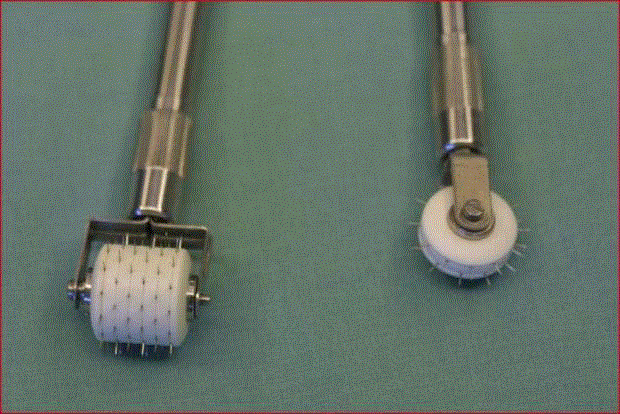
Figure 1
Needling device.
Figure 2
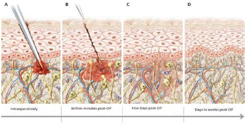
Figure 2
Schematic illustration of Medical Needling and the effects on the
wound. The needle pieces the epidermis and the blood vessels of the dermis
and when the needle is withdrawn the needle tract closes and by the next
day it cannot be detected histologically. Within days’ collagen I and elastin
are generated.
Figure 3
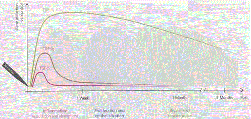
Figure 3
Microarray analyses of TGF-ß1, -2 and -3 expression level in
treated animals. The induction of TGF-ß3 gene expression continues even
beyond the initial wound healing phase whereas TGF-ß1 and -2 are downregulated
during the second week post-treatment.
Figure 4
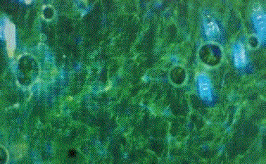
Figure 4
Immunofluorescence visualization of collagen I: Staining with
antibodies directed against Collagen I (Alexa488) and DAPI. Un-needled
animal of the dermis failed to react with the antibodies.
Figure 5
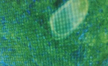
Figure 5
Immunhistochemical staining, anti-collagen I. Needled animal with
8 weeks of skincare stained without primary antibody. The amount of type
I collagen was qualitatively increased in treated group compared to their
controls judged by the brighter fluorescence.
Figure 6
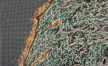
Figure 6
Masson’s Trichrome staining. (a) Untreated animal (control).
Figure 7
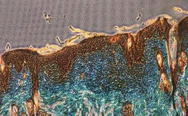
Figure 7
Needled animal with 8 weeks of skincare Collagen fiber bundles
were increased, thickened, and more loosely woven in both the papillary and
reticular dermis most prominently in the needled plus skincare group (D).
Elastin fibers in the dermis highly linear and the epidermal dermal interface
showed regular dermal papillae; cellular polarity and normal epidermal
differentiation appeared to be maintained; and the elastin network within the
reticular dermis was regularly thickened and organized in all groups.
Figure 8
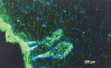
Figure 8
Immunhistochemical staining, anti-elastin. Untreated animal
Immunofluorescence visualization of elastin: Staining with antibodies directed
against elastin (Alexa488) an
Figure 9
Figure 9
Immunhistochemical staining, anti-elastin. Needled animal with 8
weeks of skincare stained without primary antibody. The amount of elastin
was qualitatively increased in treated group compared to their controls
judged by the brighter fluorescence.
Conclusion
Medical Needling offers anew alternative treatment for burn micro punctures into a scar with normal collagen. When we prick skin, we puncture blood vessels and the release of platelets signals
the release of growth factors which then improve dermal collagen,
vascularization and epidermal thickening. Medical Needling liberates
growth factors like VEGF and TGF-ß3 which initiate the replacement
of parallel, packed scar collagen by physiological collagen I in a lattice
orientation, with added elastin and glycosaminoglycans. The scars
after Medical Needling tend to appear smoother, softer and less itchy
and much less obvious. The skin becomes altogether more elastic and
as a result contractures are also softened. And in some cases, invasive
surgery to treat contractures becomes un-necessary. Skin needling is
showing us that it relieves tensions in tissues and minimizes the need
for Z-Plasty and major flaps. Early skin needling could help avoid
scar contractures which is one of the most crippling features of burns.
A significant problem arises for young girls as their breast develops,
because the breast tissue is entrapped by scar tissue and does not
develop properly. Skin needling is worth doing for these patients and
should be done as early as possible after the burn injury. There is good
reason to believe that if we can change the spectrum of tissue healing
dominated by TGF-beta 1 and 2 in the acute phase and convert it
into a regenerative phase promoted by TGF-beta-3 that we will have
long term effects and avoid contractures. Our experience at this stage
is largely on treating well established burns but the authors feel that
skin needling should become a part of the early management of
burn scars. For re-pigmentation, it offers the ideal pre-treatment for
Non-cultured autologous skin cell transplantation. Both treatments
preserve the epidermis which results in a reduced risk of new scarring
or dyspigmentation.
Medical needling offers a treatment that for the first time in
medical history can cause regeneration of tissue and soften burn
scars, reduce contractures. When done repeatedly and intensively
then burn scarred tissue can seem to be almost normal unscarred skin.
However, the timing probably is of utmost importance. The authors
believe skin needling should be done as soon after the burn injury as
is reasonable because they have had experience treating burns within
a few hours to days of the initial burn injury and it seems the earlier
the needling, the greater the chance to heal with minimal scars.
Skin needling needs to be understood by clinicians treating burns
so that this valuable technique can be offered to as many burn victims
as possible.
References
- Cancer Stat Facts: Bladder Cancer.
- Bosetti C, Bertuccio P, Chatenoud L, Negri E, La Vecchia C, Levi F. Trends in mortality from urologic cancers in Europe, 1970-2008. Eur Urol. 2011;60(1):1-15.
- Edwards BK, Ward E, Kohler BA, Eheman C, Zauber AG, Anderson RN, et al. Annual report to the nation on the status of cancer, 1975-2006, featuring colorectal cancer trends and impact of interventions (risk factors, screening, and treatment) to reduce future rates. Cancer. 2010;116(3):544-73.
- Ferlay J, Randi G, Bosetti C, Levi F, Negri E, Boyle P, et al. Declining mortality from bladder cancer in Europe. BJU Int. 2008;101(1):11-9.
- Schottenfeld D, Fraumeni JF. Cancer epidemiology and prevention: Oxford University Press. 2006.
- Burger M, Catto JW, Dalbagni G, Grossman HB, Herr H, Karakiewicz P, et al. Epidemiology and risk factors of urothelial bladder cancer. Eur Urol. 2013;63(2):234-41.
- Babjuk M, Oosterlinck W, Sylvester R, Kaasinen E, Böhle A, Palou-Redorta J, et al. EAU guidelines on non-muscle-invasive urothelial carcinoma of the bladder, the 2011 update. Eur Urol. 2011;59(6):997-1008.
- Stenzl A, Cowan NC, De Santis M, Kuczyk MA, Merseburger AS, Ribal MJ, et al. Treatment of muscle-invasive and metastatic bladder cancer: update of the EAU guidelines. Eur Urol. 2011;59:1009-18.
- Clark PE. Urinary diversion after radical cystectomy. Curr Treat Options Oncol. 2002;3:389-402.
- Hardt J, Filipas D, Hohenfellner R, Egle UT. Quality of life in patients with bladder carcinoma after cystectomy: first results of a prospective study. Qual Life Res. 2000;9:1-12.
- Krupski T, Theodorescu D. Orthotopic neobladder following cystectomy: indications, management, and outcomes. J Wound Ostomy Continence Nurs. 2001;28(1):37-46.
- Stein JP, Lieskovsky G, Cote R, Groshen S, Feng A-C, Boyd S, et al. Radical cystectomy in the treatment of invasive bladder cancer: long-term results in 1,054 patients. J Clin Oncol. 2001;19:666-75.
- Studer U, Danuser H, Hochreiter W, Springer J, Turner W, Zingg E. Summary of 10 years' experience with an ileal low-pressure bladder substitute combined with an afferent tubular isoperistaltic segment. World J Urol. 1996;14:29-39.
- Grossman HB, Natale RB, Tangen CM, Speights VO, Vogelzang NJ, Trump DL, et al. Neoadjuvant chemotherapy plus cystectomy compared with cystectomy alone for locally advanced bladder cancer. N Engl J Med. 2003;349(9):859-66.
- Turner WH, Studer UE. Cystectomy and urinary diversion. Semin Surg Oncol. 1997;13(5):350-8.
- Wein AJ, Kavoussi LR, Novick AC, Partin AW, Peters CA. Campbell-Walsh Urology: Expert Consult Premium Edition: Enhanced Online Features and Print, 4-Volume Set: Elsevier Health Sciences; 2011.
- Konety BR, Allareddy V, Herr H. Complications after radical cystectomy: analysis of population-based data. Urology. 2006;68(1):58-64.
- Kim SP, Boorjian SA, Shah ND, Karnes RJ, Weight CJ, Moriarty JP, et al. Contemporary trends of in-hospital complications and mortality for radical cystectomy. BJU Int. 2012;110(8):1163-8.
- Albisinni S, Rassweiler J, Abbou CC, Cathelineau X, Chlosta P, Fossion L, et al. Long-term analysis of oncological outcomes after laparoscopic radical cystectomy in Europe: results from a multicentre study by the European Association of Urology (EAU) section of Uro-technology. BJU Int. 2015;115(6):937-45.
- Dybowski B, Ossolinski K, Ossolinska A, Peller M, Bres-Niewada E, Radziszewski P. Impact of stage and comorbidities on five-year survival after radical cystectomy in Poland: single centre experience. Cent European J Urol. 2015;68:278-83.
- Hautmann RE, Hautmann SH, Hautmann O. Complications associated with urinary diversion. Nat Rev Urol. 2011;8(12):667-77.
- van Hemelrijck M, Thorstenson A, Smith P, Adolfsson J, Akre O. Risk of in-hospital complications after radical cystectomy for urinary bladder carcinoma: population-based follow-up study of 7608 patients. BJU Int. 2013;112(8):1113-20.
- Kim SP, Shah ND, Karnes RJ, Weight CJ, Frank I, Moriarty JP, et al. The implications of hospital acquired adverse events on mortality, length of stay and costs for patients undergoing radical cystectomy for bladder cancer. J Urol. 2012;187:2011-7.
- Lawrentschuk N, Colombo R, Hakenberg OW, Lerner SP, Månsson W, Sagalowsky A, et al. Prevention and management of complications following radical cystectomy for bladder cancer. Eur Urol. 2010;57(6):983-1001.
- Masago T, Morizane S, Honda M, Isoyama T, Koumi T, Ono K, et al. Estimation of mortality and morbidity risk of radical cystectomy using POSSUM and the Portsmouth predictor equation. Cent European J Urol. 2015;68(3):270-6.
- Niegisch G, Albers P, Rabenalt R. Perioperative complications and oncological safety of robot-assisted (RARC) vs. open radical cystectomy (ORC). Urol Oncol. 2014;32:966-74.
- Aghazadeh MA, Barocas DA, Salem S, Clark PE, Cookson MS, Davis R, et al. Determining factors for hospital discharge status after radical cystectomy in a large contemporary cohort. J Urol. 2011;185:85-9.
- Krajewski W, Piszczek R, Krajewska M, Dembowski J, Zdrojowy R. Urinary diversion metabolic complications-underestimated problem. Adv Clin Exp Med. 2013;23:633-8.
- Fulham J. Providing dietary advice for the individual with a stoma. Br J Nurs. 2008;17(2):S22-7.
- Link RE, Lerner SP. Rebuilding the lower urinary tract after cystectomy: a roadmap for patient selection and counseling. Semin Urol Oncol. 2001;19:24-36.
- Matulewicz RS, Brennan J, Pruthi RS, Kundu SD, Gonzalez CM, Meeks JJ. Radical Cystectomy Perioperative Care Redesign. Urology. 2015;86(6):1076-86.
- Smith J, Pruthi RS, McGrath J. Enhanced recovery programmes for patients undergoing radical cystectomy. Nat Rev Urol. 2014;11:437-44.
- Asgari M, Safarinejad M, Shakhssalim N, Soleimani M, Shahabi A, Amini E. Sexual function after non-nerve-sparing radical cystoprostatectomy: a comparison between ileal conduit urinary diversion and orthotopic ileal neobladder substitution. Int Braz J Urol. 2013;39:474-83.
- Mohamed NE, Pisipati S, Lee CT, Goltz HH, Latini DM, Gilbert FS, et al. Unmet informational and supportive care needs of patients following cystectomy for bladder cancer based on age, sex, and treatment choices. Urol Oncol. 2016;34: e7-531. e14.
- Shariat SF, Sfakianos JP, Droller MJ, Karakiewicz PI, Meryn S, Bochner BH. The effect of age and gender on bladder cancer: a critical review of the literature. BJU Int. 2010;105(3):300-8.
- Mohamed NE, Herrera PC, Hudson S, Revenson TA, Lee CT, Quale DZ, et al. Muscle invasive bladder cancer: examining survivor burden and unmet needs. J Urol. 2014;191:48-53.