Research Article
Clinical Comparison of Cortical Bone Trajectory and Percutaneous Pedicle Screw in Single-Level Minimally Invasive Lumbar Fusion
Ivan Gonchar1, Yoshihisa Kotani1*, Yuki Matsui1, Takuji Miyazaki1 and Norimasa Iwasaki2
1Department of Orthopedic Surgery and Spine and Spinal Cord Center, Steel Memorial Muroran Hospital, Muroran,
Japan
2Department of Orthopedic Surgery, Hokkaido University Graduate School of Medicine, Sapporo, Japan
*Corresponding author: Yoshihisa Kotani, Department of Orthopedic Surgery and Spine and Spinal Cord Center, Steel Memorial Muroran Hospital, Muroran, Chiribetsucho, Japan
Published: 18 Jul, 2017
Cite this article as: Gonchar I, Kotani Y, Matsui Y, Miyazaki
T, Iwasaki N. Clinical Comparison
of Cortical Bone Trajectory and
Percutaneous Pedicle Screw in Single-
Level Minimally Invasive Lumbar
Fusion. Clin Surg. 2017; 2: 1568.
Abstract
Object: Percutaneous pedicle screws (PPS) have been widely used for posterior fusion. However, the
osteoporotic spine in aging population leads to high rates of loosening. Recently introduced cortical
bone trajectory (CBT) technique offers higher screw pullout strength and might be a solution to
the problem. The aim of this study was to compare clinical outcomes of CBT vs. PPS in single level
lumbar fusion.
Methods: We retrospectively reviewed 30 patients who received single-level posterior lumbar
fusion with PPS (mean age 65 y.o.) and 30 patients with CBT (mean age 64 years). The procedures
included TLIF (transforaminal lumbar interbody fusion) for L3 to L5 spondylolisthesis. The clinical
outcomes were assessed with Japanese Orthopedic Association Back Pain Evaluation Questionnaire
(JOABPEQ), Oswestry Disability Index (ODI) and VAS (visual analogue scale) scoring. We also
evaluated the rates of nonunion and screw loosening.
Results: There were no complications in both groups. The average operation time was significantly
longer in the PPS group (p=0.02). The intraoperative blood loss was significantly greater in the PPS
group (p=0.002). The average serum CRP (C - reactive protein) and CPK (creatine phosphokinase)
level on POD1 (post-operative day 1) was significantly higher in the PPS group. The length of
hospital stay was significantly longer for PPS group (p=0.03). Both groups showed significant
improvement of JOABPEQ, ODI and VAS values at follow-up. The rate of screw loosening was
statistically higher in the PPS group (p=0.009). Bony fusion rate was 90% in PPS group and 100%
in CBT group (p=0.07).
Conclusion: CBT proved to be less invasive and a stronger anchor in one level lumbar fusion for
degenerative lumbar spine disease.
Introduction
Pedicle screw (PS) technique has been the mainstay anchor for posterior lumbar fusion for many years. The technique was further perfected when a percutaneous PS (PPS) was introduced which allowed for lesser soft tissue damage and postoperative pain. Outcomes of posterior lumbar fusion are strongly dependent on the achievement of bony fusion for which a stable instrumentation is a prerequisite. However, the osteoporotic spine in aging population leads to high rates of PS loosening [1]. The reason might be not only the low bone density and deteriorating bone quality but also the trajectory of PS which leads to the screw path passing through the weakest part of lamina cortex and into the osteoporotic cancellous bone of the pedicle and vertebral body with the screw tip lodging in the most osteoporotic center of the body in aged individuals. Recently introduced cortical bone trajectory (CBT) technique offers higher screw pullout strength and thus might be a solution to the problem [2-4]. The reason for higher pullout strength in CBT is that the insertion point is situated in the densest region of lamina and the screw pathway leads through the pedicle at a medio-laterally and caudo-cranially angulated trajectory which results in screw being wedged between inferomedial corner of the pedicle and the upper corner of the vertebral body. Finally the screw tip reaches the upper endplate and may even be advanced to purchase it or the latero-anterior bony spurs for better fixation. The screw thus is in intimate contact with cortical or subchondral bone so that cortical thread can be used. This trajectory gives reliable strength of fixation even in the cases with compromised bone density because the cancellous not the cortical bone is most affected by osteoporosis. The CBT technique is very promising in cadaver studies but the clinical trials are yet very scarce. We retrospectively compared indicators of surgical invasiveness, clinical outcomes and rates of screw loosening and pseudarthrosis of single-level posterior spinal MIS (minimally invasive surgery) fusion using CBT vs. PPS techniques in a cohort of 60 patients at our center.
Figure 1
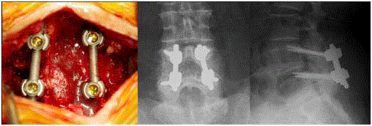
Figure 1
MIS-PLF with CBT screws. View of final construct before bone grafting and postoperative X-rays.
Materials and Methods
Patient demographics
Since December 2012 we have been using CBT screws for lumbar
spine fusion. 30 patients who received posterior decompression
with a single-level TLIF (transforaminal lumbar interbody fusion)
wereretrospectively selected for this study. The study was approved
by the Ethics Committee of the Steel Memorial Muroran Hospital
(approval number J160601).There were 24 L4 degenerative
spondylolisthesis (DS), 4 L3 DS, one L4/5 discitis in remission and
one L4/5foraminal stenosis (FS) case.
Thirty consecutive cases operated with PPS during the same
period were selected as a control group. The pathology was 12 L4 DS,
9 L3 DS, 1 L4 isthmic spondylolisthesis (IS), 7 L5 IS and 1 L4/5 FS case
in this group. Both groups were matched for gender ratio, mean age,
BMI, previous lumbar surgery, Charlson age corrected comorbidity
index [5] and duration of symptoms. There was no significant
difference in smoker/nonsmoker ratio, pre-op (preoperative) ODI
score and pre-op % slip between the 2 groups (Table 10. All cases were
followed up for at least one year post-op.
Surgical procedures
The single-level lumbar fusion with CBT was performed through
a single midline 4 cm incision with neural decompression by bilateral
medial facetectomy. The screw insertion starting point was chosen on
the medial aspect of pars interarticularis2mm caudal at the 5 o’clock
position as verified on AP (antero-posterior) C-arm image. After the
starting point was created with a 3 mm surgical burr, the polyaxial
CBT screws were inserted through a cranio-lateral trajectory as
described by Santoni [2] and Mobbs [3]. The trajectory was chosen
so that the screw tip would lodge in the upper corner of the vertebral
body. TLIF with a PEEK cage was then performed after applying
distraction directly to the CBT screw heads. This was followed by
titanium rod application and final screw tightening in compression.
The morcellated local bone mixed with artificial bone was used for
bone graft (Figure 1).
The single-level MIS lumbar fusion with PPS was performed as
previously described [6].
Quantitative analyses of outcomes
The surgical parameters of evaluation included operation time and
intraoperative blood loss. Postoperative course was evaluated using
CRP (C-reactive protein) and CPK (creatine phosphokinase) values
on POD1 (postoperative day 1) and POD7. The Japanese Orthopedic
Association Back Pain Evaluation Questionnaire (JOABPEQ) [7],
Oswestry Disability Index (ODI), Back Pain VAS (visual analogue
scale), Leg Pain VAS, Lower Extremities Paresthesia VAS were used
for clinical outcome appraisal. All the questionnaires were filled out
by the patients without supervision by a physician. The JOABPEQ,
ODI and VAS score were evaluated pre-op and at 6 months of
follow up. The postoperative complications, length of hospital stay
in days and discharge destination (home or rehabilitation facility)
were registered for all cases. The loss of correction was evaluated by
comparing % slip and slip angle values on pre-op (pre-operative),
post-op and follow up X-rays. Screw loosening and fusion rates were
evaluated by reviewing follow up CT scans.
Statistical analysis
The interval data were compared using unpaired Student’s t test
between the 2 groups. The nominal data were compared using the
Chi-square. The significance cutoff for both tests was set at p≤ 0.05.
Figure 2
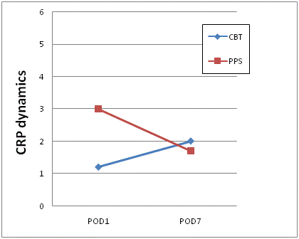
Figure 2
Postoperative dynamics of mean CRP values for CBT and PPS
groups.
Asterisk depicts the statistically significant difference between the CBT and
PPS groups (p=0.02).
Figure3
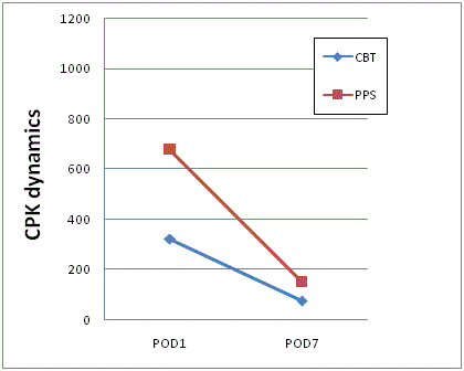
Figure 3
Postoperative dynamics of mean CPK values for CBT and PPS
groups.
Asterisk depicts the statistically significant difference between the CBT and
PPS groups (p≤ 0.002).
Figure 4
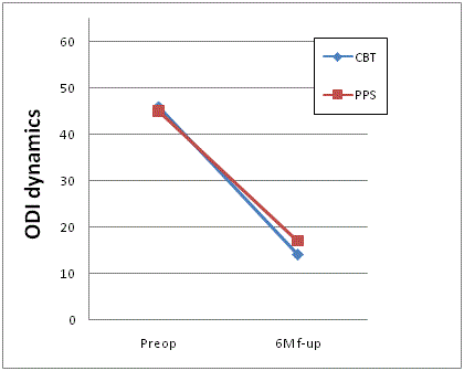
Figure 4
The ODI score dynamics.
Results
All cases were followed up until at least one year post op. The
average operation time was significantly longer in the PPS group (117
± 31 min and 137 ± 26 min in the CBT and PPS groups respectively, p=0.02). The intraoperative blood loss was significantly greater in
the PPS group (104 ± 72 ml and 280 ± 254 ml in the CBT and PPS
groups respectively, p=0.002). The average serum CRP level on POD1
was significantly higher in the PPS group (1.2 ± 0.9 and 3.0 ± 2.5 in
the CBT and PPS groups respectively, p=0.0008). The POD7 mean
CRP values were not significantly different (2.1 ± 2.0 and 1.7 ± 1.1 in
the CBT and PPS groups respectively, p=0.3). The POD1 mean CPK
level was significantly higher in the PPS group (322 ± 358 and 679 ±
410 in the CBT and PPS groups respectively, p=0.001). The POD7
mean CPK level was significantly higher in the PPS group (74 ± 37
and 151 ± 129 in the CBT and PPS groups respectively, p=0.002). The
CPK levels in the CBT group showed faster decrease (Figure 3). The
length of hospital stay was significantly longer for PPS group (16 days
for PPS vs. 13.6 days for CBT, p=0.03). Significantly more patients
in the PPS group required a transfer to a rehabilitation facility after
discharge (9 in PPS group vs. 3 in CBT group, p=0.05).
The average JOABPEQ effectiveness ratios were not statistically
different with best levels of effectiveness demonstrated for pain and
walking and lowest effectiveness for psychological disability (Table
2).
The mean preoperative ODI scores were 46% and 45% in the PPS
and CBT groups respectively which were statistically not different
(Table 1). The mean follow-up ODI scores decreased dramatically in
both groups to 17% and 15% in the PPS and CBT groups respectively
which were also statistically not different between groups (Figure 4).
The mean preoperative low back pain (LBP) VAS scores were 69.1 and
69.2 in PPS and CBT groups respectively. At follow up the mean LBP
VAS scores were 19.4 and 10.5 in PPS and CBT groups respectively
(p=0.1) (Figure 5). The mean preoperative leg pain VAS scores were
65.5 and 73.9 in PPS and CBT groups respectively. At follow up the
mean leg pain VAS scores improved to 17 and 8.8 in PPS and CBT
groups respectively (p= 0.1)(Figure 6). The mean preoperative low
extremity paresthesia VAS scores were 66 and 65 in PPS and CBT
groups respectively. At follow up the mean low extremity paresthesia
VAS scores improved to 10.6 and 8.8 in PPS and CBT groups
respectively (p=0.7) (Figure 7). No statistically significant differences
were observed between any preoperative or follow up VAS scores
between the PPS and CBT groups.
Losses of correction were evaluated by measuring % slip and slip
angles on post-op and follow up radiographs. The mean pre-op % slip
was 17% and 13% in PPS and CBT groups respectively, which was not
statistically different. Mean % slip was corrected to 5.7% and 2.6%
on post-op films in PPS and CBT groups respectively. On follow up
films mean % slip degenerated to 16% and 5% in PPS and CBT groups
respectively with mean % slip loss of correction as large as 10% in PPS
and only 2% in CBT group (p=0.0001) (Figure 8).
The mean pre-op slip angle was -12 deg (lordosis) in PPS and -7.2
deg (lordosis) in CBT group, which were not significantly different.
The mean slip angles were corrected to -16 and -7.6 deg on postop
films in PPS and CBT groups respectively. On follow up films mean slip angle changed to -12 and -6.7 deg in PPS and CBT groups
respectively with mean slip angle loss of correction as large as 5.2 deg
in PPS and only 2.3 deg in CBT group (p=0.0001) (Figure 9).
The presence of pseudarthrosis and screw loosening was evaluated
on the follow up CT scans. Screw loosening was observed in 6 patients
(20% of cases) in PPS group and in 0 cases in CBT group (p=0.009).
Bony fusion was achieved in 27 cases in PPS group (90%) and in
30 cases in CBT group (100%). This difference was not statistically
significant by Chi-square (p=0.07). Two pseudarthrosis in PPS group
required revision with CBT screws and eventually achieved fusion
and one case was asymptomatic (Figure 9). There was one case of
deep hematoma formation requiring evacuation in the PPS group
and no major complications, such as CSF leak, deep hematoma or
deep wound infection in the CBT group.
Discussion
The CBT technique for posterior lumbar spine instrumentation
has been growing popular in recent years [8,9]. There are substantial
anatomical cadaveric [2,4] and in vivo [3] studies supporting its
effectiveness. However, to our knowledge, as of yet, there are few
papers reporting the outcomes of posterior lumbar fusion with CBT
screws in human subjects. We could locate one case report of CBT
use in a long construct in combination with PS leading to a favorable
result [6]. We used CBT trajectory to insert screws for single segment
posterior lumbar fusion with TLIF. The operation was performed
through a single midline incision after spinal canal decompression
with medial facetectomy. Local bone mixed with hydroxyapatite
grafted to achieve fusion. We compared the parameters of surgical
invasion and clinical and radiological outcomes of the new CBT
procedure with the previously established MIS lumbar fusion with
PPS [6]. We found out that operation time was significantly longer
in the PPS group that might be explained by more time consuming
rod introduction through a percutaneous route, time needed for
image intensifier manipulation during the PPS insertion, longer time
needed for neural decompression through a smaller incision. The
intraoperative bleeding was also significantly greater in the PPS group
which could be explained by incisions through the muscle tissue for
PPS insertion as opposed to single midline incision in the CBT group
with only minimal muscle tissue violation.
Postoperative day one CRP and CPK values were both
significantly higher in the PPS group which could be accounted
for by the additional invasion of the additional incisions through
the paravertebral muscles for the PPS insertion in the PPS group
as opposed to paravertebral muscle preservation achieved with the
medial incision required for the CBT insertion. Less muscle damage
probably also accounts for lower POD7 levels of CPK and faster
CPK subsidence in the CBT group. Despite the mean preoperative
Charlson comorbidity indices, BMI, duration of symptoms, previous
low back surgery, ODI and VAS scores being not different between the
PPS and the CBT group, patients in the CBT group were discharged
from the hospital significantly faster and significantly more of these
patients returned home after discharge compared to the PPS group
who needed longer hospital stays and prolonged rehabilitation. This
might be partially explained by overall milder surgical invasion of
the CBT procedure as seen from the shorter operation time, and
less intraoperative bleeding. The better fixation achieved with CBT
might be another contributor to the faster recovery of the patients in
the CBT group. The higher torque of the CBT screws compared to
traditional PS was previously reported in cadaveric and in vivo studies
[2-4]. By reviewing the follow up films we found out that loss of
correction for both % slip and slip angle were significantly greater in
the PPS group compared to the CBT. This might be explained by the
ability of the CBT screws to prevent the notorious windshield wiper
motion phenomenon due to the inherent features of the cortical bone
trajectory which places the screw in intimate contact with the cortical
bone from all sides. Also there is a great extent of possible variations
for the cortical bone trajectory itself that allows for selective screw
placement into the areas of the highest bone density especially when
used with intraoperative navigation. We observed screw loosening in
20% of PPS cases and pseudarthroses in 10% of PPS cases while there
was no incidence of screw loosening or pseudarthrosis in the CBT
group. This difference might have resulted from the better fixation
achieved with CBT screws.
During the operation the distractor can be directly applied to the
CBT screw heads without risk of loosening or cutout because of the
sufficient anchoring strength of CBT screws. This allows for better
intraoperative reduction of the slipped vertebra. Such reduction is
impossible when PS is used because of high incidence of loosening.
Table 1

Table 1
Background data comparison of two surgical groups.
Table 2
Table 2
Comparison of JOABPEQ effectiveness ratios.
Conclusion
Overall, compared to PPS, single level lumbar fusion with CBT screws proved to be associated with less surgical invasion, better clinical results (faster hospital discharge with less need for rehabilitation), better spondylolisthesis correction with less loss of correction and screw loosening in our study. CBT screws were also successfully utilized during revisions for pseudoarthroses after instrumentation with PPS. In those cases there was no need for any augmentation or bone packing into the screw tract because the CBT trajectory avoids the PS screw hole and sufficient bone stock is available for screwing even after PS removal. More research is mandatory, however, to elucidate whether CBT screws will perform as well in multiple level lumbar fusion, to what extent can CBT be used in the lower thoracic spine, can CBT screws be successfully inserted percutaneously, can CBT screws be successfully aligned for rod application in a thoracolumbar fusion construct with PSs. Also higher anchoring strength of the CBT screws might lead to higher rate of implant failure such as screw or rod breakage which should become clear in a larger cohort with a longer follow up period.
References
- Ohtori S, Inoue G, Orita S, Yamauchi K, Eguchi Y, Ochiai N, et al. Comparison of teriparatide and bisphosphonate treatment to reduce pedicle screw loosening after lumbar spinal fusion surgery in postmenopausal women with osteoporosis from a bone quality perspective. Spine (Phila Pa 1976). 2013;38(8):E487-92.
- Santoni BG, Hynes RA, McGilvray KC, Rodriguez-Canessa G, Lyons AS, Henson MA, et al. Cortical bone trajectory for lumbar pedicle screws. Spine J. 2009;9(5):366-73.
- Matsukawa K, Yato Y, Kato T, Imabayashi H, Asazuma T, Nemoto K. In vivo analysis of insertional torque during pedicle screwing using cortical bone trajectory technique. Spine. 2014;15;39(4):E240-5.
- Mobbs RJ. The "medio-latero-superior trajectory technique": an alternative cortical trajectory for pedicle fixation. Orthop Surg. 2013;5(1):56-9.
- Charlson ME, Pompei P, Ales KL, MacKenzie CR. A new method of classifying prognostic comorbidity in longitudinal studies: development and validation. J Chronic Dis. 1987;40(5):373-83.
- Kotani Y, Abumi K, Ito M, Sudo H, Abe Y, Minami A. Mid-term clinical results of minimally invasive decompression and posterolateral fusion with percutaneous pedicle screws versus conventional approach for degenerative spondylolisthesis with spinal stenosis. Eur Spine J. 2012;21(6):1171-7.
- Fukui M, Chiba K, Kawakami M, Kikuchi S, Konno S, Miyamoto M, et al. JOA Back Pain Evaluation Questionnaire (JOABPEQ)/JOA Cervical Myelopathy Evaluation Questionnaire (JOACMEQ). The report on the development of revised versions. J Orthop Sci. 2009;14(3):348-65.
- Mizuno M, Kuraishi K, Umeda Y, Sano T, Tsuji M, Suzuki H. Midline lumbar fusion with cortical bone trajectory screw. Neurol Med Chir (Tokyo). 2014;54(9):716-21.
- Ueno M, Imura T, Inoue G. Posterior corrective fusion using a double-trajectory technique (cortical bone trajectory combined with traditional trajectory) for degenerative lumbar scoliosis with osteoporosis: technical note. J Neurosurg Spine. 2013;19(5):600-7.