Research Article
Heart Rate Variability Analysis Before and During Coronary Artery Bypass Graft Surgery
Tatiana Mironova1*, Vladimir Mironov2, Elena Kuvatova3 and Vladimir Kuvatov1
1Federal Yekaterinburg Medical Scientific Center of Prevention and Health Keep Industrial Workers, Yekaterinburg,Russia
2Ural State Medical University of Health Ministry, Yekaterinburg, Russia>
3Federal State Center of Cardiovascular Surgery, Chelyabinsk, Russia
*Corresponding author: Tatiana Mironova, Federal Yekaterinburg Medical Scientific Center of Prevention and Health Keep Industrial Workers, Yekaterinburg, 620028, Russia
Published: 13 Jul 2017
Cite this article as: Mironova T, Mironov V, Kuvatova
E, Kuvatov V. Heart Rate Variability
Analysis Before and During Coronary
Artery Bypass Graft Surgery. Clin Surg.
2017; 2: 1559.
Abstract
The purpose of these researches was evaluation of the high-resolution Rhythm Cardiography (ECG)
possibilities for definition of an actual cardiovascular status of the operated patients with angina
pectoris during carrying out of the Coronary Artery Bypass Graft Surgery (CABGS) for a myocardial
revascularization. These investigations were made out by means of the hardware-software complex
KAP-RC-02 -"Micor" with a monitor record and the analyses of the Heart Rate Variability (HRV)
in Time-Domain and Frequency-Domain. Monitor records were made in various stages CABGS in
123 patients. In results, the RCG manifested oneself as the quite adequate and perspective method
of the definition of the actual cardiovascular status during carry out for cardiac revascularization
in patients with angina pectoris. Also the data of the HRV studying at the CABGS testify about
possibilities of the RCG definition of the high risk of life-dangerous cardio arrhythmias during
operation, about different changes of the Sinoatrial heart Node (SN) regulation, and concerning
the symptoms of the lethal outcome of CABGS. The loss of the peripheral autonomic sympathetic
and parasympathetic control in SN in the form of an autonomic cardioneuropathy syndrome is the
predictor of the intraoperative complications during and after CABGS. The obtained data at the
RCG monitoring HRV record assume about wide prospects of the high-resolution RCG using in the
cardiac surgery as a whole. Actual multiple deregulations of SN pacemaker activity testified to its
adequate to a pathophysiology of each period of this cardiac operation, according to initial ischemic
defeats and localization of the cardiosurgical manipulations during CABGS.
Keywords: Heart rate variability; Coronary artery bypass graft surgery; Actual cardiovascular
status; Predictors of complications; Lethal outcome
Introduction
The purpose of researches was evaluation of possibilities of the high-resolution Rhythm Cardiography (ECG) for the Heart Rate Variability (HRV) analysis and definition of actual cardiovascular status in patients (pts) with angina pectoris during the Coronary Artery Bypass Graft Surgery (CABGS). This purpose was obtained by the assumption that at cardiological surgery, as well as at cardiology [1-3], the RCG analysis of the HRV for definition of deregulations at coronary artery disease may be useful actual diagnosis of the intraoperative cardiovascular status. Earlier researches on this subject had no convincing results [4]. This, we assume, was connected with insufficient results at low sensitivity of the HRV registration, and also with ignoring of a humoral-metabolic influence on the autonomic regulation of the pacemaker activity of the SN [4- 8]. Innovative contemporaneous achievements in the HRV registration and analysis are especially important for identification of predictors and markers of the intraoperative complications [9,10], including life-threatening cardio arrhythmias [7,11,12].
Patients and Methods
Standard cardiological researches were made out before cardiosurgical operation of 256 pts. After initial standard and RCG investigations there were selected 123 pts for CABGS. Healthy 47 men’s were control (Figure 1). 123 pts were investigated for HRV analysis by the specialized computer diagnostic complex CAP-RC-01-"Micor". Initially additional RCG-symptoms, characteristic for stable angina pectoris, were defined at the intranozological diagnostics of the Coronary Artery Disease (CAD) before operation. In patients was made out CABGS on open heart with an it’s stop and passage to an apparatus of artificial blood circulation (ABC) by use of the cardiopulmonary pump. HRV was recorded during CABGS by the specialized hardware-software diagnostic complex CAP-RC-02-"Micor" of high-resolution (discretization of ECG-signal (1000 ± 3Hz) at the monitor record (The certificate No. FS 02262005/2447-06; patent No 2199945). In the software at RCG-research HRV analyses were made out in Time-Domain and Frequency-Domain with using of the Fast Fourier's transformation and Parsen's and Hamming’s spectral windows. Basic HRV-indices were considered: RR-average value of the all intrasystole intervals, their standard deviation – SDNN, average square deviations of the humoral -metabolic (σl), sympathetic – (σm) and parasympathetic – (σs) amplitudes of HRV-fluctuations. Also the spectral analysis was calculated for evaluation of correlation of regulative factors influences in the sinoatrial heart node (SN) - VLF%, LF%, HF% (Figure 1-7). The monitor Rhythmocardiogram record (Rcg) was remotely transferred to neurocardiology laboratory for the immediate analysis and recommendations to cardiac anesthesiologists. HRV registration was carried out till and after cardiac arrest and at the passage of the blood circulation to apparatus blood circulation-ABC. At the same time with Rcg record in real current time the Electro Cardio Gram (ECG) was registered too. Before shunting HRV researches were made out in rest (Ph) and 4 tests. After a premedication narcosis. The monitor Rcg was registered without tests. Rcg is a graphic image of intersystole pauses between heart contractions, as the vertical rectilinear pieces, equivalent in size of RR intervals duration with the beginning on abscissa axis and as parallel to ordinate axis (Figure 1-12). The intervals were registered during of all CABGS till the passage to ABC and after ABC. Every period of CABGS had itself peculiarities of HRV wave structure. Every Rcg consists of 300 RR- intervals. The artifacts were caused by influence of the electric knife, direct surgical manipulations on the heart tissues, and it’s were removed before analysis of the stationary Rcg-part of. Episodes of the cardiac arrest under influence of introduction of drugs and a Cardio Plegic Solution (CPS) were registered too during preparation of the passage the ABC, till to the restoration of the heart systoles after the shunting. During CABGS the real work was provided to the analysis of results of HRV monitoring and dynamics of its changes. The special program "Stat" was used in statistical evaluation of the computer material for verification of the hypothesis equality of variable rows on criterion Student (t, analogous t -z and p. Normalcy of the distribution was checked on the N. Colmogorov factor with approximation on Yu. Tyurin. For correlative analysis was used nonparametric Spirmen method with program SPSS 12.0.
Figure 1
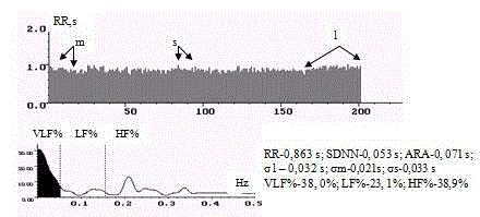
Figure 1
Rhythmocaridogram, spectrogram and middle values of RCGsigns
of healthy man.
Figure 2
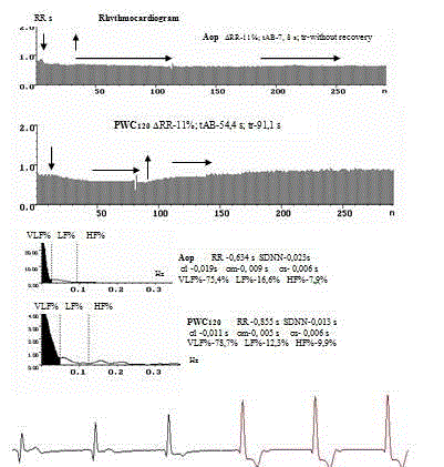
Figure 2
Here are Rhythmocaridogram, spectrogram and middle values of
the HRV indices of a patient with stable angima pectoris 2FC in Aop and
PWC120 tests.
Figure 3
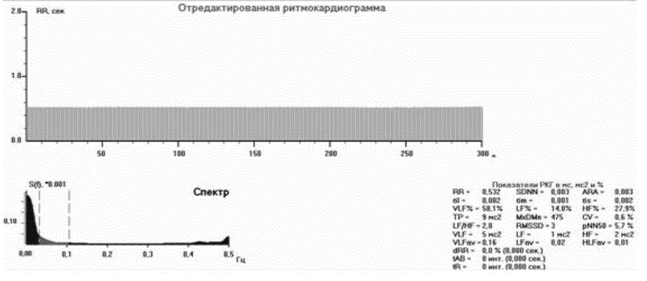
Figure 3
Here are Rcg and spectrogram and middle HRV –indices after a
premedication.
Figure 4
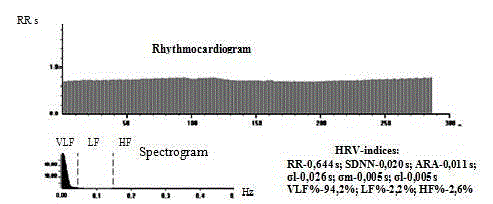
Figure 4
Rcg, spectrogram and middle value of the HRV –indices at the
intubation nacrosis.
Figure 5

Figure 5
Rcg, the spectrogram and middle of HRV indices during an
intubation nacrosis.
Figure 6
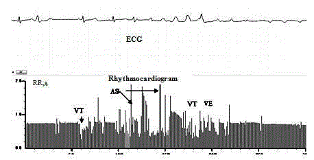
Figure 6
Rcg and the ECG at the clip installation in the aorta.
Figure 7
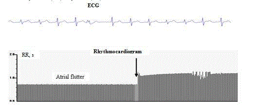
Figure 7
Rcg and ECG at the cannulation of vessels.
Table 1

Table 1
HRV-indices before (n-123) and after (n-123) premedication at the CABGS.
Table 2
Table 2
Results of HRV comparison in patients with autonomic cardioneuropathy (ACN) (n-56, first line) and in patients without ACN (n-67, second line) before
CABGS.
Results and Interpretation
Initially at primary RCG-research before CABGS HRV reduction
was defined, adequate to the CAD expression [1,2,13] at the all
patients, selected for the cardiosurgical myocardial revascularization.
Except there were before CABGS HRV fragments with HRV
stabilization without any wave structure on the Rcg during a
paroxysm of the angina pectoris. More exact computer measurements
of RR intervals demonstrated on the named fragments the differences
between the neighbouring RR intervals within 3.55 ± 1.02 ms (Figure
2). These data were obtained at measurement of each RR interval
of thousand stabilization fragments by means of the graphic cursor
and the special program. These stabilization fragments correlated
to duration of clinical and ECG symptoms of ischemic episodes. It’s
were the evidence of stenocardia in patients and could be analyzed on
the frequency, duration of ischemia, the hemodynamic importance,
the functional class of stenocardia initially before operation,arrhythmogenic background of the autonomic sinoatrial node
deregulation (Patent No. 2322942).
Premedication
Before CABGS in 30 minutes a diazepam (seducsen, relium),
atropine, promedol were introduced in patients. At the majority of
operating patients in response to these drugs there was increase of the
humoral-metabolic influence in SN and reduction of sympathetic and
parasympathetic indices in HRV regulation (Table 1). In the spectral
shares there were the humoral influences in the form of increase of
spectral density of the very low-frequency diapason of the HRV in
total spectrum (Figure 3). The HRV registration before direct made
out of CABGS and cardiac arrest is expedient for the purpose of
identification of complications risk during CABGS and registration
of the predictors of the intraoperative cardioarrhythmias, because
their development was connected with an initial arrhythmogenic
background [14]. This background were: dysfunctions of sinoatrial
heart node, extremely expressed decrease of all HRV waves (SDNN),
because of loss autonomic sympathetic and parasympathetic control
in the form of the autonomic cardioneuropathy (ACN) (Figure
2,4,5,8-10,12). There were before CABGS initial episodes of the
ischemic autonomic denervation, ECG changes in the form of
sinoatrial blockade of 1 or 2 degrees. Before CABGS ACN was defined,
as the most significant preoperative marker of life-threatening
cardioarrhythmias during CABGS. At selection of patients before
CABGS initially there were HRV stabilization and lack of reactions
in the stimulant tests. HRV data before CABGS of patients with and
without ACN were compared. Initial HRV indices had authentic and
considerable differences between groups (Table 2). In table 2 it may
be seen, that in patients with ACN (56 pts) the HRV indices were
with high authentic less, than in remaining 67 other patients, except
of the spectral share of the humoral-metabolic influence in SN and
time of achievement maximal reactions in tests and time restorative
after stimuli. HRV stabilization at the preoperative period in 100% of
cases proceeded to development of cardioarrhythmias during CABGS
(Figure 3,6-8). In the patients with ACN its testified about loss of the
autonomic control of the SN activity (Table 2). One patient from 123
died through 4 days after CABGS. RCG examination before death
showed typical ACN (stabilization of rhythm on the background
tachycardia, absent reactions on any stimuli, breaches of the atrial
conductivity) before, during and after CABGS. On this background
there were episodes of atrial fibrillation, migration of the rhythm
pacemaker, the SN dysfunction.
From initially low level of the Rcg during an anesthesia the
amplitude of HRV waves decreased even less. At first HRV waves
of the fast and sufficient autonomic parasympathetic regulation
disappeared, then sympathetic, and in the last humoral-metabolic
fluctuations. Atrial and ventricular extrasystoles proceeded to total
disappearance of HRV waves (n-56, p< 0,001). In these cases the
HRV regulation transferred up to humoral-metabolic low-amplitude
level, which is uneffective and not adequate. In the spectral analysis
this correlated to VLF%, which was 72.2-91.2% on the background
extremely low share of the autonomic influence in SN (LF% and
HF%=2.4% - 15%). The HRV indices of the sympathetic and
parasympathetic periodicity-σm, σs- were expressed in units of ms
-0,001-0,006 sec., therefore the HRV record, keeping in operative
memory and analysis it must be registered with the high exactness,
it is necessary. Then the rhythm was completely stabilized, that
corresponded to a full anesthesia. In 38 (78,1%) cases were episodes
of the rapid rhythm, there were registered signs of breaches of
conductivity between atrials because of blockade of the Bachmann
bunch, migration of the rhythm pacemaker on SN and atrials, (there
were reduction of P wave and change of its form), this corresponded
to the high Makruz index.
One of key role in the development of the cardioarrhythmias
was the ischemic dysfunction of the myocardium at the surgical
myocardial revascularization in the patients operated by CABGS.
Frequency of the cardioarrhythmias in these patients significantly
negatively with average degree correlated to the background of the
myocardial contraction ability reduction (r=0.584-0.638). At the
subsequent continuous registration of Rcg the stages of preparation
and carrying out of narcosis were registered (Figure 4 and 5). By
results of Rcg-record only in 17 (13.8%) patients, which had before
CABGS rather safe HRV wave structure, there were registered the
supra ventricular extrasystoles and paroxysms of the tachycardia
(Figure 6). The supra ventricular extrasystoles were characterized by
an uncompensated post ectopic interval on the Rcg and changes of an
atrial P wave on the synchronous ECG. This was recognizable visually at the RCG analysis, though possibility of more exact definition pre-and post ectopic intervals in milliseconds always was preferable. In the
others 106 (76.2%) cases the idioventricular rhythm was consistently
in the form of migration of the rhythm pacemaker on SN and atrials,
with ECG signs of the sinoatrial blockade and changes of the sizes
and forms of the P wave, R alternation, usually against extremely
expressed HRV decrease in 56 (46.5%) patients with initial ACN,
which was registered on Rcg before CABGS. After the premedication
and the intubation narcosis the Rcg continuous registration was
carried out on the hardware-software complex CAP-RC-02 -"Micor",
specially was created for monitor intraoperative record. These records
were carried out at stages of the intubation narcosis, the cannulation
(Figure 7), installation the clips on vessels (Figure 6), introduction of
cardioplegic solution (CPS) (Figure 8), connection up to apparatus of
ABC (Figure 9 and 10), restoration of the heart activity after CABGS
end (Figure 12).
Cannulation
The cannulation of hollow veins and an aorta was carried out for
connection with the apparatus of the ABC (Figure 7). Primary volume
of filling made 1.5-2 l. The structure perfusion solution besides protein,
glucose, salt solutions included potassium for stopping of the heart.
Transference of the perfusion solution to the blood flow was carried
out under control of the Central Venous Pressure (CVP), the blood
pressure (BP), and ECG for the purpose of prevention strain in the
heart cavities and development of cardioarrhythmias. The beginning
of the artificial circulation was accompanied by fast decrease in BP,
CVP with prevalence of deposition of the venous blood over receipts
it in aorta, decrease in a hemodilution by the blood stream without
pulse. At first it was introduction of the 10 mg/kg thyopental, then
fentanyl, and at the beginning of the warming - arduan 0.1 mg/kg.
For decrease in blood loss was used an ultra filtration. In 97 (80.5%)
operated patients there was registered the idioventricular rhythm,
it was more often at those patients, which had considerable HRV
reduction in the preoperative period, bradycardia and/or delay of
the conductivity function (Figure 7). During carrying out CABGS
episodes of the accelerated idioventricular rhythm were registered
during introduction of a Cardio Plegic Solution (CPS) at the heart
arrest.
Cardioplegic solution (CPS) introduction
Anesthesia during of artificial circulation was supported by the
barbiturates, neuroleptanalgesia, indepolarizating myorelaxants.
The normotermic cardioplegy was applied for myocardial protection
against ischemic damage. After achievement of the necessary
temperature the clip in aorta was installed, and heart activity stopped.
After the cannulation of the veins and aorta, introduction of CPS and
hypothermia, there was fibrillation of ventricles was registered and
heart stopped (Figure 8 and 9). At the initial HRV low level during
the narcosis the amplitude of HRV waves decreased even more. At
first HRV waves of the fast autonomic regulation disappeared –
protective parasympathetic fluctuations (s), then sympathetic (m),
and in the last the humoral-metabolic waves (l). Atrial fibrillation
and ventricular extrasystoly preceded to the total disappearance
of the HRV (n-48, p < 0,001). HRV regulation transferred over on
humoral-metabolic level with low HRV waves (VLF%=67.1% -
8.4% in the spectral analysis), then the heart rhythm was completely
stabilized, that corresponded to the full narcosis (Figure 4). In 38
(30.9%) cases the tachycardia appeared, because of Bachmann bunch
blockade and there were registered signs of breaches conductivity
between atrials, migration of the rhythm pacemaker on auricles
and nodal complexes, an atrial asystole in the form of reduction P
waves, its lengthening and diphasic P wave, increase of an Makruz
index. At cardioplegic solution introduction on the background of
HRV oppression there was decreased spreading of excitement in the
heart ventricles, and amplitude of R waves decreased on the ECG,
but still some time R remained in gradual decrease, manifesting the
any minimal heart activity [15]. The hemodynamic significant atrial
flutter (Figure 7) - AF during CABGS was in the patients with the
initial pathological P wave and PQ interval (r = -0.397-0.456) and
the other cardioarrhythmias. The more positive result of CABGS was
in patients without AF and initial other arrhythmias - in 17 (25.3%)
patients. The postoperative restoration in these patients was without
complications and it occurred in 1.6 times quicker.
Connection to the apparatus of the artificial blood
circulation (ABC)
AF (Figure 6) during CABGS, as variant of atrial tachycardia,
appeared much more often than other breaches of the heart rhythm
- in 106 (86.1%) of the patients (Figure 9 and 10). Life-threatening
AF obligatory was registered at cannulation of the atrials and veins,
preparation and carrying out the cardioplegia, before connection
to the ABC. At the subsequent cardiac manipulations the AF was
registered obligatory in patients with initial ACN – 56 (46.5%) with
the HRV stabilization and absent reactions in tests (Table 2 and
3) before CABGS. According to the spectral analysis in the named
patients the humoral-metabolic influence on pacemaker cells of
SN was considerable prevailing (Figure 3 and 4). These patients
had 3 and 4 FC of stenocardia and were most clinically heavy
group that confirmed ACN syndrome, as the predictor of AF and
the cardioarrhythmias during CABGS (Figure 6,7,10 and 11). The
pathogenesis of these breaches was explained by loss of the autonomic
control, surgical manipulations, corresponding to their localization,
and also dystrophic changes in the SN pacemaker cells. Last was proved
in researches with electron microscopy of the autopsy materials of SN in deceased patients [1]. During of CABGS intraoperative record of
Rcg (15-18 thousand intervals in each of 123 patients) was carried
out, that allowed to register of all changes of the HRV. At the during
of the connection to the ABC and gradual cardiac arrest (Figure 6
and 8) the surgical manipulations on the heart auricles and ventricles
tissues were accorded by the atrial and ventricular breaches of rhythm
in forms of decrease of Rcg level, single and group cardioarrhythmias
in 89 (72.2%) patients (Figure 6-11). There were wide ventricular
complexes without P waves on the background of normal rhythm, or
on the accelerated rhythms (more 100/min.), but it was obligatory in
the all patients with ACN. In each case there were the synchronous
communication of the rhythm breaches with localization of surgical
manipulations was obvious. For example, at the cannulation of
aorta and veins the ventricular arrhythmias were registered during
manipulations with heart ventricles, atrial arrhythmias were at the
aorta clipping in 106 (86.1%) patients at the installation and/or
removal of the vessel clips (Figure 6 and 11 ). Ventricular tachycardia
(Figure 8), fibrillation of ventricles appeared before cardiac arrest
just before CPS introduction with potassium, transfer of perfusion
to apparatus ABC and carrying out CABGS. Direct procedure of the
shunting was carried out within 4-10 min and could proceed on the
background of the ventricular tachycardia or fibrillation.
The pathological auricles may be manifested by the ECG changes
of the P waves at the initial investigation of patients before CABGS,
expressive electric alternation, an orientation of the diphasic P waves,
changes of duration of PQ, and also height of R. At these cases there
were the breaches of the heart rhythm, as a rule, in patients with the
background of expressed HRV decrease or its stabilization and the
absence of reactions in tests or with slow achievement of the minimal
reactions and slow restoration after stimuli in tests in the preoperative
period. That corresponded to RCG characteristic to CAD symptoms
(Table 2) and before cardiac arrest and passage to ABC. That is, the
expressed decrease or stabilization of the HRV and absence of the
reactions in tests before CABGS are predictors of the rhythm breaches
during operation, including life-threatening cardioarrhythmias.
The entire periods – introduction in an anesthesia, the cannulation
of the vessels, the hypothermia, CPS introduction with potassium
had characteristic in the most cases with visually recognizable form
of changes on Rcg. These periods had mathematical Rcg-indices’
(Table 4) But the constant HRV –indices manifested the loss of the
autonomic control (LF%, HF %) and predominant of the humoralmetabolic
influence (VLF %), slow and inadequate in all stages at
narcosis.
Before heart arrest, and also at the beginning of the independent
heart contractility there was a true atrial flutter (AF) 1:2, 1:3, 1:4 on
the macro re-entry pathogenesis round the right atrioventricular
valve on the background anesthesia in 86 (69.9%) patients (Figure
7). The differential diagnostics between AF and intsizion tachycardia
assumes from these two variants of arrhythmia after all AF. To
istmus-dependence of AF, as to the main difference from intsizion
tachycardia, in the discussed cases may be add ECG changes,
which initially connected with auricles, and also connected to AF
with localization of surgical manipulations in the named atrials.
Declared prevalence of sympathetic influences at the forming AF,
as the autonomic pathogenesis link of AF, apparently needs in the
evidences. Here are the same contrarguments - the insufficient
accuracy of registration and the HRV analysis, and also ignoring of
a humoral influence on the SN regulation, when forming potentials
in the pacemaker cells of the SN. In the Table 3 there is shown the
connection of AF with low HRV. The meaning of SDNN in patients
without atrial flutter was reliability more, than in patients with this
arrhythmia. This is testimony of pathogenic influence of the HRV
reduction at the appearance of these cardioarrhythmias during
CABGS and surgeon manipulations.
If there was no independent restoration of the heart activity after
removal of the clips from aorta and on the monitor Rcg and ECG the
fibrillation of ventricles or the migration of rhythm pacemaker were
registered, in this case the electric defibrillation (ED) of heart was
carried out (Figure 11). In 15 cases at the development of asystolia
direct massage of the heart was carried out. In compliance with HRV
indices the extremely lowered autonomic regulation in SN remained
during all CABGS. The lowest indices belonged to parasympathetic
fluctuations - in all stages CABGS their amplitude consists only 1-2
ms on the background permanent prevailing influence in SN of the
humoral-metabolic factor, slow, insufficient, sometimes causing
paradoxical reaction. The cardioarrhythmias registered mainly
during the stages, being accompanied by surgical manipulations on
the heart tissues in patients with extremely low HRV.
Figure 8
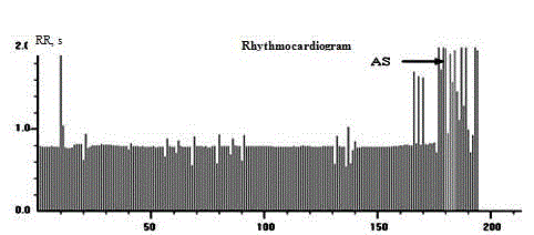
Figure 8
Rcg after introduction of cardioplegic solution.
Figure 9
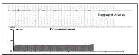
Figure 9
Rcg and ECG during the sinoatrial arrest and connection to
apparatus of artificial circulation.
Figure 10
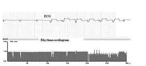
Figure 10
Rcg and ECG.
Figure 11
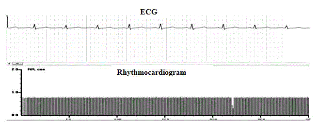
Figure 11
Rcg and ECG during removal of the clip from aorta.
Figure 12
Figure 12
Rcg and ECG during ECG restoration.
Table 3
Table 3
Analysis of SDNN in patients with atrial flutter (n-86) and without it during CABS.
Table 4
Table 4
Analysis of SDNN in patients with atrial flutter (n-86) and without it
during CABS.
Conclusion
1. The high-resolution rhythmocardiography with HRV
analysis are the adequate and perspective method of evaluation of
the actual cardiovascular status before and during carrying-out of the
cardiosurgical myocardial revascularization in patients with angina
pectoris.
2. The HRV analyzed in time- and frequency-domains consists
the supplementary symptoms for the intranosological diagnostics
at the selection of patients for the cardiosurgical treatment of the
stenocardia, and mainly it consists HRV symptoms of the high risk
of complications during CABGS, including the cardioarrhythmias.
3. The results of HRV researches testimonies about
the possibilities of definition of the high risk of life-dangerous
arrhythmias and also lethal outcome at the CABGS. The loss of peripheral autonomic control in the sinoatrial node, as syndrome of
the autonomic cardioneuropathy, is the predictor of complications at
the cardiosurgical operation.
4. The receiving data about multitude variants of deregulations
of the sinus heart node pacemaker activity corresponded to stages of
the cardiosurgical operation, connecting with ischemic breaches of
the coronary arteries, as soon as with localization of the cardiosurgical
manipulations during CABGS.
5. There was proved, that coronary artery disease obligatory
accompanied by the deregulations of the pacemaker activity of the
sinus heart node. Its dynamics corresponded to actual cardiovascular
status of patients and may be used at their cardiosurgical treatment.
References
- Mironova T, Mironov V. Heart rate Variability at the Coronary Artery Disease. / Second publication.. – Cnelyabinsk Russia: Recpol. 2008;173.
- Teplakov A, Lukinov A, Livshin A. Possibility of noninvasive diagnosis of the coronary restenosis by evaluation of dynamic heart rate variability indices. Journal “Clinical Medicine. 2010;21-6.
- Mironova T, Bavykin M, Kuvatov V. HRV-analysis in patients with ischemic cardioarrhythmias. Annales of Arrhythmology? 2, 2011. Mat. of 4 All Russian Congress of Arrhythmologists. Moscow. 2011;16-8.
- Lakusic N, Slivnjak V, Baborski F, Sonicki Z. Heart Rate Variability after Off-Pump versus On-Pump Coro nary Artery Bypass Graft Surgery. Cardiol Res Pract. 2009.
- Dao TK, Youssef NA, Gopaldas RR, Chu D, Bakaeen F, Wear E, et al. Autonomic cardiovascular dysregulation as a potential mechanism underlying depression and coronary artery bypass grafting surgery outcomes. Cardiothorac Surg. 2010;5:36.
- McHugh GJ, Sleigh JW, Bo H, Henderson JD. Heart rate variability following cardiac surgery fails to predict short-term cardiovascular instability. Anaesth Intensive Care. 1997;25(6):621-6.
- Kalisnik JM, Avbelj V, Trobec R, Ivaskovic D, Vidmar G, Troise G, et al. Effects of beating- versus arrested-heart revascularization on cardiac autonomic regulation and arrhythmias. Heart Surg Forum. 2007;10(4):E279-87.
- Min SY, Park DW, Yun SC, Kim YH, Lee JY, Kang SJ, et al. Major predictors of long-term clinical outcomes after coronary revascularization in patients with unprotected left main coronary disease: analysis from the MAIN-COMPARE study. Circ Cardiovasc Interv. 2010;3(2):127-33.
- Brener SJ, Galla JM, Bryant R 3rd, Sabik JF 3rd, Ellis SG. Comparison of percutaneous versus surgical revascularization of severe unprotected left main coronary stenosis in matched patients. Am. J. Cardiol. 2008;101(2):169-72.
- Task Force on Myocardial Revascularization of the European Society of Cardiology (ESC) and the European Association for Cardio-Thoracic Surgery (EACTS)1; European Association for Percutaneous Cardiovascular Interventions (EAPCI), Wijns W, Kolh P, Danchin N, Di Mario C, Falk V. Guidelines on myocardial revascularization. Eur Heart J. 2010;31(20):2501-55.
- Kazemi B, Ahmadzadeh A, Safaei N, Jodati A, Sohrabi B, Afrasiabi A. Influence of anterior periaortic fat pad excision on incidence of postoperative atrial fibrillation. Eur J Cardiothorac Surg. 2011;40(5):1191-6.
- Ksela J, Kalisnik JM, Avbelj V, Suwalski P, Suwalski G, Gersak B. Ventricular arrhythmic disturbances and autonomic modulation after beating-heart revascularization in patients with pulmonary normotension. Wien Klin Wochenschr. 2009;121(9-10):324-9.
- Mironova T, Mironov V, Calmikova A, Socolova T, Davidova E, Safronova E, et al. Possibilities of high-resolution HRV analysis. Anadolu Kardiyol Derg. 2007;7Suppl 1:135-8.
- Carney RM, Blumenthal JA, Freedland KE, Stein PK, Howells WB, Berkman LF, et al. Low heart rate variability and the effect of depression on post-myocardial infarction mortality. Arch Intern Med. 2005;165(13):1486-91.
- Laitio T, Jalonen J, Kuusela T, Scheinin H. The role of heart rate variability in risk stratification for adverse postoperative cardiac events. Anesth Analg. 2007;105(6):1548-60.