Research Article
The Free Radial Forearm Flap versus the Pedicled Pectoralismajor Myocutaneous Flap for Quality Reconstruction of Oesophageal and Hypopharyngeal Defects and Literature Review
Athanasios Karonidis1*, Dimosthenis Tsoutsos1, Sotiris Papouliakos2, Evangelia Zaharioudaki2, Athanasia Marinou2 and Zissis Pappas2
1Department of Plastic Surgery, General Hospital of Athens G Gennimatas, Greece
2Department of Otorhinolaryngology, General Hospital of Athens G Gennimatas, Greece
*Corresponding author: Athanasios Karonidis, Department of Plastic Surgery, Microsurgery and Burns Unit, General Hospital of Athens G Gennimatas, Sifnou 33 Agia Paraskevi 15343, Athens, Greece
Published: 11 Jul 2017
Cite this article as: Karonidis A, Tsoutsos D, Papouliakos
S, Zaharioudaki E, Marinou A, Pappas
Z. The Free Radial Forearm Flap
versus the Pedicled Pectoralismajor
Myocutaneous Flap for Quality
Reconstruction of Oesophageal and
Hypopharyngeal Defects and Literature
Review. Clin Surg. 2017; 2: 1553.
Abstract
Background: The aim of this study is to assess the efficacy of free radial forearm flap and pectoralis
major flap for the reconstruction of hypopharyngo-oesophageal defects, highlighting the technical
feasibility, complications, functional outcome and literature review.
Methods: From 2012 to 2016, twelve patients with laryngeal carcinoma underwent laryngectomy,
hypopharyngeal esophagectomy, and reconstruction of pharyngo-oesophageal defects with free
radial forearm flapin eight cases andpedicled pectoralis major myocutaneous flap in four cases.
Ten patients had partial defect, and two patients had total circumferential defect. The flap selection
was based on the general condition of the patient. The patients with better general condition
underwent microvascular free RFF reconstruction in contrast to debilitating patients who had PM
reconstruction.
Results: All flaps survived. The successful outcome was confirmed by swallowing test with
gastrographin, and associated with oral alimentation, good uncomplicated swallowing and improved
quality of life. Two patients with radial forearm flap microvascular reconstruction developed
fistula formation andone stricture. Two patients with pectoralis major reconstruction developed
fistula formation and stricture. Two patients died due to tumour recurrence. Oral alimentation
was achieved in eleven patients. The patients who underwent radial forearm flap microvascular
reconstruction reported better quality of life in terms of swallowing and cosmetic appearance
comparing to pectoralis major reconstruction, whereas there were equal results in terms of speech.
Conclusion: The radial forearm flap and the pectoralis major flap safeguard a feasible reconstruction
of hypopharyngo-oesophageal neo-tube and provide satisfactory and consistent functional results.
However the free radial forearm flap offers better quality reconstruction and is considered a superior
reconstructive option for the patients who can tolerate major surgery.
Keywords: Radial forearm flap; Pectoralis major flap; Pharyngoesophageal defects; Cervical
oesophagectomy, Laryngeal cancer, Microsurgery
Introduction
Regional pedicled flaps and free flaps have been employed to reconstruct pharyngoesophageal defects following major cancer resection. Among the flaps most commonly used are the ‘skin’ flaps such as the pedicled Pectoralis Major (PM) musculocutaneous flap and the free Radial Forearm Flap (RFF). The choice of the best reconstructive option is still controversial. The aim of this study is to assess the efficacy of RFF and PM flap for the reconstruction of hypopharyngo-oesophageal defects, highlighting the technical feasibility, complications, functional outcome and literature review.
Patients and Methods
From 2012 to 2016, twelve patients (eleven males, one female) aged 66 years in average with
laryngeal carcinoma, underwent laryngectomy, esophagectomy, hypo pharyngectomy, and neck
dissection. The oesophageal and hypopharyngeal defects were reconstructed with free RFF in eight patients and with pedicled PM flap in four patients (Table 1). In
nine cases the resection and the reconstruction were performed at
the same operating time, whereas in three cases the reconstruction
was performed a few weeks after the resection. Nine patients had
preoperative radiotherapy. Ten patients had partial defect ranging
from 50% to 80% of the circumference (Figures 1a and 2a), which
were reconstructed with free RFF in six cases and with PM flap in
four cases. Two patients had total circumferential defects and were
reconstructed with free RFF (Figure 3a). In cases where the resection
and reconstruction were performed at the same operating time,
the average operating time was 13 h for the RFF reconstruction,
whereas for PM reconstruction was 9 h. The patients who underwent
microvascular RFF reconstruction were admitted postoperatively in
the ICU for the first two or three days and then they were transferred
to the ward. We performed gastrostomy only in four patients at
the same operating time, whereas all the other patients were fed via
nasogastric tube. Maximum follow-up was 30 months.
Flap dissection and neo-tube reconstruction
The musculocutaneous PM flap is a type V Mathes-Nahai
classification flap, based on the pectoral branch of thoracoacromial
artery and venae comitantes as the dominant pedicle [1]. The PM flap
is used for partial hupo pharyngoesophageal defects. The skin island is
used for the ‘lining’ and a 6 - 8 × 10 cm skin ‘patch’ is usually sufficient.
The muscle can provide wound cover and a bed for skin grafting
(Figures 1a-1e). The RFF is a fasciocutaneous type B Mathes-Nahai
flap based on the radial artery and venae comitantes and cephalic vein
as the dominant pedicle [1]. The flap design should be tailored to the
size of the defect. For partial defects a ‘patch’ 6 - 8 ×10 cm of skin
island is usually sufficient (Figures 2a,b). For circumferential defects
a larger flap up to 10×12 cm may be required, if the defect involves
the hypopharynx from the tongue base and the cervical oesophagus
(Figures 3a, 3b). A suprafascial dissection from distal to proximal is performed. During inset, the flap is sutured to the oesophageal
stump inferiorly, preferably before vascular anastomosis (Figures
2b, 3b and 3c). The flap edges are de-epithelialized for 0.5 cm - 1.0
cm and sutured double layered to ‘seal’ the tract, aiming to prevent
the fistula formation [2,3] (Figures 2b and 3c). A nasogastric tube is
passed through the neo-tube for decompression, stenting and feeding
(Figures 2a, 2b and 3c). After the completion of vascular anastomosis
in partial defect, the skin island flap is sutured to the remaining
posterior wall. In circumferential defect the vertical line is sutured.
Then the flap is anastomosed to hypopharyngeal stumps superiorly
(Figures 2c and 3d). In cases of total circumferential defects the lateral
edges of the flap/neo-tube are sutured to the raw prevertebral fascia
to enhance the immobility of the neo-tube and reduce potential
complications (Figure 3d). The neck skin flaps are sutured and closed primarily, reconstructing the tracheostoma (Figures 2d and 3e).
Figure 1a
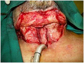
Figure 1a
Partial oesophageal defect where the anterior wall has been
resected and the posterior wall was present.
Figure 2
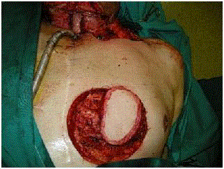
Figure 1b
The dissection of PM flap has been completed
Figure 1c

Figure 1c
The PM flap has been inset partially at the oesophageal defect.
Figure 1d
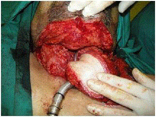
Figure 1d
The inset of PM flap has been inset completed.
Figure 1e
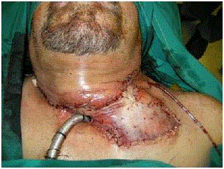
Figure 1e
A skin graft was placed over the PM muscle.
Figure 2a
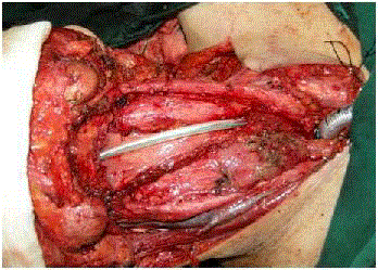
Figure 2a
Partial hypo pharyngoesophageal defect where the anterior and
lateral walls have been resected and the posterior wall was present. The
nasogastric tube has been placed.
Figure 2b
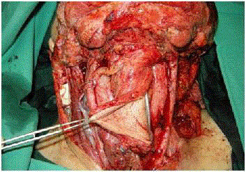
Figure 2b
The radial forearm flap has been sutured with the oesophageal
stump inferiorly and oropharynx superiorly, whereas the flap pedicle was
anastomosed with the superior thyroid vessels.
Figure 2c
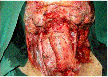
Figure 2c
The flap inset was completed and the neo-tube was created.
Figure 2d
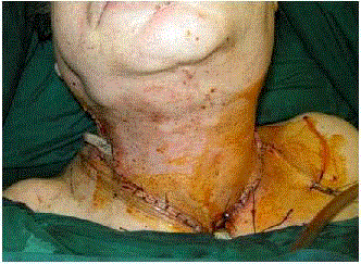
Figure 2d
The neck skin flap was closed primarily.
Figure 3a
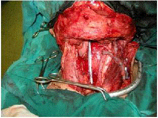
Figure 3a
Circumferential hypopharyngoesophageal defect. Note the
hypophageal stump superiorly and oesophageal stump inferiorly.
Figure 3b
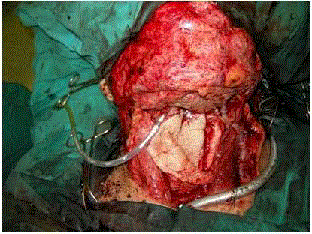
Figure 3b
A large skin island was required to reconstruct the neo-tube.The
radial forearm flap has been sutured with the oesophageal stump inferiorly.
Figure 3c
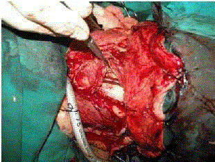
Figure 3c
The flap edges were de-epithelialized and sutured double layered.
Figure 3d
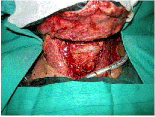
Figure 3d
The reconstruction of the circumferential defect was completed
and the neo-tube was created.
Figure 3e
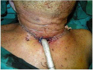
Figure 3e
The neck skin flap was closed primarily.
Figure 4a
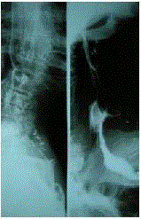
Figure 4a
The reconstruction of a partial defect and continuity of the
alimentary tract was confirmed by swallowing test with gastrographin.
Figure 4b
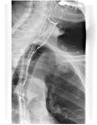
Figure 4b
The continuity of the alimentary tract in reconstruction of a
circumferential defect was confirmed.
Figure 4c
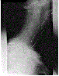
Figure 4c
The neo-tube was clearly defined.
Table 1

Table 1
Demographics and descriptive.
Table 2
Table 2
Flap comparison.
Results
All flaps survived. The integrity and continuity of the alimentary
tract was confirmed by swallowing test with gastrografin (Figure 4a-
4c). The successful outcome was associated with oral alimentation,
good uncomplicated swallowing and improved quality of life. Two
patients with RFF reconstruction had flap anastomotic dehiscence
and fistula formation. The first was treated conservatively, fed
via gastrostomy and finally healed. The second patient who had
undergone reconstruction of a circumferential defect was re-explored
(Figure 5a) and the flap anastomosis was re-sutured primarily (Figure
5b), whereas he was fed via nasogastric tube for almost four weeks.
Stricture was reported in one case and confirmed via endoscopy. A
pouch formation at the distal anastomosis was documented in two
cases, as there was a diameter discrepancy between the neotube
(higher diameter) and the oesophageal stump. Two patients with PM
reconstruction complicated with fistula formation and stricture. The
first was treated conservatively, fed via gastrostomy and finally healed.
The second patient was re-explored and the flap was re-sutured
primarily. Two patients died in five and thirteen months. The first
died due totumour recurrence and the second due to lung metastasis
respectively. Oral alimentation was achieved in eleven patients. One
patient, who underwent PM reconstruction and had radiotherapy preoperatively, was unable to achieve oral diet due to severe stenosis.
Patients reported episodes of swallowing dysfunction occasionally,
with food sticking at the pouch or the area of distal anastomosis and
attacks of hawking, where a degree of stenosis might be developed
but not documented in radiographs. All patients were advised to have
frequent small meals.
However the patients with RFF microvascular reconstruction
reported better quality of life in terms of swallowing and cosmetic
appearance comparing to those with PM reconstruction. In cases
with PM reconstruction skin grafting was applied over the PM
muscle, which might had an adverse effect to the neck motility
due to additional scarring and downgraded the cosmetic result.
However there were equal results in terms of speech for both types
of reconstructions. All the patients were satisfied with the outcome
as their quality of life was improved dramatically. All these cases were
discussed at the oncologic meeting for further adjuvant treatment.
Discussion
The reconstruction of pharyngoesophageal defects is a common
sequela following major cancer resection in the cervical region. The
aims and priorities of reconstruction include: the wound closure, in
terms of restoration of continuity of the alimentary tract and skin
wound cover, the function such as swallowing and speech issues,
cosmesis, and the overall morbidity. The ideal flaps for head and
neck reconstruction should fulfill the following criteria: versatility in
design, adequate tissue volume, superior texture, availability of diverse
tissue types on one pedicle, potential for re-innervation, large and
long pedicle with consistent anatomy, easy and safe flap dissection,
feasibility of a 2 team approach, no need for position change, and
negligible donor site morbidity [4]. Microsurgery has offered superior
reconstructive options and has superseded the regional flaps. The
latter are considered as salvage procedure or for patients with a poor
state who cannot tolerate major surgery [5]. There is no uniform
agreement for the optimal reconstructive method. It should satisfy as
many of the following attributes as possible: adequate surgical margins,
especially the inferior margin; single stage procedure; low donor site
morbidity; high rate of successful tissue transfer; low rate of stenosis
or fistula formation; simultaneous transfer to reduce operating time;
and surgeons who are experienced with the procedure and handling
complications [6]. If all options are available then the flap which best
satisfies the reconstructive aims should be the primary choice. Other
considerations such as the surgeon’s preference, tumour location
and size, general condition of the patient, technical feasibility, speech
issues, minimum associated morbidity, and patient’s preference must
be taken into account and guide the decision making. The advantages
of fasciocutaneous flaps include the large amount of extra skin paddle
with prolonged ischaemia time, safety, without the violation of the
abdominal cavity [7]. The radial forearm flap and the ALT flap fulfill
most of the aforementioned criteria.
The advantages of the PM flap include the standard anatomy,
ease dissection, single stage procedure, low perioperative mortality,
high success flap rate, and no need for microsurgical training [8].
The disadvantages are related to the geometry of the flap, making it
a suboptimal reconstructive choice. The bulkiness of the flap does
not match with the thin and pliable tissue of the hypopharyngeal
and oesophageal wall. The large volume and downward traction of
the flap may impair the mobility of the tongue, causing problems
in swallowing and speech articulation, and the quality of voice
rehabilitation is generally poor. This also associated with higher rate
of partial flap necrosis, wound dehiscence, pharyngocutaneous fistula
formation (average 27%), distal stenosis and stricture (average 17%)
comparing to other flaps [2]. The PM musculocutaneous flap is used
as a salvage procedure in debilitating patients, for the reconstruction
of partial circumferential pharyngoesophageal defects, when a free
flap reconstruction is contraindicated [2].
The RFF has many advantages. In surgical practice flap necrosis
is rare and therefore it is considered a safe and reliable procedure that
provides very well vascularized tissue and rapid healing potential.
Although microsurgical training is required, the standard flap
anatomy with long and large pedicle aids the speedy dissection. This
thin and pliable ‘skin’ flap is ideal for tubed-shaped reconstruction,
or ‘patch’ cover for partial circumferential defects, a technical
consideration that is important for the flap inset. Although the ALT
can be thinned, in our experience the RFF is more pliable and flexible
and can be better adjusted to proximal and distal stumps. Functional
swallowing with free RFF of 90% has been described with prolonged
nasopharyngeal transit time [2,9]. The RFF has higher donor site
morbidity than ALT, but the peri-operative morbidity and mortality,
function, and speech outcomes are similar for both flaps and are
superior of those attributed to jejunal flap [9]. Voice rehabilitation is
certainly superior by the RFF and other fasciocutaneous flaps such as
ALT comparing to visceral flaps [10]. RFF seems that it is best suited
for partial defects up to 50% of the circumference of the alimentary
tract [11]. Others support this choice even for circumferential
defects [12]. In our practice it is the procedure of choice for partial
and circumferential defects due to flap geometry and characteristics
comparing to the other ‘skin’ flaps, the ALT and PM. The ALT is
preferably used as chimeric with two skin paddles in cases where
reconstruction is required for hypopharyngeal and neck skin defects
simultaneously. The PM although technically is safer and dissected
faster, it is bulky and heavy and associated with higher rate of long
term complications such as stenosis and fistula formation. It can be used only for partial defects and usually requires skin grafting at
the neck for skin cover. The corresponding author has performed
hypopharyngeal reconstructions with all type of ‘skin’ flaps. We feel
that RFF is the preferable option for a quality reconstruction (Table
2).
Potential complications may be considered as disadvantages
but are justified due to the nature of the reconstruction. The
RFF donor site morbidity has been always an issue. Suprafascial
dissection and/or applicati on of skin-dermal substitute (integra)
and skin graft ameliorate the quality of donor site reconstruction.
Pharyngocutaneous fistula (10 to 20%) is a potential complication,
but edge de-epithelialization and double layered suturing ‘seals’ the
tract and may reduce the rate of fistula formation [2,3]. In cases of
circumferential defects the ‘horseshoe’ confirmation by suturing
the lateral edges of the flap to the raw prevertebral fascia further
reduces the fistula rate (Figure 2d) [12]. Stricture (10%) may also
occur, but careful flap design and inset, well prepared recipient site
with adequate surgical margins, ‘Z-plasty’ interdigitation techniques,
and judicious use of salivary bypass stent, all enhance the healing
process. Murray et al. [7] have shown that there was no statistical
significant difference in fistula and stricture rates between RFF and
ALT flap. Additionally there was no statistical significant difference
in fistula rate between partial and total circumferential defects with
the extra vertical suture line. However the partial reconstruction
was associated with lower rate of stenosis. In case of total defect
and circumferential reconstruction, fistula may occur along the
‘T-junction’, the area where the vertical suture line joins the proximal
or distal anastomosis and less likely along the vertical line only. The
‘T-junction’ is considered as an area vulnerable to wound dehiscence
due to less vascularity [7]. Although studies have shown almost equal
rates for fistula rates between proximal and distal anastomosis [13],
we have experienced fistula in proximal anastomosis, a finding which
was attributed to the fact that this area in adjacent to the tongue area,
more ‘mobile’ and stretched due to the head and tongue movements,
in addition to the corrosive effects of saliva.
Postoperatively our aim was to protect the proximal and distal
anastomosis from regurgitation of gastric contents but also to feed the
patients adequately and enhance the healing process. We tent to keep
a nasogastric tube for stenting, gastric decompression and feeding for
2 to 3 weeks. At that time we assessed the efficacy and integrity of
reconstruction by performing swallowing test with gastrografin. Then
the patient started oral liquid and soft diet for the first few days. In
cases where gastrostomy was performed for feeding, we still used the
nasogastric tube for stenting and even decompression if needed, and
performed the swallowing test at 2 to 3 weeks as well.
Figure 5a
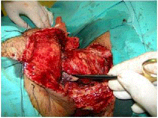
Figure 5a
Dehiscence of the proximal anastomosis and fistula formation
at a patient who underwent RFF reconstruction for a circumferential defect.
Figure 5b
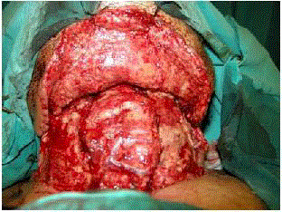
Figure 5b
The proximal anastomosis was re-sutured primarily.
Conclusion
A consensus treatment strategy for reconstructing the defect following hypo pharyngectomy and cervical oesophagectomy has not been established yet. The procedure of choice depends upon the location and size of the carcinoma as well as the patient's characteristics and the doctor's experience [6]. The RFF and the PM safeguard a feasible reconstruction of hypopharyngo-oesophageal neo-tube and provide satisfactory and consistent functional results. However the RFF offers better quality reconstruction and is considered a superior reconstructive option for patients who can tolerate major surgery. The successful outcome is associated with oral alimentation, good uncomplicated swallowing and improved quality of life. Lower complication rates can be achieved by applying technical modifications.
References
- Mathes SJ, Nahai F. Reconstructive Surgery: Principles, Anatomy & Technique. St.Louis, MO: Quality Medical Publishing. 1997:441-65.
- PiazzaC, Taglietti V, Nicolai P. Reconstructive options after total laryngectomy withsubtotal or circumferential hypopharyngectomy and cervical esophagectomy. Curr Opin Otolaryngol Head Neck Surg.2012;20:77-88.
- ChenYD, Chen HC, Vranckx JJ, Schneeberger AG. Edge deepithelialization: a method toprevent leakage when tubed free skin flap is used for pharyngoesophagealreconstruction. Surgery.2001;130:97-103.
- LutzBS, Wei FC. Microsurgical workhorse flaps in head and neck reconstruction. Clin Plast Surg. 2005;32:421-30.
- KimEK, Evangelista M, Evans GRD. Use of free tissue transfer in head and neck reconstruction.J Craniofac Surg. 2008;19:1577-82.
- XiaoQ, Hu GH, Zhong SX, Qian Y, Zeng Q, Hong SL. Reconstruction of Hypopharynx andCervical Oesophagus for Treatment of Advanced Hypopharyngeal Carcinoma andRecurrent Laryngeal Carcinoma. Asian JSurg. 2010;33:14-9.
- Murray DL, Novak CB, Neligan PC. Fasciocutaneousfree flaps in pharyngoesophageal reconstruction: a critical review of theliterature. J PlastReconstrAesth Surg.2008;61:1148-56.
- Vartanian JG, Carvalho AL,Carvalho SM, Mizobe L, Magrin J, Kowalski LP. Pectoralis major and othermyofascial/myocutaneous flaps in head and neck cancer reconstruction:experience with 437 cases at a single institution. Head Neck. 2004;26:1018-23.
- Scharpf J, Esclamado RM. Reconstruction withradial forearm flaps after ablative surgery for hypopharyngeal cancer. HeadNeck. 2003;25:261-66.
- RobbGL, Lewin JS, Deschler DG, Haughey BH, Brown DH, Langmore SE, et al. Speech andswallowing outcomes in reconstructions of the pharynx and cervical esophagus. Head Neck. 2003;25:232-44.
- DisaJJ, Cordeiro PG. Reconstruction of the hypopharynx and cervical esophagus. ClinPlast Surg. 2001;28:349-60.
- VarvaresMA, Cheney ML, Gliklich RE, Boyd JM, Goldsmith T, Lazor J, et al. Use of theradial forearm fasciocutaneous free flap and montgomery salivary bypass tubefor pharyngoesophageal reconstruction. HeadNeck. 2000;22:463-68.
- Li KK, Salibian AH, Allison GR, Krugman ME,Armstrong W, Wong B, et al. Pharyngoesophageal reconstruction with the ulnarforearm flap. Arch Otolaryngol Head Neck Surg 1998;124:1146-51.