Case Report
A Rare Presentation: Parathyromatosis
Orhan Yalcin1, Semra Gunay1, Arzu Akan1*, Caglar Cakir2, Mine Adas3 and Tamer Ozulker4
1Department of Breast Endocrine Surgery, Okmeydani Training and Research Hospital, Turkey
2Department of Pathology, Okmeydani Training and Research Hospital, Turkey
3Department of Endocrinology, Okmeydani Training and Research Hospital, Turkey
4Department of Nuclear Medicine, Okmeydani Training and Research Hospital, Turkey
*Corresponding author: Arzu Akan, Department of Breast Endocrine Surgery, Okmeydani Training and Research Hospital, Turkey
Published: 06 Jul 2017
Cite this article as: Yalcin O, Gunay S, Akan A, Cakir
C, Adas M, Ozulker T. A Rare
Presentation: Parathyromatosis. Clin
Surg. 2017; 2: 1540.
Abstract
Parathyromatosis is a rare condition which occurs after parathyroidectomy and it is a situation that is difficult to diagnose and treat in hyperparathyroidism. Parathyromatosis would result from seeding after the fracture of the parathyroid gland capsule during parathyroid surgery as well as there are some other etiological factors either. We here aimed to mention about etiology, diagnosis and treatment options of parathyromatosis with presented our case.
Introduction
Parathyromatosis is a rare cause of hyperparathyroidism. When we look at literature; two causes were defined for its occurance. In primary type, it might be due to hyperplasia of the parathyroid residues and on the other hand in secondary type generally it might be due to planting after parathyroid surgery. In secondary type residual thyroid tissues stimulate due to existing chronic renal failure. In some studies as a third theory; it was said that it is a low grade malignancy [1,2]. In this case; we aimed to present a parathyromatosis case with surgery history due to parathyroid adenoma and who had hypercalcemia in examinations which were conducted prior to the pilonidal sinus surgery.
Case Presentation
Thirty two year old male patient applied to our hospital general surgery policlinic for pilonidal sinus surgery. In his history; there was only weakness complaint and discharge from presacral region. It was learned that he had surgery with the diagnosis of parathyroid adenoma and pathology results were parathyroid adenoma. As his routinely performed preoperative laboratory test result was Ca++: 13.2 mg/dl [8.4-10.2]; parathyroid hormone levels were checked. Parathyroid hormone was high as 338 pg/ml [15-65]. Kidney function tests were normal. In his neck ultrasonography; about 24 mm × 10 mm × 13 mm sized solid lesion (parathyroid adenoma? carcinoma?) was detected which was localized beyond thyroid borders at inferior of right thyroid lobe and in Doppler ultrasonography it was hypervascular, with lobule contours and heterogeneous hypoechoic structure. In his parathyroid scintigraphy; pathological activity retention was described in favor of parathyroid adenoma at inferior part of right thyroid lobe (Figure 1). Urinary tract ultrasound and bone densitometry were found to be normal. In computed tomography of the neck and mediastinum; 11 mm × 10 mm sized hypodense lesion at posteroinferior of right thyroid lobe was reported. In September 2014, the patient was taken to surgery with these findings and as a result of 1cm sized adenoma at lower right are of parathyroid and micronodular view around it in the surgery; unilateral thyroidectomy (right), right lower parathyroidectomy, and bilateral central neck dissection were made with possible diagnosis of parathyroid carcinoma. Control parathyroid hormone after surgery was 7.94 pg/ml [15-65]. Pathological result was Parathyromatosis (Figure 2).
Discussion
Parathyromatosis is a rare condition which occurs after parathyroidectomy and it is a situation
that is difficult to diagnose and treat in hyperparathyroidism. After being described by Palmer for
the first time in 1975, about 35 cases have been reported in the literature [3]. Although parathyroid
residues can be seen in thyroid gland and cervical lipid tissue; they generally located in thymus
in parathyromatosis cases due to embryological residues. Although Parathyromatosis is a benign
disease with slow growing rate in the area that it’s located at; you should be protected from effects of
long term exposure to hyperparathyroidism [4].
In necessary cases; Parathyroid can be fed by diffusion when
they are planted to the muscle or fat tissue, can make growth by
angiogenesis and they can survive at where they are [4]. Rupture
of capsule during parathyroidectomy, can lead to dissolution of
tissue and proliferation of that tissue in time. In this sense, surgeon
can be effective in etiology. In case of renal failure, this spitted
parathyroid’s can be hyperplasia under stimulation of secondary
hyperparathyroidism. If capsular rupture happens during surgery,
parathyromatosis can happen at a certain point during surgical plan.
When we look at the literature; parathyromatosis occurance after
parathroid surgery is defined between a time intervals of 5 months to
19 years [5]. In 22 of 35 patients with Parathyromatosis, chronic renal
failure was detected [6].
Parathyromatosis mostly occurs in women and in the fifth decade
and in studies the difficulty for diagnosis is mentioned. As a cause
of this difficulty; being a diagnosis which is usually unthinkable is
specified. Only 4 cases were reported in literature which had diagnosis
before the surgery [7]. Hyperparathyroidism can cause kidney stones,
bone pain, gastrointestinal and psychiatric symptoms. In laboratory
tests; blood calcium levels can be high (1-2 mg/dl and above), in
patients with chronic renal failure normal calcium levels can be
found. In imaging methods which were used before neck exploration;
less than 50% of the patients can have diagnosis, thus ultrasonography
is one of the most helpful method in diagnosis. In the differential
diagnosis of Parathyromatosis; colored Doppler ultrasonography can
be helpful for distinction of other cervical malignancies and gray scale
ultrasonography may show similar characteristics. MIBI SPECT, can
be usually used for localization of hyperactive parathyroid tissue [8].
In differential diagnosis; parathyroid adenoma and carcinoma
should be considered. In all of these clinical conditions; there are
persistent and high levels of parathyroid hormone. In patients
with parathyroid carcinoma, calcium levels are markedly higher.
Definitive diagnosis is established histologically. There are studies in
literature about sowing while making fine needle aspiration biopsy
ultrasonographically with preliminary diagnosis of parathyroid
adenoma in order to make histopathological diagnosis or look for
parathyroid hormone but currently there is no proven information
[9,10]. Macroscopically; it can be soft, whitish yellow, in size ranging
from microscopic size to 2 cm. Adenoma and carcinoma can be
observed as a single structure and on the other hand Parathyromatosis
can be observed in a multi glandular form. Histological difference
of Parathyromatosis from cancer or adenoma is the lack of real
capsule of the parathyroid tissue [11]. In carcinoma there can be
invasion to adjacent tissues, lymph node or distant organ metastasis.
In Immunohistochemical studies, we can use some markers
(retinoblastoma expression, parafibromine loss, Galectin-3 over
expression) for the differential diagnosis of parathyroid carcinoma
[12].
While it is difficult, the treatment of Parathyromatosis surgery.
All of the possible foci can be removed with an ultrasonography which
is used during surgery. However, if differential diagnosis cannot be
made during surgery, it is suggested to behave as a carcinoma and
make the surgery. Medically, calcium and parathyroid hormone
levels can be decreased by using calcimimetic drugs (Cinacalcet®) and
bisphosphonates [13,14].
In conclusion; Parathyromatosis, is a rare cause of
hyperparathyroidism. It should be considered in patients with
previous parathyroid surgery, high levels of calcium and PTH.
For diagnosis; Histopathologic examination is necessary and in
treatment first surgical procedure should be made carefully and
if parathyromatosis is developed, surgery should be made with
extensive exploration. Medical treatment can be tried if surgery is not
effective or there is a contraendication for surgery.
Figure 1
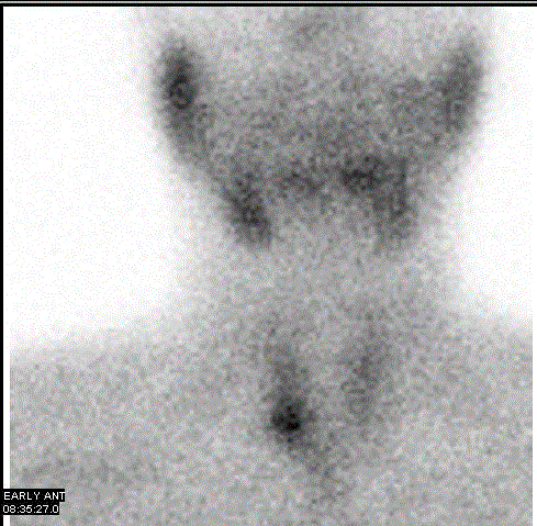
Figure 1
Patients MIBI scan.
Figure 2
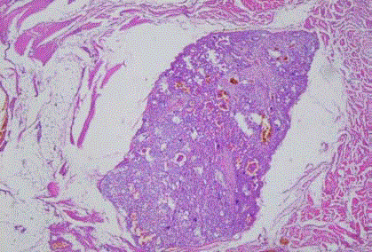
Figure 2
Striated muscle tissue and fat tissue nodules in parathyroid tissue
(H&Ex40).
References
- Fernandez-Ranvier GG, Khanafshar E, Jensen K, Zarnegar R, Lee J. Parathyroid carcinoma, atypical parathyroid adenoma, or parathyromatosis. Cancer. 2007;15;110(2):255-64.
- Lentsch EJ, Withrow KP, Ackermann D, Bumpous JM. Parathyromatosis and recurrent hyperparathyroidism. Arch Otolaryngol Head Neck Surg. 2003;129(8):894-6.
- Tublin ME, Yim JH, Carty SE. Recurrent hyperparathyroidism secondary to parathyromatosis. J Ultrasound Med. 2007;26(6):847-51.
- Sim IW, Farrell S, Grodski S, Jung C, Ng KW. Parathyromatosis following spontaneous rupture of a parathyroid adenoma: natural history and the challenge of management. Intern Med J. 2013;43(7):819-22.
- Wu TJ, Wang YT, Chang H, Lin SH. Parathyromatosis. Kidney Int. 2012;82(10):1140.
- Scorza TA, Moore AG, Terry M, Bricker LE. Secondary parathyromatosis in a patient with normal kidney function: revıew of diagnostic modalities and approaches to management. Endocr Pract. 2014;20(1):4-7.
- Matsuoka S, Tominaga Y, Sato T, Uno N, Goto N, Katayama A, et al. Recurrent renal hyperparathyroidism caused by parathyromatosis. World J Surg. 2007;31(2):299-305.
- Torregrosa JV, Fernández-Cruz L, Canalejo A, Sergio Vidal, Astudillo E, Almaden Y, et al. (99mTc)-sestamibi scintigraphy and cell cycle in parathyroid glands of secondary hyperparathyroidism. World J Surg.2000;24(11):1386-90.
- Kendrick ML, Charboneau JW, Curlee KJ, van Heerden JA, Farley DR. Risk of parathyromatosis after fine-needle aspiration. Am Surg. 2001;67:290–93.
- Baloch ZW, Fraker D, LiVolsi VA. Parathyromatosis as cause of recurrent secondary hyperparathyroidism: A Cytologic Diagnosis. Diagn Cytopathol. 2001;25:403-5.
- Altınboga AA, Sari AA, Rezanko T, Haciyanli M, Calli AO. Parathyromatosis: Critical diagnosis regarding surgery and pathologic evaluation. Korean J Pathol. 2012;46(2):197-200.
- Mohammadi A, Ghasemi-Rad M. Parathyromatosis or recurrent multiple parathyroid adenomas? A case report. Maedica (Buchar). 2012;7(1):66-69.
- Edling KL, Korenman SG, Janzen C, Sohsman MY, Apple SK, Bhuta S, et al. A pregnant dilemma: primary hyperparathyroidism due to Parathyromatosis in pregnancy. Endocr Pract. 2014;20(2):15-7.
- Hage MP, Salti I, El-Hajj Fuleihan G. Parathyromatosis: a rare yet problematic etiology of recurrent and persistent hyperparathyroidism. Metabolism. 2012 ;61(6):762-75.