Research Article
Necrotizing Bacterial Necrotizing Dermatitis with Necrotizing Fasciitis Anterior Cervico-Thoracic Complicating a Tooth Abscess: Clinical Case
Traoré A1*, Traoré A1, Dembélé BT1, Togo A1, Diakité I1, Kanté L1, Konaté M1, Karembé B1, Bah
A1, Sidibé Y1, Koné T1, Diango DM2 and Diallo G3
1Department of General Surgery, CHU Gabriel TOURE, France
2Department of Anesthesia and Intensive Care Unit, CHU Gabriel TOURE, France
3Department of Pediatric Surgery, CHU Gabriel TOURE, France
*Corresponding author: Traoré Amadou, Department of General Surgery, Faculty of Medicine and Odonto-Stomatology (FMOS), Hospital Practitioner C.H.U Gabriel Touré, Bamako (MALI) BP: 267, France
Published: 28 Jun, 2017
Cite this article as: Traoré A, Traoré A, Dembélé BT, Togo
A, Diakité I, Kanté L, et al. Necrotizing
Bacterial Necrotizing Dermatitis with
Necrotizing Fasciitis Anterior Cervico-
Thoracic Complicating a Tooth Abscess:
Clinical Case. Clin Surg. 2017; 2: 1523.
Abstract
Necrotizing bacterial dermohypodermitis with necrotizing fasciitis (DHBN-FN) is a necrotizing
infection of the hypodermis, the muscular aponeurosis and secondarily the dermis. The infection
sometimes spreads in a fulminating manner along the aponeuroses. It constitutes a medical-surgical
emergency. It is a rare and serious infection. The mortality remains high reaching 19% to 41%,
despite an improvement in surgical techniques and resuscitation. The anterior cervical and thoracic
involvement complicating a tooth abscess is exceptional. We report the case of a young woman of
35 years without comorbidity and discuss the clinical, therapeutic and evolutionary aspects through
the literature.
Keywords: Necrotizing bacterial dermohypodermitis; Necrotizing fasciitis; Dental abscess;
Cervico-thoracic; Surgery
Introduction
Necrotizing fasciitis is a severe, rapidly progressive, mutilating, often fatal infection. It is a rare condition caused by contamination with germs with a high toxinogenic potential, represented mainly by the Group a hemolytic Streptococcus. It is a therapeutic emergency with a mortality varying between 19% to 41% according to the literature [1-4]. Cervical and thoracic involvement are rare [3,5].
Patient and Observation
A 35-year-old female housewife admitted to the emergency room for anterior cervico-thoracic
pain with edema. The onset of the disease was 18 days after an abscess of dental caries. She
undertook a traditional herbal treatment and fumigation that did not improve. The pain evolved
becoming more intense, insomniante, pulsatile, permanent and irradiating towards the anterior
cervical and thoracic regions, the left breast then that of right. The pain was accompanied by fever
at 39°C, vomiting, hypersialorrhea, anorexia and slimming with weight loss of 8 kg. The patient
had a history of tooth decay that she had never treated in a medical setting. On examination, there
was a putrid odor of exudates, dehydration folds, and cyanic spots which were poorly limited in
geography. An induration of the edema extending from the left cheek to the anterior cervicothoracic
region of the two breasts was shown. There was cutaneous hypoesthesia, necrosis plaques
and fluctuations that were felt under the skin. The chest X-ray showed no pleurisy or focus of
pneumonia. A surgical exploration included a hypodermic melt with a greenish creamy appearance,
no pus frank, a large area of
1 and 2). We performed comskpinle tdee tdaecbhrmideenmt,e nnet corof taizlli nnge cfaroscticit itsi,s sruarees balneded dinragi,n Magyeo. sAiti ss p(Fecigimureens
for histopathological, bacteriological and antibiogram examination was carried out. The operative
sequences were simple. Postoperatively, we performed a cutaneous autograft.
Discussion
Necrotizing fasciitis is a rare condition in our daily practice. It usually occurs on debilitated land. The infection begins with necrosis of the hypodermis with vascular thrombosis and then extends secondary to the underlying superficial aponeurosis and the dermis. Its evolution is lightning resulting in the death of the patient by multi-visceral failure. Few data exist on epidemiology. In countries such as Canada, where we recur between 90 and 200 cases annually [6]. For invasive infections with Streptococcus A, 5% to 10% of forms with fasciitis have been reported in the USA [5,7], in France 1.7/100,000 in 2002 and 2.7/100,000 in 2004 [8]. One case of breast DHBN-FN on HIV positive soil has been described by Kourouma et al. [9]. The Group A haemolytic Streptococcus and Clostridium perfringens are the germs responsible for this pathology [5,10], which is most often polymicrobial in 40-90% of patients with risk factors: Age >50 years, obesity, diabetes (25% to 30%), immunosuppressive treatments, chemotherapy, chronic alcoholism (10% to 20%), immunosuppressed, addictions, cancers, peripheral vascular disorders (36%) [2]. In contrast, streptococcal fasciitis can occur in the young patient with no prior history as in our patient A number of local risk factors have been reported: ulcers, perforating ailments Plantar, surgery 0[11], IV injection in drug addicts. The identified germs were β hemolytic streptococcus group A and E. coli. On histopathological examination, the hypodermis is the site of oedematous inflammation, rich in polymorphonuclear cells, and extensive necrosis of the muscular aponeurosis. There is thrombosis of the subcutaneous vessels. Cervical involvement is rare and the incidence of this disease is 2.6% of all infections in the head and neck. The most common primary cause is dental infection [3] as in our patient. Isolated thoracic forms are extremely rare [6,12]. The cases reported in the literature are secondary to thoracic drainage,pulmonary, oesophageal or purulent pleurisy [13,14]. Pain is at the forefront of clinical symptomatology at the onset of fever, followed by signs of sepsis and skin lesions with hypoesthesia [7]. Anti-biotherapy supplements the surgery. Its aim is to limit the progression of the infection, but it is insufficient because of the vascular thromboses responsible for deep tissue necrosis [5]. We used a triple antibiotic initially: Ceftriaxone, Gentamycin and Metronidazole then adapt the antibiotic to the result of the antibiogram. We performed a large and dilapidated surgery. It consisted of an excision of all the necrotic tissues: the skin, the aponeurosis, and the muscles to the healthy tissues well vascularized in order to avoid a resumption of the infection leading to one of the iterative interventions (Figure 3). Restorative surgery was considered after disappearance of the infectious signs and wound budding at D30 (Figure 4). A cutaneous autograft was performed with an anterior cut of the thigh (Figure 5). The operative sequences were simple (Figure 6). The prognosis of DHBN-FN depends on two factors: early diagnosis and initial management. A diagnosis delay is associated with a mortality of 36% to 70% [5].
Figure 1
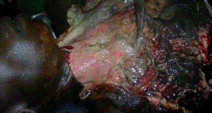
Figure 1
DHDBN-FN anterior cervico-thoracic with vascular thrombosis,
absence of pus.
Figure 2
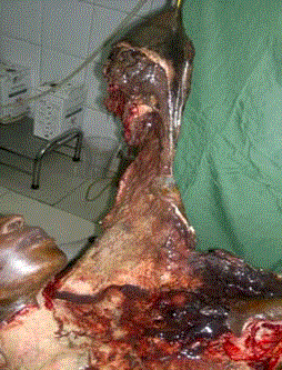
Figure 2
Breast necrosis.
Figure3
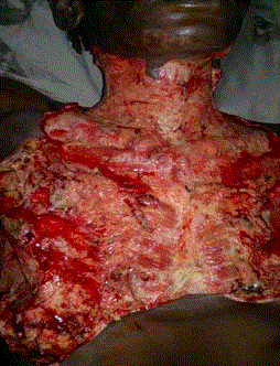
Figure 3
After extensive necrosisctomy.
Figure 4
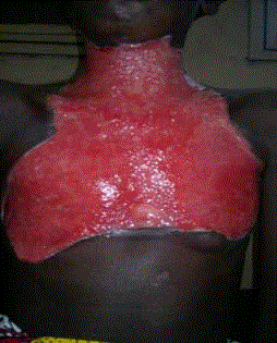
Figure 4
Budding of the wound J30
Figure 5
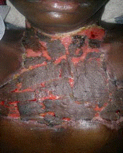
Figure 5
Thin skin autograft at D30.
Figure 6
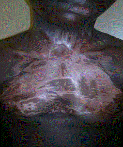
Figure 6
After autograft.
Conclusion
Necrotizing dermohypodermitis with necrotizing fasciitis is a serious infection. Management is multidisciplinary. Early diagnosis and timely and appropriate treatment can reduce mortality.
References
- Khamnuan P, Chongruksut W, Jearwattanakanok K, Patumanond J, Yodluangfun S, Tantraworasin A. Necrotizing fasciitis: risk factors of mortality. Risk Manag Healthc Policy. 2015;16(8):1-7.
- Vayvada H, Demirdover C, Menderes A, Karaca C. Necrotizing fasciitis in the central part of the body: diagnosis, management and review of the literature. Int Wound J. 2013;10(4):466-72.
- Suarez A, Vicente M, Tomás JA, Floría LM, Delhom J, Baquero MC. Cervical necrotizing fasciitis of nonodontogenic origin: case report and review of literature. Am J Emerg Med. 2014;32(11):1441.
- Sarna T, Sengupta T, Miloro M, Kolokythas A. Cervical necrotizing fasciitis with descending mediastinitis: literature review and case report. J Oral Maxillofac Surg. 2012;70(6):1342-50.
- David Jérémie Birnbaum, Xavier Benoit D’Journo, Jean Philippe Avaro, Delphine Trousse, Roger Giudicelli, Pierre Fuentes, et al. Fasciite nécrosante de la paroi thoracique. Chirurgie Thoracique Cardio-Vasculaire. 2009;13:49-52.
- Conférence de Consensus. Erysipèle et fasciite nécrosante: prise en charge. Méd Mal Infect. 2000;30:252-72.
- Hervé Dupont. Fasciite Nécrosante. Pathologies infectieuses. France. MAPAR. Loughlin R. CID. 2007;45:853-62.
- Kourouma HS, Sangaré A, Kaloga M. Fasciite nécrosante du sein: une observation chez une personne vivant avec le VIH en Côte d’Ivoire. Médecine tropicale. 2010;70:281-2.
- Sonali Prabhakar Khadakkar, Vivek V, Harkare. Necrotizing fasciitis of the Neck and anterior Chest Wall. Indian J Otolaryngol Head Neck Surg. 2011;63(1):87-9.
- Médar de Charbon V, Guevara N, Lattes L, Converset-Viethel S, Riah Y, Lebreton E, et al. Dermohypodermite bacterienne necrosante avec fasciite necrosante: complication d’une installation opératoire? J Anplas. 2007;07:009.
- Safran DB, Sullivan WG. Necrotizing fasciitis of the Chest Wall. Ann Thorac Surg. 2001;72(4):1362-4.
- Losanoff JE, Jones JW, Richman BW. Necrotizing Soft Tissue Infection of the Chest Wall. Ann Thorac Surg. 2002;73(1):304-6.
- Narindra Njarasoa Mihaja Razafimanjato, Lalaina Elianah Rasoamampianina, Manjakaniaina Ravoatrarilandy, Auberlin Felantsoa Rakototiana, Francis Allen Hunald, Luc Hervé Samison, et al. Fasciite necrosante de la paroi thoracique compliquant un empyème. Pan Afr Med J. 2013;16:108. Kim SH, Roh KH, Yoon YK, Kang DO, Lee DW, Kim MJ, et al. Necrotizing fasciitis involving the chest and abdominal wall caused by Raoultella planticola. BMC Infect Dis. 2012;12:59.