Clinical Image
Entrapment of Swan-Ganz Catheter during Heart Surgery
Imad F Tabry*
Department of Cardiac Surgery, Holy Cross Hospital Fort Lauderdale, FL 33308, USA
*Corresponding author: Imad F Tabry, Department of Cardiac Surgery, Holy Cross Hospital Fort Lauderdale, FL 33308, USA
Published: 12 Jun 2017
Cite this article as: Tabry IF. Entrapment of Swan-Ganz
Catheter during Heart Surgery. Clin
Surg. 2017; 2: 1501.
Clinical Image
The Swan-Ganz catheter (SGC) is widely used particularly during valvular surgery. Multiple
complications related to its insertion, placement and removal have been recorded. We hereby call
attention to the characteristic finding on the chest radiography of a SGC caught in a suture line
during mitral valve surgery.
After an uneventful mitral valve repair through a minimally invasive right thoracotomy incision
a routine chest radiograph showed an acute angulation of the SGC within the right atrium (RA)
(Figure 1). Despite a satisfactory pulmonary artery tracing, attempts at mobilizing it were met with
resistance and thus immediately discontinued. These findings confirmed entrapment of the catheter.
Back in the operating room femoral cardiopulmonary bypass was re-instituted. With mild
traction on the SGC by the anesthesiologist a dimple appeared at the caudal end of the left atriotomy
(LA) continuous suture line. In order to avoid its disruption, it was reconstituted along its entire
length using 5 pledgeted sutures of Prolene left untied. A RA purse string suture was then inserted
close to the Inferior vena cava junction. Under Inflow occlusion, the RA was incised within the
purse string suture and the SGC visualized, transfixed by the LA suture line. The retaining suture
was cut and the liberated catheter removed in its entirety. The RA opening was then repaired and
the previously placed pledgeted sutures tied, ensuring a hemostatic LA closure. The remainder of the
operation and the post-operative course were uneventful.
As reported in this case acute angulation of the catheter on simple chest radiograph is
pathognomonic of its entrapment. Prevention of this rare accident consists of simply pulling the
SGC a few centimeters by the anesthesiologist immediately upon completing the atrial suture line.
Any resistance would then compel the surgeon to identify and correct the cause of entrapment.
Figure 1
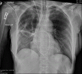
Figure 1
SGC within the right atrium (RA).