Research Article
Difficulties in the Choice of Surgical Approach in Cervicothoracic Goiters
Sani Rabiou1*, Fatima Z Ammor1, Sani M Aminou2, Harmouchi Hicham1, Layla Belliraj1, Ibrahim Issoufou1, Marwane Lakranbi1, Yassine Ouadnouni1,3 and Mohamed Smahi1,3
1Department of Thoracic Surgery, CHU Hassan II, Morocco
2Department of Endocrinology, Diabetology and Metabolic Diseases, CHU Hassan II, Morocco
3Faculty of Medicine and Pharmacy, Sidi Mohamed Ben-abdellah University, Morocco
*Corresponding author: Sani Rabiou, Department of Thoracic SurgeryC1, CHU Hassan II, Morocco
Published: 20 Apr, 2017
Cite this article as: Rabiou S, Ammor FZ, Aminou SM,
Hicham H, Belliraj L, Issoufou I, et al.
Difficulties in the Choice of Surgical
Approach in Cervicothoracic Goiters.
Clin Surg. 2017; 2: 1419.
Abstract
Introduction: Indications for thoracotomy or sternotomy are very rare or exceptional in surgery
plunging goiter. Through our experience about 10 patients we wanted to discuss the difficulty in
choosing the surgical approach to this rare condition.
Materials and Methods: This is a retrospective descriptive study covering the period January 2009
to June 2016, in the thoracic surgery department in CHU Hassan II of Fes, the inclusion criteria
were: the location of the cervico thoracic goiter, his preoperatively euthyroid character but also its
benign nature based on the results of the radiological assessment including the CT scan.
Results: In total 8 women and 2 men were involved in the study. A notion of prior thyroid surgery
was noted in 3 patients. Computed tomography confirmed the plunging goiter character in among
10 patients who had back-vascular goiter in 55% of cases. The incision was thoracotomy in 2 cases,
cervicotomy in 3 cases. The cervicotomy-manubriotomy and cervicotomy-thoracotomy association
was performed in 2 cases each. In one patient who had a goiter complex, an indication of a first triple
by-manubriotomy cervicotomy thoracotomy was necessary. In immediate post-operative, one
patient had a cervical hematoma requiring surgical revision. Other complications were dominated
by recurrent laryngeal paralysis in 2 cases and postoperative hypocalcemia in 5 cases.
Conclusion: The surgery of cervico thoracic goiters is conceivable that after a complete mapping of
the lesion using a suitable radiological assessment. The indication of a trans-sternal or thoracotomy
approach should be discussed in a posterior ineradicable by cervicotomy or in suspected neoplasia.
Keywords: Thyroid gland; Cervicothoracic goiter; Cervicotomy; Sternotomy; Thoracotomy
Introduction
The cervicothoracic goiter share a certain number of characteristics, in particular a slow growth, which is a character little symptomatic in the non compressive stage, like the absence of the malignancy in the majority of cases. On the other hand, the mediastinal localization give to the endothoracic form a particular severeness, linked in a way to the phenomenon of organs compression, such as the trachea, and in another way to the additional difficulties of the surgical management. A very large number of technics allow in the majority of cases, with a simple cervical approach, a control with satisfactory homeostasis [1-3]. However, in other situations, the thoracic approach is necessary so that the resection could happen safely without exposing the patient to the preventable operational incidents. We report the experience of a thoracic surgery department in the surgical management of the cervicothoracic goiter.
Material and Methods
It is a retrospective and descriptive study taking from January 2009 to June 2016, in the thoracic surgery department in CHU Hassan II Fes. Some of the characteristics that includes cases were the cervicothoracic localization of the goiter, its euthyroid character before surgery, and also its benign nature based on the radiological exams' results especially CT scan. All the information that contains the clinical signs, the thyroid function test, The radiological evaluation, the surgical technique and it's complications, and the follow up of the patients, were reported on an investigation sheet previously established then analyzed by Pac office excel software 2016 for Mac.
Results
Patients description
Observation 1: 43 years old patient who already undergone
cervical goiter surgery. The mediastinal tumor was discovered while
doing the preoperative tests for a vesicular lithiasis. The radiological
evaluation was in favor of an intra thoracic component of a diving
goiter neglected in the first thyroidectomy. Viewing the very
posterior localization of the mass, a right posterolateral thoracotomy
was necessary. Complete resection was obtained after opening the
mediastinal pleura and releasing the adhesions between this mass
and the trachea. The surgical follow-up was marked by the occurrence
of a hypocalcemia requiring a transitory supplementation with oral
calcium. The anatomopathological examination of the excision piece
was in favor of a multi hetero nodular goiter without any sign of
malignancy.
Observation 2: 57 years old Patient, with no particular
pathological history that was addressed to us for a cervicothoracic
mass (Figure 1). The cervicothoracic CT scan showed a multi-heteronodular,
complex, retrotracheal and prevascular diving goiter with
an isolated retrosternal component. The management consisted of a
cervicotomy first allowing controlling and performing the excision
of the cervical component with inability to deliver the endothoracic
component. A right posterolateral thoracotomy at the same operative
time allowed the removal of the posterior mediastinal plunging
component but not sufficient to reach the retrosternal component.
After 1 month evolution, the complete resection of the retrosternal
component was obtained by manubriotomy. The surgical sequences
were marked by the occurrence of a recurrent paralysis responsible
for a transitory dysphonia after short course of corticoid. The
anatomopathological analysis confirmed the diagnosis of a goiter on
all surgical specimens.
Observation 3: A 54-year-old woman with no pathological
antecedents who had been suffering from isolated chest pain for two
months. The CT scan showed an image in favor of an endothoracic
goiter whose lower pole descends lower than the hull. On the
exploration by a right posterolateral thoracotomy, the mass repressed
the azygos stock at the bottom, the superior vena cava and the right
subclavian artery onward. The resection was obtained after opening
the mediastinal pleura and releasing all the adhesions. The hemostasis
was satisfactory and the postoperative follow-up was simple except for a moderate hypocalcaemia. The histological examination of
the piece confirmed the diagnosis by showing a vesicular adenoma
without any signs of malignancy.
Observation 4: A 44 years old patient, former smoker with an
antecedent of a cervical goiter that disappeared overnight with recent
appearance of a dyspnea. The radiography (Figure 2A) and the thoracic
CT scan (Figure 2B) showed a cervicothoracic mass measuring
15 cm long, compatible with a diving goiter, with an endothoracic
component occupying the apical part of the right hemithorax. The
Exploration with a right posterolateral thoracotomy had found a
mass pushing down the azygos stock at the bottom, the superior vena
cava onwards and very glued to the trachea and the esophagus at the
back. Opening the mediastinal pleura and releasing the adhesions
allowed the mediastinal mass to be extirpated (Figure 2C), leaving
in place the cervical component which was inaccessible. The surgical
follow-up was marked by the persistence of a right pneumothorax
requiring a second drainage. The patient had been reviewed 2 months
later then a cervicotomy made it possible to carry out the excision
of the cervical component with a satisfactory haemostasis. The
occurrence of a postoperative dysphonia at Day +1 should be noted.
Nasofibroscopy was in favor of paralysis of the vocal cord requiring
short-term corticosteroid therapy with good evolution. The analysis
of the surgical specimen was in favor of a multi-hetero-nodular
thyroid hyperplasia reshaped without signs of malignancy.
Observation 5: A 49-year-old patient with no pathological
history, admitted for treatment of cervical goiter for which she
had undergone a thyroidectomy, with incidental discovery of an
endothelial component inaccessible by cervicotomy. Postoperatively,
a thoracic CT scan confirmed the thyroid nature of the mass, and its location in the posterior mediastinum (Figure 3A-C). The
thoracotomy exploration found a posterior mediastinal mass pushing
the trachea and the esophagus forward. The total resection was
obtained after a release of the attachments between the mediastinal
structures and the mass. The surgical follow-up was simple except for
a transient biological hypocalcaemia. The anatomopathological study
of the specimen confirmed the diagnosis of a goiter.
Observation 6: 47 year’s old patient operated 23 years earlier for
a thyroid nodule. She had a right lobectomy. She currently consults
for cervical mass whose radiological assessment in particular the
cervicothoracic CT was in favor of a prevascular diving goiter. The
exploration with a primary cervicotomy had made it possible to
control the cervical component with the impossibility of delivering
the endothoracic component. A second operative stage consisting
of a manubriotomy had made it possible to dissect and resect
the endothoracic component. The evolution was simple and the
anatomopathological examination was in favor of a benign goiter.
Observation 7: Patient 83 years without significant pathological
history hospitalized for cervicothoracic mass. Computed tomography
(CT) of the cervicothoracic region showed a multi-nodular,
retrovascular diving goiter with bilateral development descending
down to the carina. A cervicotomy made it possible to control the
cervical and thoracic component with the impossibility of releasing
the lower pole of the left lobe. The release and excision of the latter
was made possible by manubriotomy with satisfactory surgical
follow-up outside of a slight biological hypocalcaemia. The diagnosis
was confirmed by the anatomopathological analysis of the excision
piece.
Observation 8: A 64-year-old patient with a history of
hysterectomy 18 years ago with a cervical mass with progressive
evolution for 10 years, becoming more and more uncomfortable
by the dyspnea it causes. Cervicothoracic computed tomography
(CT) showed multi-hetero-nodular diving goiter (Figure 4A-C).
The complete excision of the cervical and thoracic component was
obtained by exclusive cervicotomy using the maneuver of hyper
extension of the neck allowing delivering the whole mass without
difficulty (Figure 3C). Postoperative evolution was satisfactory except
for transient hypocalcemia. The histological analysis confirmed the
diagnosis of goiter.
Observation 9: 70 years old patient, with no pathological history,
who has for 3 years an irritative cough associated with left chest
pain. Thoracic tomodensitometry revealed an anterior mediastinal
mass lateralized to the left, associated with a diving goiter. She had
benefited from a thymectomy by a left posterolateral thoracotomy leaving the thyroid mass in place because it was impossible to reach it by this approach. After 3 months of evolution, a cervicotomy
allowed to realize a total thyroidectomy, after delivering the plunging
component that was at the expense of the right thyroid lobe. The
operative follow-up was simple and the histological analysis of the
thymectomy and thyroidectomy pieces was in favor of an AB type
thymoma (T1N0M0) and a goiter with no signs of malignancy.
Observation 10: 60 years old patient, former smoker at the rate
of 10 packages per year, weaned 20 years ago, who was admitted to
our department for the management of a pulmonary aspergilloma.
The radiological assessment, in particular the thoracic computed
tomography (CT), revealed a diving goiter. The patient had initially
received a right upper lobectomy for his aspergilloma. A cervicotomy
in a 2nd time had allowed a total thyroidectomy after delivery of the
endothoracic component of the goiter by the maneuver of hyper
extension of the neck. The surgical sequences were marked by the
occurrence of a cervical hematoma requiring immediate secondary
surgical revision with satisfactory haemostasis. The diagnosis was
confirmed by anatomopathological examination.
Figure 1
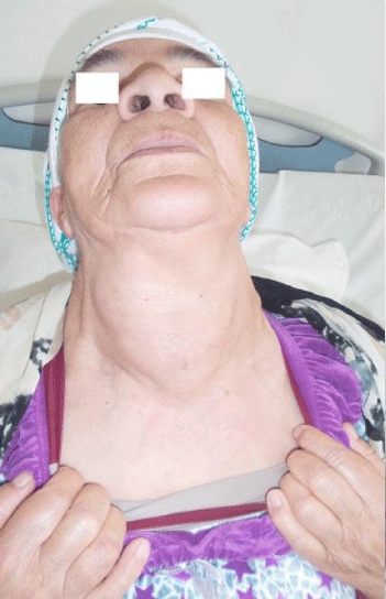
Figure 1
Anterior basicervical mass with lower pole in the thoracic cavity.
Figure 2
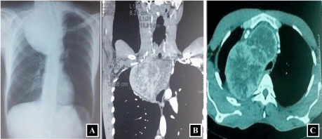
Figure 2
(a) Chest X-ray of the face showing an enormous right upper
mediastinal opacity responsible for a repression of the left trachea. (b)
Thoracic CT-scan demonstrating an enormous heterogeneous diving goitre
repressing the pulmonary parenchyma outside and mediastinal structures
within. (c) The same CT scan in axial section showing the anterior and
posterior mediastinal location of goiter.
Figure 3
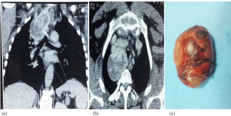
Figure 3
(a) and (b) Cervicothoracic CT scan shows an enormous goitre
plunging with repression of the mediastinal vessels downwards. (c) Piece of
the total thyroidectomy in the same patient.
Case Analysis
Age and sex
A total of 8 women and 2 men were involved in the study. The
average age was 57.1 years with extremes ranging from 43 to 83 years.
Circumstance of discovery
A concept of anterior thyroid surgery was noted in 3 patients.
Apart from a fortuitous discovery in 1 patient, the reason of
consultation was dominated by respiratory signs such as chest pain
and / or dyspnea in 6 cases. The appearance of an anterior cervical
mass was noted in 3 cases, which disappeared spontaneously after 2
years of evolution in a patient.
Radiological results
The CT scan confirmed the plunging goitre in 10 patients.
Retrovascular localization was noted in 55% of cases. The size of the
thyroid mass varied between 5 to 23 cm, exceeding the plane of the
carina at the bottom in 45% of cases. Cervical ultrasound performed
in 63% of cases, showed a multi-hetero-nodular goitre, without sign
of malignancy.
Preparation for surgery
An endocrinological opinion was sought to verify thyroid
hormone status. A total of 8 patients were in euthyroidism and 2
patients in hypothyroidism, for whom euthyroidism was obtained
before surgery.
Surgical approaches
The approach was an exclusive thoracotomy in 2 cases, an
exclusive cervicotomy in 3 cases. Cervicotomy-manubriotomy,
cervicotomy-thoracotomy was performed in 2 cases each. In a
patient with a complex goitre, the indication of a triple approach by
a cervicotomy-thoracotomy-manubriotomy was necessary to ensure
safe excision. In 3 patients the thyroidectomy was obtained in 2
operative sessions with a variable delay of 1 to 2 months between the
2 surgeries.
Post-operative complications
In the immediate postoperative period, one patient presented
a cervical hematoma requiring secondary surgical revision.
Postoperative dysphonia was noted in 2 patients, requiring naso-fibroscopic control, which showed transient unilateral recurrent
paralysis following a short course of oral corticosteroid therapy. The
postoperative biological assessment showed hypocalcaemia requiring
oral supplementation in 5 patients.
Evolution
Hormone replacement therapy was systematic in all cases with
good progression under levothyroxine. With an average follow-up of
10.75 months no case of death was noted in relation to surgery.
Figure 4
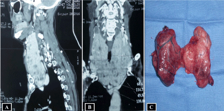
Figure 4
(a) and (b) Cervicothoracic CT scan showing an enormous goitre
plunging which located in the posterior mediastinum. (c) Piece of the total
thyroidectomy in the same patient.
Table 1

Table 1
The various surgical approach pathways of diving goitres, reported in the literature.
Table 2
Table 2
Results according to the authors of the various recurrent and metabolic complications in the surgery of the diving goitres.
Discussion
The diving goitre is defined as a goitre with cervical and mediastinal development; Endothoracicgoitre is most often defined as a goitre with essentially mediastinal development where only the superior poles emerge on the cervical area. The large volume of a diving goiter, its repercussion on the noble elements of the mediastinum and its anatomical type, are all pre-operative arguments that can guide towards a thoracic approach by sternotomy. However, many maneuvers allow the majority of patients to control the cervical and thoracic component by simple classical cervicotomy [1-3]. It is about the first dissection-release of the superior pole, then the one of the entire cervical component of the goitre. This surgical procedure generally leads to the gradual ascent and then to the controlled delivery of the diving component of the goitre. We can help ourselves by the section of the thyroid isthmus favoring the exteriorization of the gland. The indications of sternotomy appear therefore exceptional. These indications are mostly justified for controlling the vessels of endothoracic goiters which present themselves as a voluminous mediastinal tumor with its own mediastinal vascular pedicles [4]. Often, the presence of endothoracic goitre in a patient with a marked brevilineal morpho type makes a predictive argument for the indication of a sternotomy. In all cases, the preoperative diagnosis of these lesions and the programming of a probable transsternal approach are possible thanks to a simple frontal radiography analysis by comparing the diameter of the thorax's upper orifice with the largest Diameter of the goitre. In this context, cervical and thoracic computed tomography is a key examination. It allows evaluating the size, nature, pre-, retro-vascular or complex topography while specifying the depth of the endothoracic prolongation as well as the relations of the goitre with mediastinum elements [5,6]. The technique of this computed tomography must take into account the thyroid status of the patient by avoiding injections of iodinated contrast agent in order not to trigger hyperthyroidism or hinder the administration of radioactive iodine in case of unknown carcinoma until intervention. In our experiment, no indication of magnetic resonance imaging has been made since it does not provide any additional information compared to computed tomography [4]. In our study, in all patients, CT scans not only clarified the topography of the lesion, but also guided us in the choice of the approach. In fact, if the choice between total and partial sternotomy has little interest, that's because some complex goiters with mediastinal component are inaccessible by simple sternotomy. In our series, a posterolateral thoracotomy was required in 4 patients. It was an exclusive thoracotomy in two cases, a thoracotomy after an insufficient cervicotomy and a case of a thoracotomy after cervicotomy followed by a manubriotomy because of the complex nature of the goitre. The indications of this approach unusually carried out in this context, are dominated by the posterior mediastinal localization of the goitre making it impossible to undertake dissection, mobilization and therefore total thyroidectomy. In fact the type of excision has long been variable from one team to another, ranging from loboisthmectomy, to subtotal or even total thyroidectomy. At present, total thyroidectomy is the technique of choice for the removal of goiters [7], whether cervical or endothoracic. This technique prevents recurrence and avoids secondary surgical revision for totalization in case of accidental discovery of a thyroid carcinoma on the operative specimen of an initially benign goitre [7,8]. Although sternotomy is the common approach for endothoracic goitre surgery, some authors increasingly advocate a thoracotomy for a diving goiter in the posterior mediastinum or in case of a hesitant diagnosis [9-11]. This thoracotomy has been little described in classical studies. The anterior or anterolateral thoracotomy, in the third intercostal space in general, is a very good approach, easy to perform, aesthetically not very visible especially in women. It does not oblige to change the position of the patient and gives a view on the whole thickness of the upper mediastinum [12]. Its only interest is to be able to recline upwards the lower pole of large right lateralized cervicothoracic goitres with a mass effect on the trachea, the esophagus, and compressing the vena cava, the pulmonary vein and the azygos vein. It is therefore a disproportionate gesture compared to the benefit expected and to the advantages of the sternotomy [9]. Thus, in front of a voluminous thoracic goitre which cannot be removed by the neck, whatever the choice of the approach, it seems preferable to start with a cervicotomy, which ensures immediate control of the vascular pedicles and the recurrent and confirms the inextricable character of the lower lesion. Extraction by the anterolateral thoracotomy is then easy. As is the case with one of our patients, in the presence of intra-thoracic thyroid mass not accompanied by cervical goitre, a first thoracotomy may be considered, the purpose of which is to confirm the diagnosis and allow a complete treatment in case of autonomous goitre. The posterolateral thoracotomy of the fifth right intercostal space remains exceptional. It is long to execute because it necessitates the change of position of the patient [12]. According to Levasseur, it is reserved for mediastinal tumors isolated without diagnosis, no fixation at the scintigraphy and very lateral [9]. In our series, the indication for posterolateral thoracotomy is justified by a concern for complete resection, while minimizing the risk of post-operative complication, especially recurrent paralysis in patients with voluminous posterior mediastinal post goitres (Table 1) [1,4,9,13-15]. Whatever the progress in choosing the approach in cervicothoracic goitre surgery, the surgery follows up can be enameled from the complications. These latter depend on the surgeon's experience but also on the characteristics of the lesion to be treated [16]. Complications specific to the cervical approach are mainly hemorrhagic. These haemorrhages are generally secondary to defects of haemostasis in the thyroid chamber, or lesions of the elements of the neck's vascular axis elements even if this remains exceptional. This is the clinical picture of a cervical hematoma often impressive, some days after the gesture. The management consists of a secondary surgical revision without delay, allowing a complete evacuation of the hematoma as the case of one of our patient. As for the tracheal wounds, they are less frequent and easy to diagnose by simple sealing test by pouring saline serum into the thyroid chamber. These wounds are usually small in size and easy to suture [17,18]. Esophageal wounds can be prevented by placing a large oeso-gastric tube, allowing the surgeon to locate the esophagus during the dissection. As found in 2 patients of our series, recurrent paralysis occupies the first rank of the postoperative complications in the surgery of the cervicothoracic goitres. Its frequency, difficult to specify, oscillates around 2 to 10% according to the authors [1,12,19]. The risk of recurrent lesion in cervicothoracic goitre surgery appears to be significantly higher (2 to 7% of definitive recurrent paralysis for cervicothoracic goitre compared to 0.3 to 2% for cervical goiters) [19-21]. Beyond all these complications, thoracotomy may be specifically responsible for post-thoracotomy pain which is considered one of the most intense compared to other approaches. Post-thoracotomy chronic pain is most often attributed to an intercostal lesion secondary to direct surgical trauma; its intensity is variable and it is localized at the level of the path of the incision and of the dermatome of distribution of the intercostal nerve. Its management is cumbersome because of the chronic nature of the phenomenon requiring long-term treatment with analgesics, and sometimes the use of antiepileptics, antidepressants or strong opioids [22-24]. It is in the concern to minimize the occurrence of this post thoracotomy pain, that we perform in our daily practice a conservative thoracotomy without any section of the muscles of the chest wall [25], and no case of chronic pain was reported in our study. The morbidity and specific complications of the sternotomy are not negligible. It may be a pneumothorax due to accidental opening of the pleura, a chylothorax by a lesion of the thoracic duct often requiring a secondary surgical revision, a subcutaneous abscess with bone contamination that may progress to a pseudarthrosis, not to mention the dreadful mediastinitis potentially life-threatening [4]. The overall mortality of thyroid surgery remains low, estimated at 0 to 3% [26]. However, it is important to emphasize the importance of non-specific complications that can sometimes be disabling. These complications, which are not specific to the endothoracic prolongation of the goitre, are dominated by postoperative hypocalcemia related to parathyroid gland involvement (Table 2) [1,15,27,28]. This hypoparathyroidism may occur acutely postoperatively in the form of a major hypercalcemia, or silently several months after the thyroidectomy. As in the case of our two patients, the hypocalcaemia may be transitory or definitive and more frequent as the goitreis voluminous, with a posterior extension, or in case of secondary surgical revision. The thyroid surgery requires careful monitoring. It includes the search for an argument in favor of postoperative hypocalcemia and the adaptation of levothyroxine treatment by the TSH test 1 to 2 times a year.
Conclusion
The treatment of cervicothoracic goitres is surgical. This surgery is conceivable only after a complete mapping of the lesion by an adapted radiological assessment. Computed tomography (CT) plays an important role in the planning of surgical strategy, particularly in the choice of the approach. The cervicotomy is the classic approach allowing the excision of the majority of cervicothoracic goitres. The indication of a trans-sternal or thoracotomy approach must be discussed in the presence of a very posterior goitre ineradicable by cervicotomy or in case of a suspicion of neoplasia.
References
- Makeieff M, Marlier F, Khudjadze M, Garrel R, Crampette L, Guerrier B. [Substernal goiter. Report of 212 cases]. Ann Chir. 2000;125(1):18-25.
- Hedayati N, McHenry CR. The clinical presentation and operative management of nodular and diffuse substernal thyroid disease. Am Surg. 68(3):245–51.
- Mellière D, Saada F, Etienne G, Becquemin JP, Bonnet F. Goiter with severe respiratory compromise: evaluation and treatment. Surgery. 1988;103(3):367-73.
- Lifante JC, Fernandez Vila JM, Mingoutaud L, Pourret D, Peix JL. [Indications for sternotomy in thyroid surgery. Evolution over 20 years' experience]. J Chir (Paris). 2007;144(3):221-4.
- Newman E, Shaha AR. Substernal goiter. J Surg Oncol. 1995;60(3):207-12.
- Netterville JL, Coleman SC, Smith JC, Smith MM, Day TA, Burkey BB. Management of substernal goiter. Laryngoscope. 1998;108:1611-7.
- Agarwal G, Aggarwal V. Is total thyroidectomy the surgical procedure of choice for benign multinodular goiter? An evidence-based review. World J Surg. 2008;32(7):1313-24.
- Tezelman S, Borucu I, Senyurek Giles Y, Tunca F, Terzioglu T. The change in surgical practice from subtotal to near-total or total thyroidectomy in the treatment of patients with benign multinodular goiter. World J Surg. 2009;33(3):400-5.
- Cougard P, Matet P, Goudet P, Bambili R, Viard H, Vaillant G, et al. [Substernal goiters. 218 operated cases]. Ann Endocrinol (Paris). 1992;53(5-6):230-5.
- Borrelly J, Grosdidier G, Hubert J. [Proposal of a finer classification of diving goiters. Apropos of a series of 112 cases]. Ann Chir. 1985;39(2):153-9.
- Balawi A, Thvnet F, Gamondes JP. Goiters plongeants et endothoraciques: unesérie de chirurgicale de 55 observations. Lyon Chir. 1991;473-477.
- Merlier M, Eschapasse A. Les goitres à développementthoracique. Les cahiers Baillère. 1972.
- Viard H, Ptelat R, Barrault JF, Beurtheret G. Les goitresendothoraciques 37 casopérés. Lyon chir. 125–28.
- Blondeau P. [Substernal goiter: diagnostic and therapeutic problems (apropos of a personal series of 585 interventions)]. Bull Acad Natl Med. 1994;178(7):1257-64.
- Atoini F, Zidane A, Traibi A, Arsalane A, Elkaoui H, Tahri N, et al. [Surgical management of substernal goiters: a series of 27 patients]. J Chir (Paris). 2009;146(2):229-31.
- Brunaud L. Cancer papillaire de la thyroïde: vers un curage central systématique? Journal de Chirurgie Viscérale. 2008;145:13–16.
- Travalgli JP, Nocera M, Baudin E, Schlumberger M. Traitement de la maladieganglionnaire des cancers papillaires et vésiculaires de la thyroïde. Mt endocrinologie. 2003;2(4).340–4.
- Chevallier JM, Martelli H, Wind P. [Surgical discovery of parathyroid glands and the recurrent laryngeal nerve. Application of well known embryological concepts in the operating room]. Ann Chir. 1995;49(4):296-304.
- Rolet JP, Guibert B, Braillon G, Gilly FN. Les goitresplongeants 110 observations. Lyon Chir. 1991;87:6; 478–86.
- Goudet P, Ragois P, Guergah M, Cougard P. [Specific morbidity of substernal goiters. A comparative study with a matched series of cervical goiters]. Ann Chir. 1996;50(10):913-7.
- Judd ES, Beahrs OH, Bowes DE. A consideration of the proper surgical approach for substernal goiter. Surg Gynecol Obstet. 1960;110:90-8.
- Hazelrigg SR, Cetindag IB, Fullerton J. Acute and chronic pain syndromes after thoracic surgery. Surg Clin North Am. 2002;82(4):849-65.
- Gerner P. Postthoracotomy pain management problems. Anesthesiol Clin. 2008;26(2):355-67,vii.
- Dajczman E, Gordon A, Kreisman H, Wolkove N. Long-term postthoracotomy pain. Chest. 1991;99(2):270-4.
- Rabiou S, Ammor FZ, Issoufou I, Belliraj L, Lakranbi M, Ouadnouni Y, et al. Comment je peux faire ma thoracotomiepostéro-latérale sans sectionneraucun muscle de la cage thoracique. J Fran Viet Pneu. 2016; 20(7):1–78.
- Bazire A, Lesven S, Potard G, Leroyer C. Goitreendothoracique. EMC pneumologie. 2012:6-040-D-30.
- Filho J, Kowalski L. Surgical complications after thyroid surgery performed in a cancer hospital Otolaryngol Head Neck Surg. 2005;132(3):490–4.
- Snyder SK, Lairmore TC, Hendricks JC, Roberts JW. Elucidating Mechanisms of Recurrent Laryngeal Nerve Injury During Thyroidectomy and Parathyroidectomy. J Am Coll Surg. 2008 Jan;206(1):123–30.