Research Article
Stapled Hemorrhoidopexy in Egyptian Patients with Liver Cirrhosis: Initial Single Institution Experience
Magdey Mohamed Elsebae* and Ahmed Mohamed Abdelaziz Hassan
Department of General Surgery, Theodor Bilharz Research Institute, Egypt
*Corresponding author: Justin M. Sacks, Department of Plastic Surgery, The Johns Hopkins Hospital Outpatient Center, 601 N Caroline St., Suite 8161, Baltimore, MD 21287, USA, Tel: 443-287-6012; Fax: 410-955-7060; E-mail: [email protected]
Published: 31 Mar, 2017
Cite this article as:Walia GS, BS, Broyles JM, Christensen
JM, Lo AY, Rochlin DH, et al. Pedicled
Anterolateral Thigh Flaps for Salvage
Reconstruction of Complex Abdominal
Wall Defects. Clin Surg. 2017; 2: 1298.
Abstract
Introduction: symptomatic internal hemorrhoids in liver cirrhosis patients in Egypt, with its
associated bleeding diathesis, would favour a transanal-stapled hemorrhoidopexy precluding
the need to excise either anoderm or perianal skin in those patients with potential advantages of
reduction of operating time, postoperative pain, hospital stay and time to return to work. The
aim of this work was to assess the efficacy, safety, pitfalls and the surgical outcome of stapled
hemorrhoidopexy in liver cirrhosis patients in Egypt.
Patients and Methods: Thirty patients with symptomatic prolapsed hemorrhoids comorbid
with liver cirrhosis who had intractable response to other non-surgical interventions underwent
stapled hemorrhoidopexy. The efficacy outcomes measures were operative time, post-operative
pain, analgesia requirement, and length of hospital stay, patient satisfaction and return to normal
activities. The safety outcomes measures were post-operative bleeding, urinary retention, anal
stenosis and sphincter damage.
Results: The average operative time was 27 min (range 20-45 min). Bleeding from the staple
line after removal of the hemostatic gauze occurred in seven patients. VAS sore was ≤ 3 in 80%
and 93.3% of patients at 1st and 2nd postoperative days respectively 73.3% of patients required
two doses of parenteral analgesia (Ketolac®) in first postoperative day, which reduced to a single
dose in 60% of patients in second postoperative day. Post-operative hospital stay was 2-4 days.
Postoperative complications were urinary retention (10%), Minor delayed postoperative stapleline
bleeding per rectum, which did not require any intervention, (46.7%) of patients. All patients
received the procedure without symptom relapse except six of patients complained of prolapse of
mass per rectum during defecation at 1 week -1st month (20%) both however, recovered and became
symptom free at 3 months of follow-up. No patient reported incontinence to flatus or stool and
none developed anal stenosis.
Conclusion: Stapled hemorrhoidopexy is a feasible and safe approach for prolapsed hemorrhoids
concurrent with liver cirrhosis however; a larger scale controlled trials needed to support our results.
Keywords: Stapled hemorrhoidopexy; Liver cirrhosis; hemorrhoids
Introduction
Although Excisional Hemorrhoidectomy (EH) remains the mainstay operation for advanced and/or complicated internal hemorrhoids, several minimally invasive operations such as Ligasure hemorrhoidectomy and Stapled Hemorrhoidopexy (SH)have been introduced into surgical practice with potential advantages of reduction of operating time, postoperative pain, hospital stay and time to return to work [1-3]. The frequently found symptomatic internal hemorrhoids in liver cirrhosis patients in Egypt, with its associated bleeding diathesis, would favour a transanal-stapled hemorrhoidopexy precluding the need to excise either anoderm or perianal skin in those patients [4-6]. The procedure could eliminate the obstacle of postoperative pain because the excision does not extend to the somatic innervation area as with excisional hemorrhoidectomy. Few reports have described this novel technique to treat severe hemorrhoidal bleeding or anorectal varices bleeding for patients with liver cirrhosis [6,7]. The aim of this work was to assess the efficacy, safety, pitfalls and the surgical outcome of stapled hemorrhoidopexy in liver cirrhosis patients in Egypt.
Patients and Methods
During the period from April 2016 to January 2017, thirty patients with symptomatic prolapsed
hemorrhoids comorbid with liver cirrhosis at Theodor Bilharz Research Institute enrolled in this study.
Inclusion criteria
Include patients with circumferential hemorrhoids grade III and
IV, who had intractable response to other non-surgical interventions
and aged 18 years or older.
Exclusion criteria
Include previous surgery for hemorrhoids, symptomatic
incontinence, peri-anal sepsis, anal fissure, previous radiation of the
immediate area and known inflammatory bowel disease. Patients with
rectal and anal varices proved by preoperative colonoscopy excluded.
Ethical approval
All of patients gave a written informed consent. The Ethical
Committee of Theodore Bilharz Research Institute (TBRI) approved
the study.
Preoperative evaluation
All patients had classified according to Child-Pugh-Turcotte
(CPT) classification. A detailed pre-operative local evaluation
including Symptom scores of bleeding and prolapse, anal inspection
during straining, digital rectal examination and anoscopy had done.
Colonoscopy was carried out for all patients. Visual Analog Scale for
recording post-operative pain was explained to all patients and cooperation
sought in recording it (A score of 10 represents the worst
pain experienced, and 0 indicates no pain). Preoperative full bowel
preparation with fleet enema and rectal wash out had done.
Surgical technique
All procedures carried out with spinal anesthesia combined with
local anesthesia under cover of peri-operative intravenous plasma
infusion and ciprofloxacin and flagyl antibiotic administration. The
patients had placed in lithotomy position and the surgical table
adjusted at a height appropriate for the surgeon to sit during rectal
suturing and standing during implementation of the stapler. All
procedures were done using the Covidien EEA™
Hemorrhoid and Prolapse Stapler Set 3.5 mm with DST Series™ Technology (Figure 1). Anal dilator supplied with the set applied for 3-5 minutes then
the suture port sutured into place (Figure 2 and 3). After which the
anoscope of the set introduced for examination. With the suture
port in place, a circumferential purse string suturing with no gaps
between sutures, at 2 cm to 3 cm above the hemorrhoid pedicle the,
approximately 4 cm from dentate line was carried out (Figure 4). The
depth of the sutures was ensured not extend beyond the submucosal layer. A careful manual and visual inspection of the purse string
performed to assure the suture line is complete and not spiraled
within the rectum. A surgical lubricant applied to the anvil prior
to its insertion. The anvil inserted gently at an angle, its post then
straightened within the canal when its head is past the purse string.
In female patients, a digital and visual vaginal exam done to confirm
the vagina is not involved in the sutured tissue. Gentle movement
of the anvil during the manual exam helped to determine vaginal
involvement. Prior to anchoring the anvil, the purse string inspected
for location and accuracy of suturing. The purse string cinched prior
to anchoring, and then anchored to the center rod by tying three
or four tight square knots. Both ends of the suture line inserted, in
opposite directions, through the center rod hole (the second hole on
the anvil post) that is proximal to the tissue to be removed (Figure
5). After attaching the anvil to the stapler, the device closed. Before
firing the device, the surgeon was positioned appropriately for singlesqueeze
firing by shifting from a seated position (during suturing
and insertion) to standing for firing the stapler (Figure 6). The device
handle closed completely in one uninterrupted squeeze when the
green color on the device indicator appeared. After firing, the stapler
removed following one full turn of the black handle. After removal
of the fired stapler, careful inspection of the stapled suture line was
carried out and the sutures anchoring the suture port was then
removed (Figure 7). Gauze with hemostatic agent (Surgicel®
Original Absorbable Hemostat, ETHICON) was inserted in the anal canal and
put in place for 5 min following removal of the device (Figure 8-10).
After surgery follow up
Post-operative stool softening agent (oral magnesium oxide,
500 mg at night) and parenteral analgesia (Ketolac®
) as required prescribed. Follow-up consisted of clinical follow-up at 1 week,
1 month and 3 months. Operative time, post-operative pain and
analgesia requirement, hospital stay, return to normal activities and
any post procedure complications within 3 months of the surgery
recorded.
Assessment criteria
The safety outcomes measures were post-operative bleeding,
urinary retention, anal stenosis and sphincter damage. The efficacy
outcomes measures were operative time, post-operative pain,
analgesia requirement, and length of hospital stay, patient satisfaction
and return to normal activities.
Table 1

Table 1
Patient’s demographics and preoperative data.
Table 2
Table 2
Intra-operative data and Hospital stay.
Figure 1
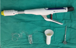
Figure 1
Covidien EEA™ Hemorrhoid and Prolapse Stapler Set 3.5 mm with
DST Series™ Technology.
Figure 2
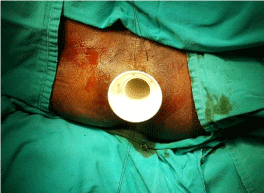
Figure 2
Application of anal dilator.
Figure 3
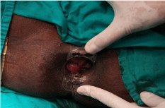
Figure 3
Suture port in place.
Figure 4
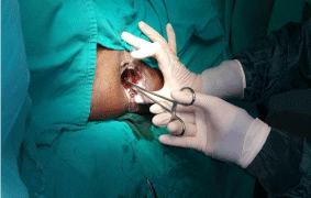
Figure 4
Circumferential purse string suturing of the subucosal layer above
the dentate line.
Figure 5
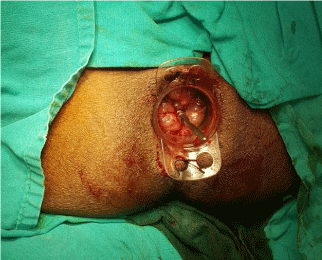
Figure 5
Anvil application and suture fixation.
Figure 6
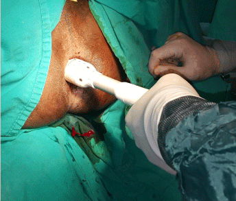
Figure 6
Firing of the device.
Figure 7
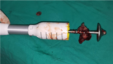
Figure 7
Excised tissue after device removal.
Figure 8
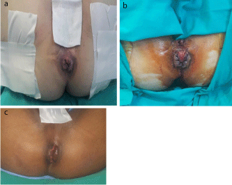
Figure 8
Before the procedure.
Figure 9
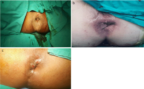
Figure 9
Immediate post-operative results.
Figure 10
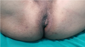
Figure 10
Three months post-operative.
Graph 1
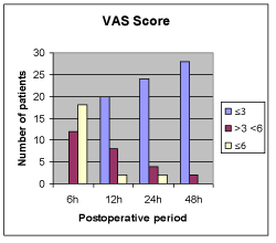
Graph 1
Post-operative VAS scores.
Graph 2
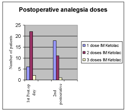
Graph 2
Post-operative analgesia requirement.
Results
Table 1 summarizes the demographic and preoperative data of
the patients. The main presenting symptoms were bleeding and /or
prolapsed mass per annum. Hemorrhoids were grades III and IV.
Liver cirrhosis classified as grade B according to Child–Turcotte’s
classification in 60%of patients.
The average operative time was 27 min (range 20-45 min).
Bleeding from the staple line after removal of the hemostatic gauze
occurred in seven patients. It required only cauterization of the
bleeding spots with re application of the hemostatic gauze for further
5 min (Table 2).
The post-operative Visual Analog Scale (VAS) for recording post operative pain at 6 h, 12 h, 24 h and 48 h is depicted (Graph 1). VAS
sore was ≤ 3 in 80% and 93.3% of patients at 1st and 2nd postoperative
days respectively. Analgesia required according to patients self pain
assessment. Post-operative analgesia requirement in the first 24 h
and the next 24 h is depicted (Graph 2). 73.3% of patients required
two doses of parenteral analgesia (Ketolac®
) in first postoperative day, which reduced to a single dose in 60% of patients in second
postoperative day. Post-operative hospital stay was 2-4 days.
There were only three patients (10%) complicated with postoperative
urinary retention that needed urinary catheterization.
Minor delayed postoperative staple-line bleeding per rectum in the
first 48 h, which did not require any intervention, noticed in fourteen
of patients (46.7%). It was still present in six of patients at 1st month
(20%) and finally disappeared altogether at 3 months of follow-up.
All patients received the procedure without symptom relapse except
six of patients complained of prolapse of mass per rectum during
defecation at 1 week -1st month (20%) Both however, recovered and
became symptom free at 3 months of follow-up. No patient reported
incontinence to flatus or stool and none developed anal stenosis.
Discussion
Stapled Hemorrhoidopexy (SH) aims to correct haemorrhoidal
prolapse by excising a ring of redundant rectal mucosa above the
haemorrhoidal cushions with immediate re-anastomosis of the
mucosa using a circular staples- not hemorrhoids per se. By doing this,
prolapsing hemorrhoids will be repositioning (hemorrhoidopexy)
and shrinking (due to a partial interruption of blood supply to
hemorrhoid plexus). In addition, the terminal branches of the inferior
hemorrhoidal artery disrupted, and blood flow into the cushions
thereby decreased. SH, leaves the richly innervated anal canal tissue
and perianal skin intact, thus reducing the pain usually associated
with EH [8-10]. Nevertheless, uncertainties around complication
rates, recurrence of symptoms and costs preclude its widespread use
[11]. SH is simple to perform but, if not done carefully by experienced
surgeons who have undergone appropriate surgical training, it can
be associated with a number of serious complications caused by a
very low peritoneal reflection incorporated into the anastomosis.
Proper placement of the purse string suture at least 3-4 cm above the
dentate line to incorporate the redundant tissue circumferentially
examination the vagina during the application of the purse string and
insertion of the stapler to avoids rectovaginal fistula [12-18].
Liver cirrhosis with associated Portal Hypertension (PH) is
common in Egypt as a sequela to the high prevalence of hepatitis
C virus. This work elucidates the effectiveness, safety and clinical
outcomes of SH for treating symptomatic hemorrhoids in patients
with liver cirrhosis. A clinician must differentiate bleeding
hemorrhoids form bleeding anorectal varices because they are
separate and distinct entities. Hemorrhoids occur independently of
anorectal varices and their presence was unrelated to the degree of
portal hypertension. There was no relation between portal pressure
and the size of hemorrhoids; no relation found between the size of
hemorrhoids and the grade of esophageal varices. However, both can
bleed and careful examination is essential to prevent misdiagnosis
and inappropriate treatment [19-21]. Of note, rubber band ligation
contraindicated in patients with advanced cirrhosis with coagulation
disorders is generally due to the risk of profound secondary bleeding
following the procedure. For portal hypertensive patients with
internal hemorrhoids and without coagulation disorders or after the
correction of any coagulopathy SH seems to be superior to endoscopic
band ligation. However further studies are needed to evaluate EBL in
different grades of cirrhosis [5,22].
It worth notice that in this study, four patients with Child’s C
grade liver cirrhosis could be recruited following intensive medical
liver support regimen. Several studies demonstrated that stapled
hemorrhoidopexy offers less post-operative pain, and less analgesic
requirements, while providing similar control of symptoms. We have
noted similar results in our series and all our patients had a postoperative
VAS score of < three after 48 h. The operative time for SH
has been demonstrated to be shorter than EH in several trials and
is generally reported at average 20 (range 15-25) min. Our average
operating time of 27 min is a bit longer than that in other studies
[23-25]. We needed more operative time in cirrhotic cases than in
noncirrhotic cases for it was time consuming to reach hemostasis of
the staple line. There were more amounts of blood loss in our patients
with Persistent oozing from the stitch hole while undertaking pursestring
suture, which eventually controlled by mass suture ligation of
the bleeding site. Most of the patients in the study appreciated the SH
procedure and returned to normal activities which suggests that is a
feasible and safe.
Conclusion
Stapled hemorrhoidopexy is a feasible and safe approach for prolapsed hemorrhoids concurrent with liver cirrhosis however; a larger scale controlled trials needed to support our results.
References
- Lacerda-Filho A, Silva RG. Stapled hemorrhoidectomy: present status. Arq Gastroenterol. 2005;42(3):191-4.
- Shao WJ, Li GC, Zhang ZH, Yang BL, Sun GD, Chen YQ. Systematic review and meta-analysis of randomized controlled trials comparing stapled haemorrhoidopexy with conventional haemorrhoidectomy. Br J Surg. 2008;95(2):147-60.
- Burch J, Epstein D, Baba-Akbari A, Weatherly H, Fox D, Golder S, et al. Stapled haemorrhoidectomy (haemorrhoidopexy) for the treatment of haemorrhoids: a systematic review and economic evaluation. Health Technol Assess. 2008;12(8):1-19.
- Misra SP, Dwivedi M, Misra V. Prevalence and factors influencing hemorrhoids, anorectal varices, and colopathy in patients with portal hypertension. Endoscopy. 1996;28(4):340-5.
- Awad AE, Soliman HH, Saif SA, Darwish AM, Mosaad S, Elfert AA. A prospective randomised comparative study of endoscopic band ligation versus injection sclerotherapy of bleeding internal haemorrhoids in patients with liver cirrhosis. Arab J Gastroenterol. 2012;13(2):77-81.
- Huang WS, Lin PY, Chin CC, Yeh CH, Hsieh CC, Chang TS, et al. Stapled hemorrhoidopexy for prolapsed hemorrhoids in patients’ with liver cirrhosis; a preliminary outcome for 8-case experience. Int J Colorectal Dis. 2007;22(9):1083-9.
- Biswas S, George ML, Leather AJ. Stapled anopexy in the treatment of anal varices: report of a case. Dis Colon Rectum. 2003;46(9):1284-5.
- Shrestha S, Pradhan GB, Shrestha R, Poudel P, Bhattachan CL. Stapled haemorrhoidectomy in the operative treatment of grade III and IV haemorrhoids. Nepal Med Coll J. 2014;16(1):72-4.
- Aytac E, Gorgun E, Erem HH, Abbas MA, Hull TL, Remzi FH. Long-term outcomes after circular stapled hemorrhoidopexy versus Ferguson hemorrhoidectomy. Tech Coloproctol. 2015;19(10):653-8.
- Lohsiriwat V. Treatment of hemorrhoids: A coloproctologist's view. World J Gastroenterol. 2015;21(31):9245-52.
- Jayaraman S, Colquhoun PH, Malthaner RA. Stapled hemorrhoidopexy is associated with a higher long-term recurrence rate of internal hemorrhoids compared with conventional excisional hemorrhoid surgery. Dis Colon Rectum. 2007;50(9):1297-305.
- Porrett LJ, Porrett JK, Ho YH. Documented complications of staple hemorrhoidopexy: a systematic review. Int Surg. 2015;100(1):44-57.
- Bilgin Y, Hot S, Barlas IS, Akan A, Eryavuz Y. Short- and long-term results of harmonic scalpel hemorrhoidectomy versus stapler hemorrhoidopexy in treatment of hemorrhoidal disease. Asian J Surg. 2015;38(4):214-9.
- Zanella S, Spirch S, Scarpa M, Ricci F, Lumachi F. Long-term outcome of stapled transanal rectal resection (STARR) versus stapled hemorrhoidopexys (STH) for grade III-IV hemorrhoids: preliminary results. In Vivo. 2014;28(6):1171-4.
- Wong LY, Jiang JK, Chang SC. Rectal perforation: a life-threatening complication of stapled hemorrhoidectomy: report of a case. Dis Colon Rectum. 2003;46(1):116-7.
- Ripetti V, Caricato M, Arullani A. Rectal perforation, retropneumoperitoneum, and pneumomediastinum after stapling procedure for prolapsed hemorrhoids: report of a case and subsequent considerations. Dis Colon Rectum. 2002;45(2):268-70.
- Molloy RG, Kingsmore D. Life threatening pelvic sepsis after stapled haemorrhoidectomy. Lancet. 2000;355(9206):810.
- Cipriani S, Pescatori M. Acute rectal obstruction after PPH stapled haemorrhoidectomy. Colorectal Dis. 2002;4(5):367-70.
- Le Quellec A, Bories P, Rochon JC, Garrigues JM, Poirier JL, Michel H. Portal hypertension and hemorrhoids. Cause effect relationship? Gastroenterol Clin Biol. 1988;12(8-9): 646-8.
- Hosking SW, Smart HL, Johnson AG, Triger DR. Anorectal varices, haemorrhoids, and portal hypertension. Lancet. 1989;1(8634):349-52.
- Zuberi FF, Zuberi BF, Khan MA, Khan MH. Frequency of rectal varices in patients with cirrhosis. J Coll Physicians Surg Pak. 2004;14(2):94-7.
- Zaher T, Ibrahim I, Ibrahim A. Endoscopic band ligation of internal haemorrhoids versus stapled haemorrhoidopexy in patients with portal hypertension. Arab J Gastroenterol. 2011;12(1):11-14.
- Pescatori M. Prospective randomized multicentre trial comparing stapled with open haemorrhoidectomy. Br J Surg. 2002;89-122.
- Wilson MS, Pope V, Doran HE, Fearn SJ, Brough WA. Objective comparison of stapled anopexy and open hemorrhoidectomy: a randomized, controlled trial. Dis Colon Rectum. 2002;45(11):1437-44.
- Pavlidis T, Papaziogas B, Souparis A. Modern stapled Longo procedure vs. conventional Milligan–Morgan hemorrhoidectomy: a randomized controlled trial. Int J Colorectal Dis. 2002;17(1):50-3.