Clinical Image
Stump Dilatation after Ileocolic Resection in Crohn’S Disease
Enrique Colás Ruiz*, Paula Dujovne Lindenbaum, Federico Ochando Cerdán, Jose Antonio Rueda Orgaz and Jose María Fernández Cebrián
Department of General and Gastrointestinal Surgery, Hospital Universitario Fundación Alcorcón, Madrid, Spain
*Corresponding author: Enrique Colás Ruiz, Department of General and Gastrointestinal Surgery, Hospital Universitario Fundación Alcorcón, Calle Budapes, Alcorcón, Madrid, Spain
Published: 30 Mar, 2017
Cite this article as: Ruiz EC, Lindenbaum PD, Cerdán
FO, Orgaz JAR, Cebrián JMF. Stump
Dilatation after Ileocolic Resection in
Crohn’S Disease. Clin Surg. 2017; 2:
1384.
Abstract
Ileal stump dilatation is very rare. We present a patient who presented a calcified mass in right lower quadrant that looked like an intraabdominal foreign body.
Keywords: Stum dilatation; Bezoar; Crohn´s disease
Introduction
Ileal stump dilatation after ileocolic resection with side to side anastomosis is really rare [1-3]. We present a case report which ileal stump presents inside a bezoar [4].
Clinical Image
A 39 year-old woman underwent an ileocolic resection for appendicitis involving ileum and cecum with intraoperative suspicion of Crohn´s disease ten years before. The reconstruction was made side to side anastomosis.
The anatomopathological findings were compatible with Crohn’s disease.
She consults emergency service for abdominal pain, nausea and emesis. Her abdomen is
lightly distended and with a mass in right lowers side. Laboratory workup was normal. Abdominal
radiography showed in the right side a zone with increased density that we could see retrospectively
in other radiography three years ago. CT scan showed a concentric calcified lesion in right lower
quadrant approximately with 8 cm, 7 cm x 4 cm, 5 cm that force us to discard an intraabdominal
foreign body because it seems outside of intestinal tract (Figure 1). Three years before, the last colonoscopy didn’t report the existence of large ileum blind intestinal loop. During the exploratory
laparoscopy we found a big ileal stump dilated with hard consistency and increased vascularization
because chronic inflammation. We cut the ileal stump preserving the old anastomosis with endo
GIA. The specimen opened presented a rock-hard mass compatible with ileal bezoar (Figure 2).
Conclusion
A bezoar in small intestine affected by Crohn’s disease appears to be very rare, but sometimes can cause a small bowel obstruction. Ileal stump dilatation is a rare find. Maybe, in this case is due to excessive length of ileal stump, but it is impossible to be sure. We have to remember the possibility similar compress and bezoar image.
Figure 1
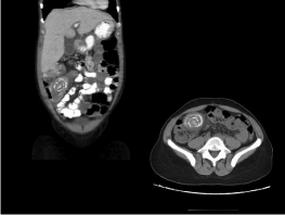
Figure 1
Outside of intestinal tract.
Figure 2
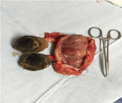
Figure 2
Rock-hard mass compatible.
References
- Harrington S, Mohamed S, Bloch R. Small bowel obstruction by a primary phytobezoar in Crohn’s disease. Am Surg. 2009;75(1):93-4.
- Chary S, Crisp JC. Ileo-vesical fistula with phytobezoar in Crohn’s disease of ileum. Br J Urol. 1975;47(2):160.
- Mmeje C, Bouchard A, Heppell J. Pharmacobezoar: A rare complication after ileal pouch-anal anastomosis for ulcerative colitis. Clin Gastroenterol Hepatol. 2010;8(6):A28.
- Koktener A, Dilmen G, Turkayc, Erbayrak M. Small bowel obstruction caused by phytobezoar and associated with vitamin B12 deficiency. South Med J. 2006;99(9):1013-4.