Case Report
Lower Limb Salvage at any Cost – A Complex Case
Amir K Bigdeli*, Frederick J Hernekamp, Florian Neubrech, Nico Leibig, Chrisitan Radu, Emre Gazyakan, Ulrich Kneser and Thomas Kremer
Department of Hand and Plastic Surgery, University of Heidelberg, Heidelberg, Germany
*Corresponding author: Amir Khosrow Bigdeli, Department of Hand, Plastic and Reconstructive Surgery, Burn Centre, BG Trauma Centre Ludwigshafen, Ludwig- Guttmann-Strasse 13, Ludwigshafen, 67071 Hand and Plastic Surgery, University of Heidelberg, Heidelberg, Germany
Published: 30 Mar, 2017
Cite this article as: Bigdeli AK, Hernekamp FJ, Neubrech
F, Leibig N, Radu C, Gazyakan E, et
al. Lower Limb Salvage at any Cost –
A Complex Case. Clin Surg. 2017; 2:
1380.
Abstract
Lower limb salvage for osteomyelitis of the tibia after open fracture presents an ongoing challenge.
In the field of modern reconstructive surgery, we increasingly find ourselves asking “What can we
salvage?” instead of “What should we salvage?” We report the case of a 37-year-old male patient,
who sustained an open tibial fracture in a motorcycle accident. He underwent more than thirty
operative procedures for lower limb reconstruction over a five-year period. Complicated by
osteomyelitis, limb salvage was finally achieved with a free contralateral vascularized osteocutaneus
fibular transfer after 13-hour surgery. The combination with an arterio-venous loop was necessary
for and created through the cephalic and small saphenous vein as unusual feature due to sacrifice
of autologous vein grafts for previous reconstructive procedures. Two years later, the patient is
satisfied with his limb function and has returned to work. The free osteocutaneous vascularized
fibula transfer prevented lower limb amputation but still remains complex and invasive. We want
to discuss aspects of decision-making involving clinicians as well as the patient and recommend the
installation of multidisciplinary extremity boards.
Keywords: Free fibula; Distal tibia fracture; Limb salvage; Arteriovenous loop
Introduction
Advances in reconstructive surgery nowadays allow impressive lower limb salvage after severe trauma. More and more, clinicians find themselves asking “What can be salvaged?” instead of “What should be salvaged?” Especially, free microvascular bone transfers have revolutionized reconstruction of large osseous defects since the first free fibular transplantation was described [1,2]. However, one should not ignore the risk of complications at the donor site that may lead to functional impairment of the healthy leg. Other potential adverse effects are moderate to severe pain at the donor site, delayed wound healing, sensory changes and the sacrifice of a major vascular axis [3]. The alternative is below-knee amputation, whereas amputation index scores have been developed in order to facilitate decision-making between a salvage attempt and amputation [4]. Nevertheless, the prediction of successful limb salvage with a good functional result remains difficult and the decision is still hard to make. We report the case of a 37-year-old male patient, who underwent more than 30 surgical procedures over a five-year period for lower limb reconstruction of osteomyelitis-associated tibial non-union after open fracture. The procedures included revision osteosyntheses, cancellous bone grafts, bone transports and two free tissue transfers. We address the main question, whether or not limb salvage is beneficial to certain patient groups. Furthermore, we critically discuss the reconstructive approach focusing on aspects of decision-making involving a multidisciplinary team of clinicians as well as the patient. Additionally, different therapeutic aspects including complication rates, donor-site morbidity, procedure complexity, failure rates and potential limb function are discussed.
Case Presentation
A 37-year-old male sustained an open fracture of the left tibia and fibula in a motorcycle
accident. In an external hospital, open reduction and internal fixation through an unreamed
intramedullary tibial nail was performed immediately. Three months later, implant removal and
re-osteosynthesis with a cannulated tibial nail and plate osteosynthesis of the fibula were performed
due to bony non-union. Seven months after trauma, infectious pseudarthrosis of the tibia and
fibula were suspected, and re-osteosynthesis was performed by the reamed technique after implant
removal. Systemic test-appropriate antibiotic treatment was initiated. In the further course, bony
union of the tibia did not occur and tibial osteomyelitis was diagnosed. To cut a long story short,
the patient underwent more than 30 surgical procedures, including many attempts of bony repair
with re-osteosyntheses, cancellous bone grafts, and bone transports. During the reconstructive course, soft tissue defect reconstruction became necessary and was
performed using a free anterolateral thigh (ALT) flap, that was lost
due to arterial thrombosis and finally achieved using a free latissimus
dorsi flap that was anastomosed via an arteriovenous loop (AVL)
using the contralateral saphenous vein to the femoral artery and
vein. Finally, the patient was admitted to our clinic for limb salvage.
A multidisciplinary team of clinicians (plastic and reconstructive
surgeon, trauma surgeon, vascular surgeon, psychologist, and
microbiologist) was involved. A salvage attempt versus below-knee
amputation was extensively discussed with the patient. From the
medical point of view, amputation and prosthetic provisioning was
advised. However, the patient insisted on limb salvage and after a risk assessment examination in an elaborate explanatory meeting, the decision for a salvage attempt through a free contralateral vascularized osteocutaneous fibula transfer was made.
Surgical technique
After more than 30 surgical procedures within five years, the
patient was referred to our center with non-union of the tibia.
Preoperatively, stable skin and soft-tissue were given (Figure 1a and b)
and Magnetic resonance angiography revealed one-vessel flow to the
distal leg through the anterior tibial artery (ATA). After debridement,
a large segmental bone defect was present (Figure 2a and b). In the
absence of systemic signs of infection, the vascularized bone transfer
was initiated with a two-team approach. The ATA was not dissectable
due to scar formation. Therefore, the superficial femoral artery (SFA)
and vein were dissected proximal to the anastomosis of the AVL that
was used for the latissimus dorsi flap. Simultaneously, the following veins were harvested for an additional AVL: both cephalic veins
(from the infraclavicular fossa to the dorsum of the hand on the
left side, and from the shoulder to the anticubital fossa on the right
side with a length of approximately 80 centimeters). The AVL was
anastomosed to the SFA and the accompanying vein in an end-toside
fashion. Simultaneously, the vascularized osteocutaneous fibular
graft was harvested from the contralateral leg with a skin paddle of
15 cm × 5 cm through a standard posterolateral approach (Figure 3
and 4). The donor-site wound was closed primarily. The transported
fibula was fitted tightly into the medulla of the tibia and fixed through
plate osteosynthesis. The AVL was cut in the middle. A low flow was
observed at the arterial part and attributed to fibrosis of the vessel’s
wall. Fibrotic parts were excised and replaced by segments of the
contralateral small saphenous vein. Lastly, the anastomoses were
carried out in end-to-end fashion to the peroneal artery and vein
of the fibula graft (Figure 5a and b). After the 13-hour surgery, the
patient was kept on a test-appropriate antibiotic therapy for four
weeks and the left lower leg was immobilized in a leg cast for three
months. Thereafter, a long leg brace was applied for an additional
three months with non-weight bearing crutch ambulation. After
six months, gradual weight bearing under protection of a brace
was allowed. During regular follow-up in our outpatient clinic,
bone union was indicated by the clinical absence of pain and callus
formation on X-Ray at six months (Figure 6). Full weight bearing was
allowed after 6 months (Figure 7a-c). The patient underwent routine
follow-up visits and is still disease free after three years. He is fully
recovered and has returned to all his activities of daily living and work
without restriction.
Figure 1
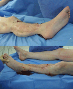
Figure 1
(a and b) Clinical appearance at the beginning of the operation with
intact skin and soft- tissue over the left tibia.
Figure 2
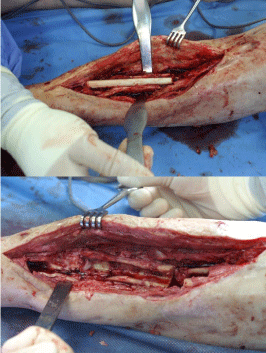
Figure 2
(a and b) Intraoperative view of the segmental tibial bone defect
after debridement.
Figure 3
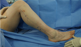
Figure 3
Intraoperative design of the vascularized osteocutaneous fibular
graft with a skin paddle (15 × 5 cm).
Figure 4
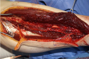
Figure 4
Intraoperative preparation of the vascularized osteocutaneous
fibular with perforator-based skin paddle.
Figure 5
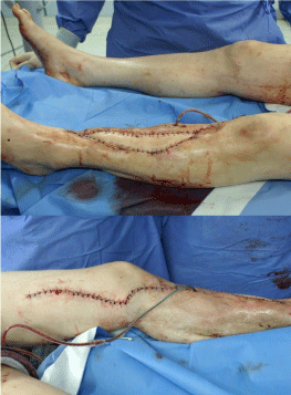
Figure 5
(a and b) Intraoperative view after successful free fibula transfer.
Figure 6
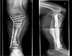
Figure 6
Postoperative X-Ray with ossification of the periostal tissue at six
months.
Figure 7
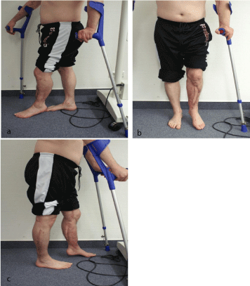
Figure 7
(a-c) Clinical appearance after 6 months.
Discussion
Advances in reconstructive surgery nowadays allow impressive lower limb salvage procedures. A major contribution was the introduction of the free fibular microvascular transfer [1,2]. It provides not only bone with a sufficient length and biological properties, but also enables the reconstruction of accompanying tissue defects, as it may include skin, muscle, tendon, and even nerve tissue. However, donorsite morbidity remains a concern, but is inconsistent throughout the literature with rates between 2 and 38% [3-7]. The technique requires an experienced team of microsurgeons, as failure of surgery is a serious loss to the patient, not only in the failed reconstruction but also the risk of morbidity of the healthy leg. To evaluate donor-site morbidity in free fibular microvascular transfer, Momoh and colleagues conducted a prospective cohort study including 157 patients [7]. The perioperative donor-site complication rate was 31.2% in this largest series. However, they did not find a significant association between perioperative complications and long-term morbidity. 26 patients (16.6%) experienced leg weakness (8%), ankle instability (4%), great toe flexion contracture (9%), and decreased ankle range of motion (2%), 37% of the patients were able to run or engage in athletic activity after surgery. Focusing on functionality, most studies found, that nearly all patients were able to perform their activities of daily living without significant limitations [7-10]. Our patient underwent more than 30 operative procedures for lower limb reconstruction after open fracture of the tibia. He was referred to our center with osteomyelitis related bony non-union of his tibia five years after the trauma. A multidisciplinary team was involved and amputation and prosthetic provisioning advised. However, the patient insisted on limb salvage and therefore a last salvage attempt through a free contralateral vascularized osteocutaneous fibula transfer was indicated. As described above, an AVL was necessary to extend the pedicle of the flap. Threlfall “et al.” [11] first suggested the use of a temporary AVL prior to flap harvesting in 1982. When recipient vessels for free tissue transfer become difficult to find, an AVL facilitates the use of normal recipient vessels for microvascular anastomosis far away from the zone of injury [12]. In our case, a venous graft around 80 centimeters in length was necessary. In absence of the great saphenous vein, the cephalic vein had to be harvested from both arms. The experienced flow problems in the arterial side of the loop were attributed to segmental vein wall fibrosis. We think that it may be caused by prior damage through blood drawing procedures. Finally, the contralateral small saphenous vein was harvested and successfully used for loop salvage. Generally, we agree with Pelissier and colleagues who state, that decision-making involves clinicians and the patient in equal measure [13]. Here, a detailed explanation of the operative procedure including the duration and expected results is absolutely necessary. Furthermore, such cases should be discussed in multidisciplinary extremity boards. Retrospectively, we think that an interdisciplinary approach would have initiated the reconstruction through a free contralateral vascularized osteocutaneous fibula transfer earlier and thus reduced morbidity as the operation took more than 13 hours. Also, patients who generally ask for reconstruction instead of amputation should be aware, that the procedure comes with a long operation time and an unpredictable functional result [13]. Further disadvantages may include moderate problems at the donor site (e.g., pain, delayed wound healing, sensory changes, sacrifice of a major vascular axis) [13]. In order to compare functional outcomes and disability between amputation and limb salvage after major lower extremity trauma sustained in the US military, the Military Extremity Trauma Amputation/Limb Salvage (METALS) Study was conducted [14]. The retrospective cohort study included 324 patients and showed that amputation had better functional outcomes when compared with limb salvage. However, the main question is, whether or not salvage is beneficial to certain patient groups. Some authors suggest that early amputation and rehabilitation with prosthesis provides a better outcome [15,16]. In contrast, other groups have found that a majority of patients with severe lower extremity injuries preferred their salvaged extremity, even when delayed amputation was required. Rehabilitation has the greatest influence on postoperative outcome [17]. Here, amputees may receive more focused and earlier rehabilitation. Furthermore, weight bearing is allowed as soon as there is sufficient wound healing. In contrast, patients may need to wait three or more months after limb salvage for healing of fractures and bone defects before full weight bearing is allowed. Furthermore, rehabilitation after limb salvage may be less organized due to the individual type of procedure and healing capabilities [14,17].
Conclusion
Decision-making on limb salvage versus amputation continues to be difficult. However, the medical literature suggests similar functional outcomes and rates of return to work after salvaged and amputated lower extremities, but the latter ones are plagued by higher complication and reoperation rates. The free vascularized osteocutanous fibula transfer is capable to prevent lower limb amputation. However, the procedure remains complex and invasive with a significant complication rate, moderate donor-site morbidity, and failure rates. To facilitate decision-making, we recommend the installation of multidisciplinary extremity boards and have implemented it at our Center. Lastly, more work will need to be done to help surgeons to consistently and reliably predict which patients will benefit from limb salvage or an early amputation.
References
- Taylor GI, Miller GD, Ham FJ. The free vascularized bone graft. A clinical extension of microvascular techniques. Plast Reconstr Surg. 1975;55(5):533–44.
- Ueba Y, Fujikawa S. Nine years’ follow up of a vascularized fibular graft in neurofibromatosis: a case report and literature review. Jpn J Orthop Trauma Surg. 1983;26:595–600.
- Vail TP, Urbaniak JR. Donor–site morbidity with use of vascularized autogenous fibular grafts. J Bone Joint Surg Am. 1996;78(2):204–11.
- Dagum AB, Best AK, Schemitsch EH, Mahoney JL, Mahomed MN, Blight KR. Salvage after severe lower–extremity trauma: are the outcomes worth the means? Plast Reconstr Surg. 1999;103(4):1212–20.
- Malizos KN, Nunley JA, Goldner RD, Urbaniak JR, Harrelson JM. Free vascularized fibula in traumatic long bone defects and in limb salvaging following tumor resection: comparative study. Microsurgery. 1993;14(6):368–74.
- Minami A, Usui M, Ogino T, Minami M. Simultaneous reconstruction of bone and skin defects by free fibular graft with a skin flap. Microsurgery. 1986;7(1):38–45.
- Momoh AO, Yu P, Skoracki RJ, Liu S, Feng L, Hanasono MM. A prospective cohort study of fibula free flap donor–site morbidity in 157 consecutive patients. Plast Reconstr Surg. 2011;128(3):714–20.
- Bodde EW, de Visser E, Duysens JE, Hartman EH. Donor–site morbidity after free vascularized autogenous fibular transfer: subjective and quantitative analyses. Plast Reconstr Surg 2003;111(7):2237–42.
- Hidalgo DA, Rekow A. A review of 60 consecutive fibula free flap mandible reconstructions. Plast Reconstr Surg. 1995;96(3):585–96.
- Shpitzer T, Neligan P, Boyd B, Gullane P, Gur E, Freeman J. Leg morbidity and function following fibular free flap harvest. Ann Plast Surg. 1997;38(5):460–4.
- Threlfall GN, Little JM, Cummine J. Free flap transfer––preliminary establishment of an arteriovenous fistula: a case report. Aust N Z J Surg. 1982;52(2):182–4.
- Silveira LF, Patricio JA. Arteriovenous fistula with a saphenous long loop. Microsurgery.1993;14(7):444–5.
- Pelissier P, Boireau P, Martin D, Baudet J. Bone reconstruction of the lower extremity: complications and outcomes. Plast Reconstr Surg. 2003;111(7):2223–9.
- Doukas WC, Hayda RA, Frisch HM, Andersen RC, Mazurek MT, Ficke JR, et al. The Military Extremity Trauma Amputation/Limb Salvage (METALS) study: outcomes of amputation versus limb salvage following major lower–extremity trauma. J Bone Joint Surg Am. 2013;95(2):138–45.
- Georgiadis GM, Behrens FF, Joyce MJ, Earle AS, Simmons AL. Open tibial fractures with severe soft–tissue loss. Limb salvage compared with below–the–knee amputation. J Bone Joint Surg Am 1993;75(10):1431–41.
- Hansen ST. The type–IIIC tibial fracture. Salvage or amputation. J Bone Joint Surg Am.1987;69(6):799–800.
- Momoh AO, Chung KC. Measuring outcomes in lower limb surgery. Clin Plast Surg. 2013;40(2):323–9.