Editorial
Measurement of Plantar Sole Sensitivity: A Tool for the Podiatric Diagnosis
Yves Jammes*, Bruno Vie, Patricia Griffon and Jean Paul Weber
Department of Physiology, Hôpital Européen Marseille, France
*Corresponding author: Yves Jammes, Department of Physiology, Hôpital Européen Marseille, France
Published: 06 Mar, 2017
Cite this article as: Jammes Y, Vie B, Griffon P, Weber
JP. Measurement of Plantar Sole
Sensitivity: A Tool for the Podiatric
Diagnosis. Clin Surg. 2017; 2: 1344.
Editorial
Measurement of plantar sole sensitivity could be useful to the podiatrists who have to diagnose patients at risk of diabetic neuropathy. The plantar surface is the major weight bearing surface
during gait and standing. The cutaneous mechanoreceptors of the plantar sole detect the application
of load. An electrophysiological study in humans [1] reported the presence of both slow (Merkel
and Ruffini corpuscles) and fast (Meissner and Pacinian corpuscles) adapting receptors in the
foot sole, with far greater numbers of the latter (71% of tested units). Skin mechanoreceptors were
classified into four groups based on their afferent firing properties [fast adapting (FA) vs. slow
adapting (SA)] and receptive field size [type I (small defined boundaries), vs. type II (large undefined
boundaries)]. Johansson et al. [2] found that SAI and SAII skin afferents were activated by low
vibration frequencies between 2 and 32 Hz whereas FAI were activated between 8 and 64 Hz and
FAII between 64 and 400 Hz.
There are several studies indicating that plantar cutaneous mechanoreceptors contribute to
controlling the standing balance and postural reflexes in healthy subjects [3-6]. Indeed, balance
problems are often related to cases where a reduced plantar sensitivity occurs. Electrophysiological
studies have shown that the cutaneous afferents from the plantar surface project on the somatosensory
cortex leading to a perceptual representation [7]. Examination of the vibration sense is widely used
as a clinical examination in neurology and it is usually done at the medial or lateral malleolus.
Most of studies only measured the vibration detection threshold of the sole in healthy subjects in
an attempt to compare normal values to those measured in aged individuals and diabetic patients
[8-13]. On the other hand, very few data are found on the magnitude of the vibration estimate of the
foot sole in response to the increased amplitude of the vibratory stimulus. To determine the estimate
of vibrations delivered to the skin of the hand, Verillo et al. [14] already used a psychophysical
approach giving the global gain of the sensory detection. We already used this method to determine
the sensory detection of tactile stimulation applied on the fingers [15] or the plantar sole [16]. More
recently, the global gain of vibration detection by the plantar sole was determined in young and
old healthy subjects [17]. The Stevens power function brings a psychophysical approach of the
relationship between the estimate (Ψ) of stimuli applied on the foot sole and its physical magnitude
(Φ) (Ψ = k *Φn). The exponent n in the power law was determined by a linear regression analysis
between Napierian logarithmic (Ln) transformed stimuli and estimation data. Any increase in k
value indicates an elevated sensitivity to the lowest loads and thus a lowered detection threshold. The
n coefficient measures the changes in perception between the extreme values of loads. Thus, the n
coefficient measures the gain in perception and k gives an approach of the threshold for perception.
To test tactile perception of the foot sole [16], the participant layed flat on a bed. Mechanical stimuli
were randomly applied to the heel or metatarsal area using a bespoke electronic stimulator device.
The stimulator delivered rectangular pulses of 100 ms duration which drove a mechanical probe
via a solenoid. Four mechanical stimuli (24, 41, 69 and 116 g) were delivered in random order,
producing local plantar pressures of 3.4, 5.7, 9.7, and 16.3 N.cm-2. To test vibration perception of the
foot sole [17], the participants sat comfortably. The skin sensitivity was evaluated using vibration
testing at three frequencies (25, 50, and 150 Hz) at each one of three foot plantar locations (hallux,
fifth metatarsal head, and heel). The vibration frequencies were chosen to target the activation of
three different skin receptors in the glabrous skin of the foot (25 Hz for SAI, 50 Hz for FAI, and 150
Hz for FAII) based on data obtained by Johansson et al. [2]. Vibrations were applied to the foot sole
by a minishaker and a preload force of 2N was measured with a force transducer. The vibrator device
allowed delivering different amplitudes of vertical motions of the probe. The vibration magnitude
depended on its frequency and varied in a range of 10 to 360 μm at 25 Hz, 10 to 330 μm at 50 Hz,
and 10 to 180 μm at 150 Hz. The bespoke apparatus built to deliver tactile or vibratory stimulus are fully described and shown in our previous studies [16,17]. Judgments of increased stimuli intensity were recorded on a 0-10 cm visual
analogue scale. The whole duration of the two trials to explore the
tactile and vibratory sensitivities was 10 min. We prefer to explore
the vibration sensitivity than the tactile one. Indeed, vibration testing
allows examining different skin mechanoreceptors including the
fast adapting ones. Besides, the tactile stimulation allows to only exploring the slow adapting sensory units. Figure 1 gives an example of linear regressions obtained between the vibration amplitude
and its perception at two frequencies (25 and 150 Hz). The gain of
perception was always higher with the 150 Hz vibration frequency
which activated the fast adapting units. Compared to the young
adults, the elderly had lower n values measured at this high frequency.
Figure 2 shows an example of clinical use of plantar vibration
sensitivity to assess an altered sensitivity in a patient with a reduced
clinical sensitivity on the course of the left external popliteal nerve.
Figure 1
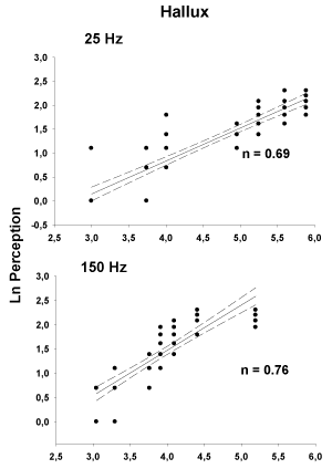
Figure 1
Graphical representation of the relationships between perception
and vibration amplitude tested at two vibration frequencies. The n coefficient
which measures the gain of sensation is significantly higher at the highest
vibration frequency which activates the fast adapting sensory endings.
Figure 2
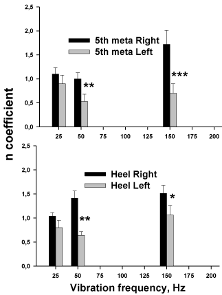
Figure 2
Altered vibration sensitivity in a 66 yr old male patient who related
posterior pinch disc of the 3rd to the 5th lumbar metamere assessed by X ray
images, leading to an alteration of clinical sensitivity on the course of the left
external popliteal nerve.
References
- Kennedy PM, Inglis JT. Distribution and behaviour of glabrous cutaneous receptors in the human foot sole. Journal of Physiology. 2002; 538: 995-1002.
- Johansson RS, Landstrom U, Lundstrom R. Responses of mechanoreceptive afferent units in the glabrous skin of the human hand to sinusoidal skin displacements. Brain Res. 1982; 244: 17-25.
- Allum JH, Bloem BR, Carpenter MG, Hulliger M, Hadders-Algra M. Proprioceptive control of posture: a review of new concepts. Gait and Posture. 1998; 8: 214-242.
- Eils E, Nolte S, Tewes M, Thorwesten L, Völker K, Rosenbaum D. Modified pressure distribution patterns in walking following reduction of plantar sensation. J Biomech. 2002; 35: 1307-1313.
- Höhne A, Ali S, Stark C, Brüggemann GP. Reduced plantar cutaneous sensation modifies gait dynamics, lower-limb kinematics and muscle activity during walking. Eur J Appl Physiol. 2012; 112: 3829-3838.
- Maurer C, Mergner T, Bolha B, Hlavacka F. Human balance control during cutaneous stimulation of the plantar soles. Neuroscience Letters. 2001; 302: 45-48.
- Kandel E, Schwartz J, Jessell T. 2012; Principles of Neural Science, 4th ed. McGraw-Hill Medical, New York.
- Kekoni J, Hämäläinen H, Rautio J, Tukeva T. Mechanical sensibility of the sole of the foot determined with vibratory stimuli of varying frequency. Exp Brain Res. 1989; 78: 419-424.
- Kowalzik R, Hermann B, Biedermann H, Peiper U. Two-point discrimination of vibratory perception on the sole of the human foot. Foot Ankle Int 1996; 17: 629-634.
- Wells C, Ward LM, Chua R, Inglis JT. Regional variation and changes with ageing in vibrotactile sensitivity in the human footsole. J Gerontol A Biol Sci Med Sci. 2003; 58: 680-686.
- Perry SD. Evaluation of age-related plantar-surface insensitivity and onset of age of advanced insensitivity in older adults using vibratory and touch sensation test. Neurosci Lett. 2006; 392: 62-67.
- Hennig EM, Sterzing T. Sensitivity mapping of the human foot: thresholds at 30 skin locations. Foot Ankle Int. 2009; 30: 986-991.
- Gu C, Griffin MJ. Vibrotactile thresholds at the sole of the foot: effect of vibration frequency and contact location. Somatosens Mot Res. 2011; 28: 86-93.
- Verillo RT, Bolanowski SJ, Gescheider GA. Effect of aging on the subjective magnitude of vibration. Somatosens Mot Res. 2002; 19: 238-244.
- Balzamo E, Burnet H, Zattara-Hartmann MC, Jammes Y. Increasing background inspiratory resistance changes somatosensory sensations in healthy man. Neurosci Lett. 1995; 197: 125-128.
- Vie B, Nester CJ, Porte LM, Behr M, Weber JP, Jammes Y. Pilot study demonstrating that sole mechanosensitivity can be affected by insole use. Gait Posture. 2015; 41: 263-268.
- Jammes Y, Guimbaud J, Faure R, Griffon P, Weber JP, Vie B, et al. Psychophysical estimate of plantar vibration sensitivity brings additional information to the detection threshold in young and elderly subjects. Clin Neurophysiol Practice. 2016; 1: 26-32.