Review Article
Image Guided Breast Oncologic Treatment
Robert L Bard*
Department of Breast Surgery, Bard Cancer Center, USA
*Corresponding author: Robert L Bard, Department of Breast Surgery, Bard Cancer Center, USA
Published: 07 Mar, 2017
Cite this article as: Bard RL. Image Guided Breast
Oncologic Treatment. Clin Surg. 2017;
2: 1334.
Abstract
3 Dimensional vascular imaging has provided a quantifiable measure of treatment success or failure in breast diseases. The ability to presurgically map the vessel distribution allows for targeted surgical planning. The vessel density correlates with activity in both the breast cancer and the locoregional disease. Postoperative pathologic analysis is assisted by focusing on tumor tissue rather than benign disease associated with a primary or recurrent malignancy. The new advances of volumetric analysis make this exam less operator dependent as the probe is fixed in position.
Introduction
Women are searching for a diagnosis on any palpable lesion that may be subdermal in location and, thus, invisible to the spatially restricted human eye. Patients are currently concerned about possible side effects from biopsy, radiation and gadolinium based MRI contrast agents. Diagnostic ultrasound meets these requirements and can perform in the office setting accurately and rapidly due to the high resolution and low cost of today’s sonographic equipment. This diagnostic technology requires extensive experience and training in interpreting the images. However, advances in the computerization of the imaging, blood flow, and tumor measures of exact volume and vessel density are now fewer operators dependent. That provides for an accurate and repeatable diagnosis, and a means to follow the individual’s unique pattern of cancer development, progress, and response to treatment. Recent technological advances also make these procedures available to much broader clinical application, without requiring years of very unique training and experience, for example, with diagnoses of cystic versus lesions. Accuracy in assessing breast [1] tumors and metastatic foci has been documented. It must be emphasized that the beginner will find many confusing artifacts and findings should be confirmed with all pertinent imaging modalities. 3D Doppler ultrasound with Dynamic Contrast Enhanced MRI is the gold standards by which cancers are initially diagnosed or confirmed and serially followed after treatment. The percentage of malignant vessels can be quantified and re-evaluated in the identical tumor volume as serial follow ups to the standard treatments using: radiation, surgery, hormones, chemotherapy, cryotherapy, watchful waiting and the non-standard treatments: ablation using focal laser, focal ultrasound, photodynamic, radiofrequency and microwave technologies. Since vessel mapping is possible, embolic treatments may be considered. 3D sonography can demonstrate the tumor volume and marginal capsule of locally affected lymph nodes more accurately than the MRI since the resolution is 100 microns at 18 MHz. The exam takes about 10 minutes and the probes are automated meaning that this is less operator dependent than other sonographic procedures. Vessel density index (VI) imaging is performed on the data set at an independent workstation and comparison made with prior exams if available. 3D power Doppler indices vary according to the tumor stage, the histologic grade, capsular disruption, and lymph node metastases. Histologic grade has been studied with this technology and the following approximation has proven useful in prostate tumor staging. This quantitative measure of neovascularity was initially applied to prostate cancer [2]. While it does not exactly correlate with histologic Gleason grading since this is a current functional measure while the microscopy is purely anatomical and may not represent current aggressive potential. It’s predictive value of aggression could be studied in the context of breast and other cancers [3,4]. Medical imaging can map the arteries, veins and nerves providing preoperative landmarks reducing postoperative bleeding and avoiding nerve damage. Tumors of low aggressive potential may be treated medically and followed by interval scans or locally reduced by radiation or laser ablation. Biopsies of certain abnormalities may be averted or postponed (Figure 1).
Breast Cancer
High tumor vessel density correlates with greater aggression. Axillary and mediastinal imaging can document lymphadenopathy. Abdominal scans simultaneously performed may detect ascites and metastases to the liver, periaortic nodes and pelvic organs. Response to neoadjuvant chemotherapy may be assessed MRI, CT, mammography, PET/CT and ultrasound. The new technology of ultrasound elastography, assessing tumor stiffness, predicts response to treatment accurately and may indicate better therapeutic strategies on a timelier basis [5]. Residual cancer burden scoring could provide better treatment options since the treatment response for evaluation of neoadjuvant chemotherapy needs a more comprehensive and authoritative standard than is currently available.
Figure 1
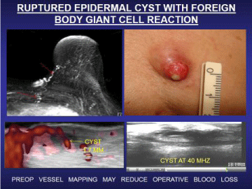
Figure 1
Vessel mapping preoperative.
Figure 2
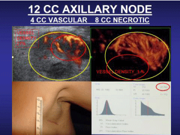
Figure 2
Mixed solid/necrotic axillary lymph node-vessel density allows
targeting of active.
Lymph Node Disease
Lymph node assessment is possible at the same time. Under sonographic guidance, biopsies may be obtained. Sonographic criteria for malignancy are published elsewhere. Image guidance of enlarged node can distinguish between active tumor and necrotic areas diminishing necessity of repeated aspirations for indeterminate findings. (Figure 2) Pathologic assessment of a large postoperative specimen may be facilitated by high frequency scanning to relocalize the suspect region for targeted study that has been removed from its previous anatomic position [6].
Image Guided Biopsy and Treatment
New computer programs use nanotechnology and cybernetic modalities for accurate image guided biopsy and treatment options. Lesions as small as 3 mm have been successfully imaged at frequencies above 14 MHz. Employing 3D sonography with Doppler, the physician manually targets the area of highest tumor neovascularity. This is critical since only part of a mass may be cancerous and missed on non targeted punch biopsies. The marriage with fusion of MRI with ultrasound permits image guided biopsies that spare the adjacent vascular channels. The same technology allows customized ultrasound or MRI guided biopsies to be performed under local anesthesia. Immediate cytologic confirmation of tumor cells permits the withdrawal of the biopsy needle and insertion of a LASER fiber or cryogenic probe immediately treating the proven tumor. MRI thermocouple sensors prevent overheating of the adjacent nerves and sensitive tissues. Following ablation, the zone of destruction is confirmed with Doppler, contrast ultrasound or DCE-MRI. Inflammatory lesions that are deeply seated may be approached by robotic image guided subdermal injections or targeted biopsies if necessary. This outpatient procedure allows the patient to return to work immediately. RF thermoprobes with temperature auto cutoffs prevent thermal skin damage [7]. Similar user friendly and cost effective modalities may replace other therapies in the near future. At the 2016 AMERICAN SOCIETY OF LASERS IN MEDICINE meeting in Boston, cutaneous melanoma with intransit metastases was successively treated by laser technologies.
Conclusion
3D sonography and power Doppler angiography are techniques that contribute new morphologic parameters and noninvasive functional tumoral angiogenic markers for evaluation and treatment follow up of hyperplastic diseases and cancers. The addition of in vivo real time volume density measurements of tumor neovascularity may prove to be of value in determining the risk of tumor recurrence noninvasively. The widespread availability of point-of-care sonography units means a cancer survivor with a new lump can have certainty that it is indeed a benign lipoma or cyst which be readily differentiated from a metastatic node or recurrent cancer. The more oncologists, dermatologists and plastic surgeons use the imaging capacities of sonography, the more they will find its attributes essential to the modern practice of skin disorder therapies.
References
- Bard R. Vascular imaging of cancer in the dense breast. 60th Journees Francaises de Radiologie 2012, Paris.
- Bard R. Image guided cancer treatment. Advances in Medical and Surgical Dermatology, 15th Annual Mt Sinai Winter Symposium New York 2015.
- Merce L, Alcazar J, Lopez C, Iglesias E, Alvarez de los Heros J, Bau S et al. Clinical usefulness of 3 Dimensional sonography and power Doppler angiography for the diagnosis of endometrial carcinoma. J Ultrasound Med. 2007; 26:1279-1289.
- Mitterberger M. Comparison of contrast enhanced ultrasound targeted biopsy. J Urol. 2007; 178; 464-472
- Jing M, Wen C, Zi L, Ying L, Wang QC, Tian JW et al. Early evaluation of relative changes in tumor stiffness by elastography predicts response. J Ultrasound Med. 2016; 35:1619-1627.
- Obek C. Core needle biopsy length affects grading and staging. J Urol. 2012; 187: 2051-2055.
- Bard R, Futterer J, Sperling (Eds) Image Guided Prostate Cancer Treatment. 2014 Springer Berlin.