Case Report
Resection of a Rare Hepatic Myxoid Liposarcoma: Case Report
Fadi Rayya*
Department of General Surgery, University Hospital Damascus, Syria
*Corresponding author: Fadi Rayya, Department of General Surgery, University Hospital Damascus, 17-Nissan Street-Damascus-Syria, P.O.Box: 10769, Syria
Published: 14 Feb, 2017
Cite this article as: Rayya F. Resection of a Rare Hepatic
Myxoid Liposarcoma: Case Report. Clin
Surg. 2017; 2: 1310.
Abstract
Background: Hepatic sarcoma is a rare malignant hepatic tumor with poor prognosis. Complete resection is the only curative therapy.
Case Presentation: We report a case of a 59-year-old male patient, who presented with nine months history of non-specific right hypochondriac pain and increased abdominal size. He was diagnosed with hepatic tumor. Tumor marker levels were within normal limits. Exploratory laparotomy
revealed a 40 cm x 26 cm x 12 cm Hepatic mass. Partial right hepatectomy was performed (Segments VI&VII)
along with right nephrectomy.
Conclusion: The early Diagnosis and Complete resection of Hepatic sarcoma in selected patients is
the only hope for prolonged survival.
Keywords: Myxoidliposarcoma; Partial hepatectomy; Nephrectomy
Introduction
Sarcomas are rare malignant tumors arising from mesenchymal cells. Hepatic Sarcomas represent 0.1% to 2% of primary hepatic cancer [1,2]. The liposarcoma, which is a kind of these sarcomas, was classified according to WHO to differentiated, myxoid, round cell, pleomorphic, mixed-type, liposarcoma, not otherwise specified [3]. We present a case of hepatic myxoid liposarcoma in a previously healthy 59 year-old man, He was admitted to our hospital with a palpable mass in the abdomen.
Case Presentation
A 59-year-old male was presented with a palpable abdominal Mass. The patient complained of increased abdominal size with weight loss (10 Kg) for the last 9 months. Previous to this he was fit and well. He had no Hepatic risk factors such as alcoholism, drug abuse or viral infection. Physical examination revealed distended, firm, and tender abdomen with palpable large mass occupied the right upper quadrant of abdomen, (Figure 1). There were no ascites, edema, or splenomegaly. He had normal blood cell counts. Standard serum liver tests including albumin, prothrombin time, and α-fetoprotein, were within the reference range. Abdominal ultrasound showed a 40x26x12 cm hepatic heterogeneous mass with necrosis and hemorrhage. Enhanced Multi-Slice Computed Tomography (MSCT) of chest, abdomen and pelvis revealed a huge cystic lesion involving the right lobe of the liver and extending from the diaphragm to the pelvis compressing on the intestine, right kidney and inferior vena cava (Figure 2). The clinical, laboratory and radiological findings supported the diagnosis of hepatic sarcoma. No other abnormality was detected in other abdominal viscera or the chest. The patient underwent an explorative laparotomy, which revealed a 40 cm x26 cm x 12 cm cystic lobulated hepatic mass occupying the right abdomen and extending from the diaphragm to the pelvis and compressing on the right colon, duodenum, small intestine, inferior vena cava and right kidney (Figure 3). Tumor resection with right nephrectomy and lateral partial right hepatectomy (LSg VI&VII) was performed. The macroscopic examination of specimen showed multilobular mass (Figure 4). The postoperative period was uneventful. The patient received no adjuvant chemotherapy and he was discharged home on the7th postoperative day. The patient has been followed for the last three months in the outpatient clinic and so far there is no evidence clinically or radio logically for any recurrence. Microscopic examination revealed that the tumor is mostly composed of edematous myxoid stroma that includes scattered large atypical cells having pleomorphic nuclei with coarse chromatin. A prominent vasculature is seen in the background, which composed of thinwalled crossed small vessels, giving chicken-wire appearance; with numerous large vessels with thick muscular walls (Figure 5). Areas of hypercellularity, adipocytic differentiation, and necrosis are also seen (Figure 6). A thin rim of compressed hepatic tissue is present at the periphery. The histologic diagnosis was hepatic myxoid liposarcoma.
Figure 1
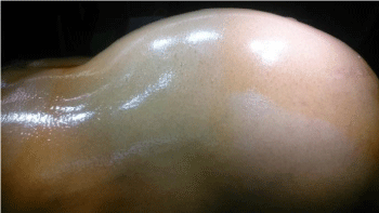
Figure 1
Abdomen of the patient.
Figure 2
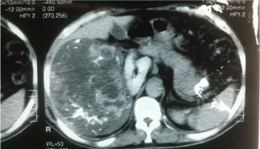
Figure 2
Abdomen MCT shows a large tumor.
Figure 3
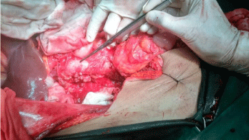
Figure 3
Intraoperative view after tumor resection shows the VCI (forceps).
Figure 4
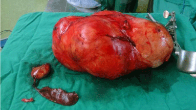
Figure 4
Shows the large mass measuring 40 cm x 26 cm x 12 cm, having smooth
outer surface, covered by an incomplete thin capsule, with right kidney and
LSg VI&VII.
Figure 5
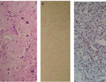
Figure 5
nuclear pleomorphism and atypia; b: tumor necrosis; c: high
MIB1 nuclear rate.
Figure 6
Figure 6
Myxoid area with chicken-wire vascular pattern (left side), area
of hypercellularity (middle), and loose edematous area (right). (H&E x100).
Discussion
Although liposarcoma is one of the most common soft tissue sarcomas in adults, primary hepatic liposarcoma is very rare. Wolloch, who described the first hepatic sarcoma in 1973 [4] reviewed 16 cases of malignant hepatic tumors, including one patient with myxoid liposarcoma who had undergone right hepatic lobectomy. This patient died 46 days postoperatively. Kim et al. [5] described a 14 X 10 X 5-cm liposarcoma of the right lobe of the liver. The patient subsequently underwent a hepatic resection and remained tumor free for 10 months. According to this literature, most patients presented with nonspecific symptoms, such as abdominal discomfort, pain, abdomen mass or weight loss [4-14]. Most of the symptoms are caused either by displacement or compression of nerves, vessels, biliary tract, and intestinal structure. Our case had abdominal mass with weight loss. Because of their various histological compositions, including areas of necrosis and hemorrhage, sarcomas present inconsistently in computed tomographic scan as well as in magnetic resonance and ultrasonography. In spite of constantly improving imaging techniques, it is still a challenge to differentiate sarcomas from other liver diseases like hepatic cysts, hemangiomas, or metastases [15,18]. Common tumor markers (such as Alfa- fetoprotein AFP, carcinoembryonic antigen CEA, cancer antigen 19-9 CA 19-9) are not elevated in patients with sarcoma. Primary liver liposarcoma should be considered in the differential diagnosis, especially in those patients who are potential candidates for hepatic resection. Liposarcoma is an absolute contraindication for hepatic transplantation. Curative and aggressive hepatectomies for this group of patients are still the best policy to achieve a long-term survival [4-14,16,17,19].
Table 1

Table 1
Characteristics of Previously Reported Cases of Primary Liposarcoma of the Liver in adults.
References
- Liver Cancer Study Group of Japan. Primary liver cancer in Japan: clinicopathologic features and results of surgical treatment. Ann Surg. 1990; 211: 277-287.
- Carriaga MT, Henson DE. Liver, gallbladder, extra hepatic bile ducts, and pancreas. Cancer. 1995; 75: 171-190.
- Fletcher CDM, Unni K, Mertens F. World Health Organization Classification of Tumours: Pathology & Genetics of Tumours of the Soft Tissues and Bones. Lyon: IARC Press. 2002.
- Wolloch Y, Dintsman M, Garti I. Primary malignant tumors of the liver. Isr J Med Sci. 1973; 9: 6-11.
- Kim TW, Reyes CV. Myxoid liposarcoma mimicking fluid density. J Surg Oncol. 1985; 30: 80-82.
- Kim YI, Yu ES, Lee KW, Park EU, Song HG. Dedifferentiated liposarcoma of the liver. Cancer. 1987; 60: 2785-2790.
- Golebiowski L, Abycht K. Primary liposarcoma of the liver. Wiad Lek. 1987; 40: 1064-1066.
- Aribal E, Berberoglu L. Primary liposarcoma of the liver. AJR Am J Roentgenol 1993; 161: 1331-1332.
- Nelson V, Fernandes NF, Woolf GM, Geller SA, Petrovic LM. Primary liposarcoma of the liver: a case report and review of literature. Arch Pathol Lab Med. 2001; 125: 410-412.
- Kuo LM, Chou HS, Chan KM, Yu MC, Lee WC. A case of huge primary Liposarcoma in the liver. World J Gastroenterol. 2006; 12: 1157-1159.
- Gajda M. Prim_res Liposarkom der. Z Gastroenterol. 2007; 45: 1241-1244.
- Nakhai B, Motabar AR. “Primary liposarcoma of the liver: a case report and review of literature.” Med J Islam Repub Iran. 2007; 21: 167-172.
- Fariba Binesh, Ali Akhavan, Saeed Kargar, Hossein Navabii. Primary liposarcoma of liver: a rare case and literature review. BMJ Case Rep. 2012.
Naik PRT, Kumar P, Kumar PV. Primary Pleomorphic Liposarcoma of Liver: A Case Report and Review of the Literature. Case Reports Hepatol. 2013; 2013: 398910.
- Murphey MD, Arcara LK, Fanburg-Smith J. From the archives of the AFIP: imaging of musculoskeletalliposarcoma with radiologic-pathologic correlation. Radiographics. 2005; 25: 1371-1395.
- Forbes A, Portmann B, Johnson P, Williams R. Hepatic sarcomas in adults: a review of 25 cases. Gut. 1987; 28: 668-674.
- Poggio JL, Nagorney DM, Nascimento AG, Kay P, Young RM, Donohue JH, et al. Surgical treatment of adult primary hepatic sarcoma. Br J Surg. 2000; 87: 1500-1505.
- Koyama T, Fletcher JG, Johnson CD, Kuo MS, Notohara K, Burgart LJ. Primary hepatic angiosarcoma: findings at CT and MR imaging. Radiology. 2002; 222: 667-673.
- Hanno Matthaei, Andreas Krieg, Moritz Schmelzle, Rogiers X, Knoefel WT, Peiper M, et al. Long-term Survival After Surgery for Primary Hepatic Sarcoma in Adults. Arch Surg. 2009; 144: 339-344.