Case Report
A Rare Cause of Intestinal Obstruction in a Male Patient: Abdominal Cocoon, Case Report
Mehmet KUBA1, Bahadir O BOZKIRLI1*, Berkay KÜÇÜK1, Mehmet Ali AKKUŞ1, Kerim TEMİZ2 and Hasan YİĞİT2
1Department of General Surgery, Ankara Training and Research Hospital, Turkey
2Department of Radiology, Ankara Training and Research Hospital, Turkey
*Corresponding author: Bahadir Osman Bozkiri, Department of General Surgery, Ankara Training and Research Hospital, Ankara, Turkey
Published: 09 Feb, 2017
Cite this article as: KUBA M, BOZKIRLI BO, KÜÇÜK B, Ali
AKKUŞ M, TEMİZ K, YİĞİT H. A Rare
Cause of Intestinal Obstruction in a
Male Patient: Abdominal Cocoon, Case
Report. Clin Surg. 2017; 2: 1295.
Abstract
Sclerosing encapsulating peritonitis (SEP) is a rare cause of intestinal obstruction which can be classified into a primary and a secondary form. The recommendation for mild cases is conservative treatment, surgical intervention reserved for the patients with severe symptoms of obstruction. The advised surgical approach is the excision of the membrane and adhesiolysis when there are no contraindications for this procedure. Here we present the case of a 58 year old male patient with SEP who was hospitalized for incomplete intestinal obstruction with a plan of conservative treatment, but had to be operated due to the situation progressing into total obstruction. In the present case, adhesiolysis and the excision of the dense and almost totally calcified fibrous capsule that tightly adhered to the viscera was not possible. Conservative treatment was continued after the operation and the intestinal obstruction resolved spontaneously in the postoperative period. Depending on the presented experience, we suggest aggressive resection and adhesiolysis may be abandoned in the absence of intestinal necrosis or perforation related to SEP, if considered to be too risky and the patient must be given another chance for spontaneous recovery.
Introduction
Sclerosing encapsulating peritonitis is a rare cause of intestinal obstruction [1]. First described by Foo et al. [2] in 1978 and named as “abdominal cocoon”, this syndrome is characterised by the encapsulation of the bowel by a fibrotic sac [1] and it has a primary (idiopathic) and a secondary form [3]. Idiopathic sclerosing encapsulating peritonitis is classically seen in adolescent female patients from tropical countries and it is the less frequently encountered form of this entity [1,4]. The secondary form, although mainly encountered as a feared complication of peritoneal dialysis, may also result from various other conditions that cause chronic peritoneal inflammation such as abdominal tuberculosis, cirrhotic ascites, peritoneal carcinomatosis, severe pancreatitis, and autoimmune diseases [5]. Here, we present the case of a 58 year old male patient who presented with intestinal obstruction caused by primary sclerosing encapsulating peritonitis.
Case Presentation
A 58 year old male patient presented to the emergency service of our hospital with a history
of six-day colicky abdominal pain and distension that worsened in the last two days. Nausea and
vomiting was accompanying these symptoms in the last day of the patient’s history. He also did not
defecate in the last two days but he had gas discharge. Anamnesis revealed that the patient had several
attacks of abdominal pain and distension in the last ten years and was hospitalised twice in this
period, his symptoms resolving spontaneously without requiring surgical intervention. The patient
also did not have a history of any other previous abdominal operations. He was not regularly using
any medications. In physical examination, the patient was hemodynamically stable. Abdominal
examination revealed distention, hyperactive bowel sounds and tenderness without rigidity or
rebound tenderness. There were no specific alterations in the laboratory tests. Abdominal X-ray
showed a few central air-fluid levels with no finding of free subdiaphragmatic air. On abdominal
ultrasound, there were not any abnormal findings except multiple dilated intestinal segments.
The patient was hospitalised for conservative treatment with an initial diagnosis of partial
intestinal obstruction. However, after medical treatment and nasogastric decompression for five
days, the symptoms of the patient worsened. An abdominal computerised tomography revealed
centralised intestinal loops encased by a partially calcified sac. These radiologic findings were
interpreted to be the signs of abdominal cocoon and possible internal
herniation (Figure 1). Exploratory laparotomy was performed with
these preoperative diagnoses. In the operation, it was seen that the
abdominal viscera was totally enveloped by a dense and mostly
calcified membrane forming a single cocoon. The operation was
terminated after taking biopsies from the non-calcified sections of
the membrane deciding on the impossibility of the resection of the
membrane without causing major visceral injuries. The conservative
treatment was continued in the postoperative period. Histopathology
revealed dense fibrous tissue with massive collagen deposition.
Intestinal obstruction spontaneously resolved on the second
postoperative day and the patient was discharged from the hospital
six days after the operation. The patient was hospitalised once again
one year later for another episode of intestinal obstruction; however,
this attack also resolved spontaneously with conservative treatment.
The patient was symptom-free on his follow-up at the end of the
second year. He gave his consent for his medical record to be used for
scientific research.
Figure 1
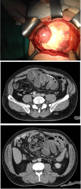
Figure 1
Abdominal cocoon and possible internal herniation.
Discussion
SEP was first documented in 1907 by Owtschinnikow and
termed as the “peritonitis chronica fibrosa incapsulata” [1,6]. Then,
in 1978 Foo et al. [2] reported the primary form of the disease in
ten adolescent female patients naming the condition the abdominal
cocoon. Today, although used as a general synonym of SEP in many
reports, abdominal cocoon is stressed by some researchers to be the
name describing the primary form of the disease [7].
The primary form of SEP, which is classically seen in young
women from tropical countries, was thought to arise from retrograde
menstruation [1]. However, this condition was also encountered in
some children and male patients, contradicting this theory [8]. In fact,
a recent review of the studies published from 2000 to 2014 revealed
that almost two thirds of the patients diagnosed with this entity were
males [7]. Therefore, the aetiology of this condition is still not clear.
In the patient presented in this report, there was no apparent reason
explaining the presence of SPE except a doubtful exposure to asbestos
25 years ago which could not be documented. Thus, we believe the
patient to have the primary form of SPE.
Since it is rarely seen and the symptoms are nonspecific, the
diagnosis of the situation is difficult in the preoperative period [9].
Contrast enhanced abdominal CT with multiplanar reconstructing
images, should be the preferred imaging technique [3]. Using this
method, it is possible to see the peritoneal thickening, signs of
intestinal obstruction, clustering, and fixation of the intestinal loops
[3]. In the present case, SEP was suspected preoperatively thanks
to such preoperative radiologic evaluation. Unfortunately, the
preoperative diagnosis of the entity did not have an influence on the
operative success in the present case because of the dense and almost
totally calcified fibrous capsule that tightly adhered to the large and
small intestine it was encapsulating, preventing any adhesiolysis
without intestinal injury. In this case we preferred not to take the risk
of organ injury and contamination of the abdomen, which could lead
to septic complications or the necessity to create an ostomy that could
result in short bowel syndrome if it had to be created on a proximal
segment. Also, intestinal resections are known to increase morbidity
and mortality in SEP cases [7].
Terminating the operation led to a more favourable outcome
in the present case, since the patient recovered with conservative
treatment in the postoperative period with gas and faecal discharge
followed by regression of abdominal distension and pain.
The recommendation for the cases of SEP with mild symptoms is
conservative therapy, surgical intervention reserved for the patients
with severe signs of intestinal obstruction [7]. In the present case,
although the patient did not develop acute abdomen during the course
of the disease, the incomplete obstruction progressed into a complete
one. Thus, the decision for an exploratory laparotomy is justified in
the present case. In the literature, the advised surgical approach is
the excision of the membrane and adhesiolysis when there are no
contraindications for this procedure [7]. Depending on the presented
experience, we suggest aggressive resection and adhesiolysis may be
abandoned in the absence of intestinal necrosis or perforation related
to SEP, if considered to be too risky and the patient must be given
another chance for spontaneous recovery.
References
- Solak A, Solak I. Abdominal cocoon syndrome: preoperative diagnostic criteria, good clinical outcome with medical treatment and review of the literature. Turk J Gastroenterol. 2012; 23: 776-779.
- Foo KT, Ng KC, Rauff A, Foong WC, Sinniah R. Unusual small intestinal obstruction in adolescent girls: the abdominal cocoon. Br J Surg. 1978; 65: 427-430.
- Karan A, Özdemir M, Bostancı MT, Bostancı EB. Idiopathic abdominal cocoon syndrome: Preoperative diagnosis with computed tomography. Turk J Gastroenterol. 2015; 26: 193-194.
- Oran E, Seyit H, Besleyici C, Ünsal A, Alış H. Encapsulating peritoneal sclerosis as a late complication of peritoneal dialysis. Ann Med Surg (Lond). 2015; 4: 205-207.
- Robert B, Hanes A. Abdominal cocoon, an uncommon cause of intestinal obstruction. Presse Med. 2015; 44: 352-354.
- Sohail MZ, Hasan S, Dala-Ali B, Ali S, Hashmi MA. Multiple abdominal cocoons: an unusual presentation of intestinal obstruction and a diagnostic dilemma. Case Rep Surg. 2015; 2015: 282368.
- Akbulut S. Accurate definition and management of idiopatic sclerosing encapsulating peritonitis. World J Gastroenterol. 2015; 21: 675-687
- Shah MY, Gedam BS, Sonarkar R, Gopinath KS. Abdominal Cocoon: An Unusual Cause of Subacute Intestinal Obstruction. Indian J Surg. 2013; 75: S391-393.
- Uzunoglu Y, Altintoprak F, Yalkin O, Gunduz y, Çakmak G, Ozkan OV, et al. Rare etiology of mechanical intestinal obstruction: Abdominal cocoon syndrome. World J Clin Cases. 2014; 2: 728-731.