Case Report
Pyogenic Granuloma or?? Beyond the Naked Eye: Merkel Cell Carcinoma; Case Report and Literature Review
Charandeep Singh* and Fuad Aftab
Department of General Surgery, Mallow General Hospital, Ireland
*Corresponding author: Charandeep Singh, Department of General Surgery, Mallow General Hospital, Cork, Ireland
Published: 05 Dec, 2016
Cite this article as: Singh C, Aftab F. Pyogenic Granuloma
or?? Beyond the Naked Eye: Merkel
Cell Carcinoma; Case Report and
Literature Review. Clin Surg. 2016; 1:
1206.
Abstract
Introduction: Merkel cell carcinoma (MCC) is a rare and highly aggressive primary cutaneous
neuroendocrine carcinoma, most often occurring at an elderly age group. Recurrence is frequent
and in 40% of cases regional and distant metastases develop. We report the case of excision of MCC
in a 85-year-old woman followed by wide excision and sentinel lymph node biopsy.
Case Presentation: An 85-year-old woman presented to surgical out-patient clinic with a firm fleshy,
protuberant lump on the superior margin of right zygomatic area of face measuring 20 x 20mm,
approximately. She was under investigation for Iron Deficiency Anemia along with high WBC. The
lesion was excised under local anesthesia and immunohistochemistry confirmed diagnosis of MCC.
Upon histological confirmation of diagnosis, the patient underwent further staging, to exclude
regional and distant metastasis, including CT scan of the neck and TAP. Further treatment included
wide local excision and sentinel lymph node mapping.
Discussion: MCC is rare carcinoma with very few known causes. Immune suppression is proposed
as an etiological factor. Surgery has a major role with radiotherapy and chemotherapy playing their
roles for palliation.
Conclusion: MCC is very aggressive cutaneous carcinoma with poor prognosis and high mortality
rate, with life expectancy of 8-10 months from the time of distant diagnosis. Radiotherapy has a role
but surgery still is the Gold Standard. Due to rarity of this disease, we feel that these patients need to
be under strict follow-up program.
Introduction
Merkel cell carcinoma (MCC) is an uncommon highly aggressive skin malignancy that
originates from the neuroendocrine and mechanoreceptor “Merkel Cells” located in the basal layer
of epidermis and neurosecretory granules [1]. It is most frequently found on skin-damaged and
sun-exposed areas particularly face, head, and neck (55%), followed by extremities (40%) and lastly
followed by truncal structures (5%) [2]. Due to its rarity and early asymptomatic clinical course,
diagnosis of MCC is fairly challenging, often delayed, or even missed [3]. Clinical features of MCC
are summed up by the acronym AEIOU (Table 1) [4].
Although, aetiology is not fully understood, there are several risk factors that are proposed
towards its pathogenesis, including UV light, sun-related skin malignancies (Squamous and
Basal Cell Carcinoma), psoriasis treatment with methoxsalen and arsenic exposure, patients on
immunosuppressive agents or patients with AIDS, chronic lymphocytic leukemia, congenital
dysplasia syndrome and organ recipients [6-11]. Most cases present as localized disease (70%–
80%) followed by regional lymph node involvement (9%–26%) and distant metastasis (1%–4%)
[9] to lung, liver, brain, bone, and skin [5]. These characteristics often raise the suspicion of a skin malignancy but confirmation of diagnosis is by excisional biopsy. The differential diagnosis
of MCC from other small cells neoplasms can be difficult, even on histological examination [11].
For a definitive diagnosis, electron microscopy is necessary [6]. Management and prognosis of
MCC is largely dependent on tumor staging at the time of presentation. Management modalities
include utilization of surgical excision with safe margins, lymphadenectomy, radiotherapy, and
chemotherapy [3]. Generally, prognosis of MCC is extremely poor with a high mortality rate [2]. Merkel Cell Carcinoma Staging (Table 2) [11].
Case Presentation
An 85-year-old Caucasian female patient was referred by general practitioner (GP) to surgical
outpatients (SOPD) at our hospital for further management of an
eight-week history of a skin lesion on right cheek. Past medical history
included hypertension, hypercholesterolemia, osteo-arthritis, peptic
ulcer disease (PUD), and diverticular disease. Patient was also under
investigation for Iron deficiency anemia and slightly raised white cell
count (WCC; 12-14.5) by bone marrow biopsy. Past surgical history
included appendectomy and right total hip replacement (THR).
Blood biochemistries (renal profile, liver profile, CEA, CA 12.5) were
normal. On physical examination, on the superior margin of right
zygomatic area around 2 x 2 cm, red coloured skin lesion was noted.
It was non-tender, firm, not fixed to underlying tissue, and with no
surrounding skin changes. Initial diagnosis was made a cystic lesion
of the right cheek or a pyogenic granuloma. She was admitted as a
day-case for excision of a relatively innocuous appearing skin lesion
under local anesthesia. Histology report revealed presence of MCC
extending into subcutaneous fat, Clarke level V and pathological
Staging of pT2 and Immunohistochemistry confirmed the above
diagnosis. Staging CT neck and TAP was negative for regional and
distant metastasis. Due to high incidence of regional disease, urgent
mole mapping was arranged which marked a sentinel lymph node at
right cervical area (Figure 1).
Same day wide re-excision of scar tissue was performed with
excision of cervical sentinel and one non-sentinel lymph node,
utilizing both the gamma probe and blue dye technique. Both the
sentinel and non sentinel lymph nodes were in the level 3 drainage
area of the neck in close proximity to the internal jugular vein (Figure
2 and 3).
The histology confirmed absence of disease in the re-excised skin
sample and also both sentinel and non sentinel lymph node specimens.
Patient was discharged home the same day and is currently awaiting
radiotherapy to the cheek.
Table 1

Table 1
Clinical features of MCC [4].
Discussion
Merkel Cell Carcinoma (MCC) is a rare and aggressive primary
anaplastic undifferentiated neuroendocrine carcinoma of the skin
[13] arising from uncontrolled proliferation of the neuroendocrine
and mechanoreceptor Merkel Cells located in the epidermis of skin
with an overall unfavorable prognosis [12]. It generally occurs in
elderly patients (age >50 years). It generally affects both sexes of fair
skin population but with a male predominance [14]. Areas of skin
mostly exposed to sun-light are at higher risk such as head and neck
(50%), upper and lower limbs (35%–40%) and less than 10% in the
trunk [3].
Due to its rarity, the aetiology of this carcinoma still remains
unclear. However, immune suppression i.e., HIV patients and
transplant recipients with 50% cases in age< 50 years) [17], ultraviolet
radiation (high sun-exposed and /or psoriasis patients treated with
UV light) [15] and Merkel Cell Polyomavirus (MCV) being put
forward as aetiological factors. The latter is a double stranded DNA
virus, which causes uncontrollable mitosis. It is present in about
80% of MCC cases [16]. But still there is a debate as there is lack of
sufficient data and evidence for the above mentioned etiology. There
are reports of MCV positive with MCC negative in patients. 20%
MCC cases are negative with MCV.
MCC is occasionally mistaken for other cutaneous neoplasms,
such as a cystic lesion, malignant melanoma, or lymphoma [19].
Electron Microscopy is ideal to exclude these lesions which also
feature intra-cytoplasmic neurosecretory/neuroendocrine granules
[1,4]. As it is a rare type of skin tumor, with a few reported cases,
there is no definite treatment protocol for such aggressive carcinoma.
Surgical excision remains the gold standard with clear margins
of 2-3 centimeters been accepted. Therapeutic modalities such as
chemotherapy, radiotherapy, are also an option, but it is dependent
on the stage of the disease [4]. A few studies have shown no significant
difference between surgeries alone vs. postoperative radiation but it
would be beneficial in cases where free surgical margins are hard to
obtain due to cosmetic reasons [11].
In case of nodal involvement, therapeutic lymphadenectomy
and postoperative radiotherapy must be considered. Due to this high
incidence of metastasis, prophylactic lymphadenectomy is advocated
in order to improve outcome [24-25]. However, prophylactic
lymphadenectomy is associated with high morbidity. In order to avoid
this, sentinel node status was evaluated and a sentinel node biopsy
was performed for diagnosis, management and further treatment
[11,22,23]. Presence of regional lymph node involvement markedly
decreases overall survival from 90% to 50%, a frequent occurrence in
50%-70% of all patients within 24 months from the time of clinical
diagnosis [20].
Regional metastases are very common followed by distant
metastases (lung, brain, liver). Treatment of MCC with distal
metastases consists of palliative radiotherapy and chemotherapy.
Overall prognosis of this disease is very poor. Mortality ensues within8 to 10 months from the time of diagnosis of metastatic disease.
Several studies have not shown a favorable response of radiotherapy
and chemo therapy on metastatic disease [21].
Table 2
Table 2
Merkel Cell Carcinoma Staging [11].
Figure 1
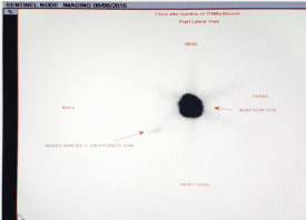
Figure 1
Sentinel lymph node (right cervical area) mapping.
Figure 2
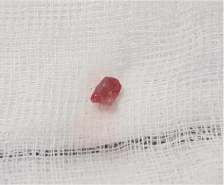
Figure 2
Sentinel lymph node.
Figure 3
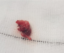
Figure 3
Scar tissue with irregular skin lesion..
Conclusion
Merkel cell carcinoma (MCC) is a very aggressive carcinoma. Therefore, in the elderly (above 50 years old) MCC must be out ruled. A detailed history, including a relatively short growth phase of a rapidly expanding, red-bluish color skin tumor, in an immunocompromised patient, with sun exposure should be taken into account. If wide excision is not possible, then biopsy is essential for diagnosis and subsequent management. Therapeutic options vary depending on the stage of the disease. Surgery remains the gold-standard procedure with 2-3 cm of clear margins. Staging includes Computerized Axial Tomography (CT scan), Magnetic Resonance Imaging (MRI) are utilized for distant metastases and lymph nodal involvement, respectively. Sentinel Lymph Node Biopsy (SLNB) should always be considered. If lymph nodes are involved, then lymphadenectomy is the procedure of choice, followed by radiotherapy/chemo therapy. The role of radiotherapy in distant metastases remains unclear. MCC has high tendency to recur locally (27%-60%), followed by lymph node involvement (45%-91%), and metastases 20%-90%). Although, less frequent than melanoma, it compromises of 0.25% of all melanomas, the mortality is 1 in 3 as compared to 1 in 6 in melanomas. Due to its poor prognosis patient survival period is 8-10 months. Therefore, we propose once diagnosed, the patient should be under strict follow-up program. Immune suppression is proposed as an aetiological factor. We further propose that immune suppression may be considered as a promoter rather as an aetiological factor.
References
- Gollard R, Weber R, Kosty MP, Greenway HT, Massullo V, Humberson C. Merkel cell carcinoma review of 22 cases with surgical, pathologic, and therapeutic considerations. Cancer. 2000; 88: 1842-1851.
- Rockville Merkel cell Carcinoma Group. Merkel cell carcinoma: recent progress and current priorities on etiology, pathogenesis, and clinical management. J Clin Oncol. 2009; 27: 4021-4026.
- Pectasides D, Pectasides M, Economopoulos T. Merkel cell cancer of the skin. Annals of Oncology. 2006; 17: 1489-1495.
- Heath M, Jaimes N, Lemos B, Mostaghimi A, Wang LC, Peñas PF, et al. Clinical characteristics of Merkel cell carcinoma at diagnosis in 195 patients: the AEIOU features. J Am Acad Dermatol. 2008; 58: 375-381.
- Peloschek P, Novotny C, Mueller-Mang C, Weber M, Sailer J, Dawid M, et al. Diagnostic imaging in Merkel cell carcinoma: lessons to learn from 16 cases with correlation of sonography, CT, MRI and PET. Eur J Radiol. 2010; 73: 317-323.
- Payne MM, Rader AE, McCarthy DM, Rodgers WH. Merkel cell carcinoma in a malignant pleural effusion: case report. Cytojournal. 2004; 18: 5-10.
- Koljonen V. Merkel cell carcinoma. World J Surg Oncol. 2006; 4: 7-10.
- Dinh V, Feun L, Elgart G, Savaraj N. Merkel cell carcinomas. Hematol Oncol Clin North Am. 2007; 21: 527-544.
- Veness MJ. Merkel cell carcinoma (primary cutaneous neuroendocrine carcinoma): an overview on management. Australas J Dermatol. 2006; 47: 160-165.5
- Boyse K, Foley EH, Bradley V, Scarborough D. Merkel cell carcinoma: a case report with treatment summary and updates. Cutis. 2004; 74: 350
- Bichakjian CK, Lowe L, Lao CD, Sandler HM, Bradford CR, Johnson TM, et al. Merkel cell carcinoma: critical review with guidelines for multidisciplinary management. Cancer. 2007; 110: 1-12.
- Rosai J. Rosai and Ackerman's Surgical Pathology. Mosby. 2004; 1: 177- 179.
- Haag ML, Glass LF, Fenske NA. Merkel cell carcinoma. Diagnosis and treatment. Dermatol Surg. 1995; 21: 669-683.
- McAfee WJ, Morris CG, Mendenhall CM, Werning JW, Mendenhall NP, Mendenhall WM. Merkel cell carcinoma: treatment and outcomes. Cancer. 2005; 104: 1761-1764..
- Fernández-Figueras MT, Puig L, Musulén E, Gilaberte M, Lerma E, Serrano S, et al., “Expression profiles associated with aggressive behavior in Merkelcell carcinoma. Mod Pathol. 2007; 20: 90-101.
- Feng H, Shuda M, Chang Y, Moore PS. Clonal integration of a polyomavirus in human Merkel cell carcinoma. Science. 2008; 319: 1096-1100.
- Koljonen V, Kukko H, Tukiainen E, Böhling T, Sankila R, Pukkala E, et al. Incidence of Merkel cell carcinoma in renal transplant recipients. Nephrol Dial Transplant. 2009; 24: 3231-3235.
- Swann MH, Yoon J. Merkel cell Carcinoma. Semin Oncol. 2007; 34: 51-56.
- Santos Gorjón P, Morales Martín AC, Blanco Pérez P, Gómez González JL, del Pozo de Dios JC, Romo Melgar A. Merkel cell carcinoma: a presentation of 5 cases and a review of the literature. Acta Otorrinolaringol Esp. 2011; 62: 306-310.
- Mehrany K, Otley CC, Weenig RH, Phillips PK, Roenigk RK, Nguyen TH. A meta-analysis of the prognostic significance of sentinel lymph node status in Merkel cell carcinoma. Dermatol Surg. 2002; 28: 113-117.
- Desch L, Kunstfeld R. Merkel cell carcinoma: chemotherapy and emerging new therapeutic options. J Skin Cancer. 2013; 9.
- Koljonen V. Merkel cell carcinoma. World J Surg Oncol. 2006; 4: 7-10.
- Mehrany K, Otley CC, Weenig RH, Phillips PK, Roenigk RK, Nguyen TH. A meta-analysis of the prognostic significance of sentinel lymph node status in Merkel cell carcinoma. Dermatol Surg. 2002; 28: 113-117.
- National Comprehensive Cancer Network. Clinical Practice Guidelines in Oncology. (Online) 2004.
- National cancer Institute. Merkell cell carcinoma treatment. (Online) 2007.