Case Report
Metachronous Bladder Metastasis from Clear Cell Renal Cell Carcinoma
Terence Chun-ting Lai and Ming-kwong Yiu*
Department of Surgery, Division of Urology, University of Hong Kong, China
*Corresponding author: Ming-kwong Yiu, Department of Surgery, Division of Urology, University of Hong Kong, 102 Pokfulam Road, Hong Kong SAR, China
Published: 28 Nov, 2016
Cite this article as: Chun-ting Lai T, Yiu M-k. Metachronous
Bladder Metastasis from Clear Cell
Renal Cell Carcinoma. Clin Surg. 2016;
1: 1178.
Abstract
Bladder metastasis, either synchronous or metachronous, is an uncommon presentation of renal
cell carcinoma. We herein report a metachronous bladder metastasis from renal cell carcinoma of
clear cell type, and review the current literature on the presentation, management and prognosis
of this kind of metastasis. A 77-year-old man was presented to Queen Mary Hospital, Hong Kong
in 2011 with hematuria, subsequently found a renal tumor invading into renal pelvis. Open right
radical nephrectomy was performed and the tumor was confirmed to be pT1b clear cell carcinoma
of Fuhrman grade 2. He remained asymptomatic all along after nephrectomy, but surveillance
ultrasonography (USG) revealed a 2.2 cm bladder neck mass in 23 months after nephrectomy.
After patient refused transurethral resection, the mass further enlarged to 3.8cm one year later.
Transurethral resection was performed and histopathological examination confirmed clear cell
carcinoma, with no fibro-muscular invasion. Multiple pulmonary metastases were also found on
computed tomography (CT) of the thorax. He refused target therapy and preferred conservative
treatment. He remained symptom-free 2 months after the transurethral resection.
Keywords: Bladder metastasis; Renal cell carcinoma; Ultrasonography
Introduction
It is uncommon for renal cell carcinoma to metastasize to bladder. In this study we presents a man with pT1b clear cell renal cell carcinoma, treated with right radical nephrectomy in 2011, and developed bladder metastasis 23 months after nephrectomy. He has been asymptomatic since the nephrectomy. Literature of on the presentation, treatment options and the prognosis of this kind of bladder metastasis is reviewed.
Case Presentation
A 77-year-old man was first presented to the Urology Division of Surgical Department, Queen
Mary Hospital, Hong Kong in 2011 for hematuria. He had hypertension, diabetes mellitus and preexisting
chronic renal disease, with glomerular filtration rate (GFR) of less than 20 ml/min/1.73 m.
Ultrasonography (USG) of the kidneys revealed a 4.7 x 5.2 x 5.9 cm heterogeneous soft tissue mass in
right renal pelvis, distorting the normal renal architecture, with intra-lesional colour Doppler flow
signals. Flexible ureterorenoscopy found a soft and sludge-like mass within renal pelvis, and biopsy
returned clear cell renal cell carcinoma. The renal mass had low-grade F18-fluorodeoxyglucose
(FDG) uptake on positron emission tomography - computed tomography (PET-CT), and no distal
metastases were found.
He subsequently underwent open right radical nephrectomy in May 2012. Final histopathological
examination of the specimen confirmed pT1b clear cell carcinoma of Fuhrman Grade 2, and the
resection margin was clear. His renal function remained stable after operation and did not require
dialysis.
He was put on regular surveillance with USG of urinary system and chest radiography (CXR).
A new 1.3 x 2 x 2.2 cm soft tissue mass was found at the bladder base on USG in April 2015, but
he refused either transurethral resection (TUR) or flexible cystoscopy (FC) as he did not have any
obstructive symptoms or hematuria. USG was repeated in April 2016, and the mass had enlarged to
3.8 x 3.7 x 2.8 cm (Figure 1). In addition, CXR taken in April 2016, showed a new 1.5 cm nodule at
right mid-zone; CT thorax confirmed multiple pulmonary nodules, measuring up to 2.1 x 1.4 cm
He finally agreed to TUR of the bladder mass, which was performed in July 2016. Intra-operatively
a 4cm soft sessile mass was noted at 7-8 O’clock of bladder neck; complete resection was achieved.
On histopathological examination, the tumour consisted of nests of
round to polygonal cells associated with rich vascular network; the
cells had distinct cell borders and possessed clear cytoplasm (Figure
2a and 2b); there was no fibro muscular invasion. Findings were
compatible with clear cell renal cell carcinoma.
Option of target therapy with tyrosine kinase inhibitor
(TKI), panzopanib, was offered by the clinical oncologist, and he
decided not for any systemic treatment. He had no symptoms, for
instance hematuria or dyspnea, from the metastases 2 months after
transurethral resection.
Figure 1
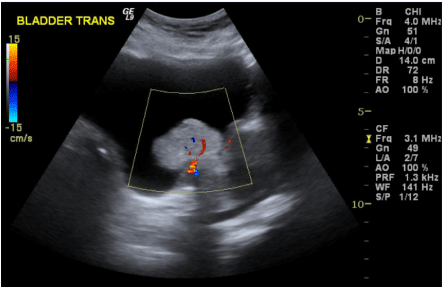
Figure 1
3.8 x 3.7 x 2.8cm mass at bladder base, with intra-lesional Doppler flow signals on USG.
Figure 2
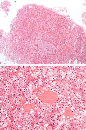
Figure 2
2a & 2b: Hematoxylin and eosin (H&E) stained sections, taken at 40x (Figure 2a) and 200x (Figure 2b) magnification respectively. The tumor consists of nests of round to polygonal cells associated with a rich vascular network. The cells have distinct cell borders and possess clear cytoplasm. Their nuclei are mostly round and hyperchromatic, some with distinct nucleoli.
Discussion
Bladder is an uncommon site of metastasis for renal cell carcinoma.
1,451 autopsy cases of renal cell carcinoma only revealed 1 case of
isolated bladder metastasis, and 23 (2%) multi-organ metastases
with bladder involvement [1]. The mode of spread to bladder is still
uncertain. Some postulated tumour spread via hematogenous route
from systemic circulation or retrograde metastasis through the
gonadal vein, from the left renal vein, to the bladder [2]. Retrograde
tumour dissemination by the lymphatic system is also possible.
Another theory is antegrade spread of renal tumours to the ureter and
bladder [3], especially for metastasis confined to the bladder mucosa.
In our case we believe an antegrade spread via the urinary tract is
the most possible route, since the initial renal tumour had invaded
into the renal pelvis, which facilitated tumour dissemination into the
collecting system, and the tumour was limited to bladder mucosa
only. Hematogenous spread was another possibility for our case as
the man also developed multiple pulmonary metastases.
Metastases were predominantly of clear cell type in reported
literature. Zhang et al. [4] reported 11 cases of bladder metastases,
and noted long disease-free survival was possible for solitary
metastasis, but prognosis was poor for those with other distal
metastases, with survival ranging from 5 -71 months. Matsumoto et
al. [5] has reviewed another 65 published cases, in which 33 of them
had metachronous metastasis, with median time to metastasis of 33
months (1-204 months).With a median follow-up time of 15 months,
34% died of renal cell carcinoma. 2 reported cases had survival more
than 5 years after resection of solitary bladder metastasis. They
identified metastasis within 1 year from nephrectomy and presence
of other distal metastases as adverse factors on survival, with hazard
ratio of 2.30 and 3.61, respectively.
Most of the bladder metastases, unlikely our case, were presented
with symptoms, mostly hematuria (75%), flank pain (15%), and fever
(5%). Only 13% were asymptomatic [5].
Due to the scarcity of reported cases, treatment is not
standardized for bladder metastasis; TUR, partial or total cystectomy
have been reported. Nonetheless, if the tumor is resectable, complete
metastectomy should be attempted as resection of metastasis of
other sites has shown improved survival. The 5-year survival after
metastectomy of pulmonary [6] or pancreatic metastasis [7] can be
up to 50% and 75%, respectively.
In conclusion, bladder metastasis from renal cell carcinoma
is rare. Solitary metastasis and latemetachronous metastasis with
complete resection may have good prognosis and survival. However,
majority are accompanied with distal metastases and the prognosis is
generally poor.
References
- Saitoh H. Distant metastasis of renal adenocarcinoma. Cancer. 1981; 48: 1487-1491.
- Abeshouse BS. Metastasis to ureters and urinary bladder from renal carcinoma: report of two cases. J Int Coll Surg. 1956; 25: 117-126.
- Hajdu SI, Savino A, Hajdu EO, Koss LG. Cytologic diagnosis of renal cell carcinoma with the aid of fat stain. Acta Cytol. 1971; 15: 31-33.
- Zhang M, Wah C, Epstein JI. Metastatic renal cell carcinoma to the urinary bladder: a report of 11 cases. Am J Surg Pathol. 2014; 38: 1516-1521.
- Matsumoto K, Hayakawa N, Nakamura S, Oya M. Bladder metastasis from renal cell carcinoma: retrospective analysis of 65 reported cases. Clin Exp Metastasis. 2015; 32: 135-141.
- Amiraliev A, Pikin O, Alekseev B, Kalpinksiy A. Treatment strategy in patients with pulmonary metastases of renal cell cancer. Interact Cardiovasc Thorac Surg. 2012; 15: S20.
- Zerbi A, Ortolano E, Balzano G, Borri A, Beneduce AA, Di Carlo V. Pancreatic metastasis from renal cell carcinoma: which patients benefit from surgical resection? Ann SurgOncol. 2008; 15: 1161-1168.