Case Report
“Apartment” Decompression for Saving Optic Nerve in Fronto-Orbital Fibrous Dysplasia: Strategy and Advantage
Yunhe Lu, Abdulsamad Ghanem, Junyi Yang Master and Xiongzheng Mu*
Division of Plastic Surgery, Fudan University, China
*Corresponding author: Xiongzheng Mu, Division of plastic Surgery, Huashan Hospital, Fudan University, No.12, Mid Wulumuqi Road, Shanghai, 200040, China
Published: 24 Oct, 2016
Cite this article as: LLu Y, Ghanem A, Master JY, Mu X.
“Apartment” Decompression for Saving
Optic Nerve in Fronto-Orbital Fibrous
Dysplasia: Strategy and Advantage.
Clin Surg. 2016; 1: 1159.
Abstract
strong>Background: During the past decades, surgical intervention is the primary treatment modality for fronto-orbital fibrous dysplasia involving optic nerve. However, controversy has surrounded the role of intra-canal decompression in a number of ways.
Objective: Our philosophy of saving optic nerve is that treatment paradigms should be tailored to the individual. Herein, we describe three patients with fronto-orbital fibrous dysplasia involving optic nerve who underwent an “Apartment” sub-craniotomy strategy with navigation for intraorbital unit optic nerve decompression.
Methods: From 2013 to 2015, three patients with fronto-orbital fibrous dysplasia were investigated in a retrospective fashion. They underwent unilateral intra-orbital optic nerve decompression with the help of “Apartment” strategy and navigation. The key procedures comprise preoperative simulation, fronto-orbital sub-craniotomy (like entering apartment), expanding cone-shaped surgical field, intra-orbital unit optic nerve decompression with navigation, correcting frontalorbital dystopias and deformities.
Results: Both at the immediate postoperative period and the 3-12months follow-up, two cases showed improvement of visual acuity in the affected eye and one case showed no deterioration. Other ocular exams including eye movement were stable. Subsequent reconstruction yielded a satisfactory cosmetic result. No postoperative complications happened.
Conclusion: In our philosophy, surgical management should be tailored to each patient, which
is based on the most possible potential etiology. We consider the intra-orbital optic nerve
decompression may be more feasible and safer with the help of “Apartment” strategy and navigation,
especially for those with exophthalmos, orbital volume decreasing, and non-acute visual loss.
Keywords: "Apartment" sub-craniotomy; Fronto-orbital fibrous dysplasia; Intra-orbital unit; Navigation; Optic nerve decompression
Introduction
During the past decades, surgical intervention is the primary treatment modality for frontoorbital
Fibrous dysplasia (FD) involving optic nerve [1]. However, controversy has surrounded the
role of optic nerve decompression: prophylactic unroofing is supported by some [2], while others
think unroofing should only be used when visual symptoms have developed [3]. Some think that
optic canal stenosis is the most common cause of visual loss [4], whereas others have doubts about
whether surgery has any positive effect on disease progression. Despite above controversy, there are
two noteworthy issues. One is that it is the intra-canal unit which is focused on and controversial
in literatures regarding the optic nerve decompression [5]. The other is that there is concern intracanal
decompression may lead to optic nerve injury due to iatrogenic cause or loss of blood supply.
Here in, we consider the intra-orbital optic nerve decompression may be more feasible and
safer with the help of “Apartment” strategy and navigation, especially for those with exophthalmos,
orbital volume decreasing, and non-acute visual loss.
Methods
Statement
The experimental protocol and informed consent were approved by the Institutional Review
Board of Huashan Hospital, and that all subjects gave informed consent.
Patients
From 2013 to 2015, three patients with fronto-orbital FD were
investigated in a retrospective fashion. We focused our attention on
those with radiologic or clinical optic nerve involvement, meanwhile
ophthalmology and neurosurgery services were co-consulted.
Fronto-orbital osteotomy and reconstruction was simulated for
each patient using “Mirror Technique” by Mimics 15.0 (Materialise
Co.). Subsequently, intra-orbital unit optic nerve decompression
was performed during surgical correction of orbital dystopias and
craniofacial deformities. We noted the preoperative and postoperative
vision as assessed by the same ophthalmologist, both at the immediate
postoperative period and during the 3-12months follow-up. The
progression of the disease was also noted.
Surgical protocol
Navigation: The Stealthstation Treon neuro-navigator
(Medtronic Sofamor Danek Co., Minneapolis, MN) was used. The
data set was transferred into the workstation by using the Digital
Imaging and Communications in Medicine protocol (DICOM). A
combined strategy for registration was adopted, more specifically,
by means of anatomical point with surface mapping. After that, the
real-time position of the probe in the surgical space was displayed
on the workstation with the corresponding location in the image.
Consequently, the real-time anatomy between the lesion and the
optic nerve was re-assessed whenever we needed intraoperatively.
“Apartment” strategy: A coronal wave-shaped incision was
outlined 2 cm behind the hairline. A subgaleal scalp flap was lifted
anteroinferiorly to expose the superior orbital rim with underlying
pericranium intact. The pericranial dissection was extended into
the orbit. The supraorbital neurovascular bundle was released. The
periorbital tissue was released from its superolateral extension.
In order to access to the deep recesses of the orbital cone, the
affected side of the supraorbital rim was first removed temporarily.
Then according to preoperative simulation, fronto-orbital subcraniotomy
was performed like “entering apartment” with the aim of
expanding surgical field (Figure 1). Subsequently, the optic foramen
and neurovascular bundle was localized, in the vicinity of which
decompression was performed within orbit. Finally, orbital dystopias
and frontal deformities were corrected by means of contouring,
osteotomy and reconstruction according to “Mirror Technique”
(Figure 2).
Figure 1
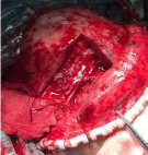
Figure 1
“Apartment” strategy followed by intra-orbital unit optic nerve decompression.
Figure 2
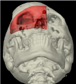
Figure 2
Mirror Technique
Results
Case presentation 1
A 15-year-old girl presented with an asymmetrical face and
right visual blurring. Visual acuity was count finger at the distance
of 1 meter in right side and 0.2 in left side, respectively. She also had
visual field defect in right side. The funduscope confirmed atrophy
of right optic nerve. Right exophthalmos measured 3 mm by Hertel
exophthalmometry. CT showed FD of the right anterior cranial base,
ethmoid bone, sphenoid bone, superolateral orbit, and surrounding
the optic canal. Ipsilateral orbital volume also decreased. The patient
underwent resection of the FD of the right fronto-orbital bones
with ipsilateral intraorbital optic nerve decompression and split
cranial bone-graft reconstruction. After therapeutic decompression,
her visual acuity in the affected eye improved markedly to 0.2.
Postoperative CT at 3-12 months showed an enlarged intraorbital
volume without optic foramen involvement.
Case presentation 2
A 16-year-old boy complained of progressive protrusion of
right forehead and supraorbital ridge with mild dystopia in the
right eye. His visual acuity was 0.6 in the right eye and 1.0 in the left
eye. He had neither visual field defect nor atrophy of optic nerve.
Right exophthalmos measured 3 mm by Hertel exophthalmometry.
CT examination exhibited the ground-glass appearance of FD
lesion involvement in the superolateral orbit, frontal and sphenoid
bones. Optic canal was surrounded and ipsilateral orbital volume
also decreased. Right intraorbital optic nerve decompression was
done and subsequent orbital reconstruction yielded a satisfactory
cosmetic result. His postoperative CT, ocular examination and orbital
positioning were stable at 3-12 months follow-up.
Case presentation 3
A 20-year-old man was admitted to our hospital with severe left
orbital dystopia, exophthalmos, and visual impairment. Visual acuity
was 0.2 in left side and 0.8 in right side, respectively. Ophthalmologic
examination was normal with no evidence of visual field loss
and optic nerve compromise. Four millimeters of proptosis was
documented by Hertel exophthalmometry. His CT scans revealed
extensive fronto-orbital FD surrounding and narrowing the left optic
canal. Ipsilateral orbital volume also decreased. Left intraorbital optic
nerve decompression was done, which was followed by fronto-orbital
contouring and reconstruction. Postoperatively, his visual acuity in
the affected eye improved slightly to 0.3. His appearance, especially
the orbital dystopia, improved markedly at 3-12 months follow-up.
CT scans and ocular examination also remained stable
Discussion
It is noteworthy the optic nerve comprises five major units:
chiasmatic, intracranial, intra-canal, intra-orbital, and intraocular
part [6]. It is the intra-canal unit decompression that has received
considerable attention. However, it has been thought to play a limited
role [5,7]. This is because patho-etiological basis of visual impairment
is controversial. Consequently, indications for optimal treatment
paradigms remain unclear.
For the prophylactic philosophy, it has been assumed FD is a
progressive disease, which will lead to blindness ultimately. Therefore,
early unroofing may prevent such disastrous consequences. We and
others have questioned whether the most common cause of visual loss
is indeed bone overgrowth-induced optic canal stenosis [8]. For the
therapeutic philosophy, it certainly has a questionable value in cases
of established blindness [9,10]. Moreover, both may be associated
with visual loss as a complication in and of itself.
Our philosophy is treatment paradigms should be tailored to the
individual. According to characteristics of our practice (exophthalmos
and orbital volume decreasing), the most possible underlying causes
may be traction and ischemia of optic nerve. As orbital volume
gradually decreases by FD involvement, rising intra-orbital pressure
would cause an increase in intra-luminal pressure of the retinal veins,
which in turn leads to a drop in perfusion of the retinal artery, with
eventual cessation of retinal perfusion. Additionally, exophthalmos
usually indicates traction, which makes the nerve more susceptible.
Furthermore, the risks of intra-canal decompression are not trivial,
which may have more probability of injury due to direct trauma,
burring, thermal damage, traction, loss of blood supply, and vascular
thrombosis.
Advantage
Finally, we consider the intra-orbital decompression may be
more feasible and safer with the help of preoperative simulation,
“Apartment” strategy and navigation. Preoperative simulation could
help us dealing with important decompression site more precisely.
Fronto-orbital sub-craniotomy has the advantage of less injury, which
was performed like “entering apartment” in order to safely expand
surgical field step by step. We also apply navigation, thereby we
could localize the optic neurovascular bundle in real-time and avoid
iatrogenic injury. Last but not least, fronto-orbital reconstruction
could be easier to achieve by this strategy.
Limitation
Our preliminary results seem favorable. However, shortcomings
are obvious because we have fewer patients and no results of long
term follow-up. We will increase sample size and update the data in
future work. And if possible, with the approval of ethic committee,
we attempt to compare the follow-up results between intra-canal and
intra-orbital optic nerve decompression in patients of fronto-orbital
FD without preoperative visual impairment.
Conclusion
Visual loss is the most feared complication. Besides canal stenosis, other causes such as ischemia, lesion degeneration and optic nerve traction appear more common. We consider the intraorbital decompression may be more feasible and safer with the help of “Apartment” strategy, especially for those with exophthalmos, orbital volume decreasing, and non-acute visual loss.
References
- Fattah A, Khechoyan D, Phillips JH, Forrest CR. Paediatric craniofacial fibrous dysplasia: the Hospital for Sick Children experience and treatment philosophy. J plast Reconstr Aesthet Surg. 2013; 66: 1346-1355.
- Mahapatra AK, Gupta PK, Ravi RR. Craniofacial surgery and optic canal decompression in adult fibrous dysplasia. Neurology India. 2003; 51: 123,124.
- Lee JS, FitzGibbon E, Butman JA, Dufresne CR, Kushner H, Wientroub S, et al. Normal vision despite narrowing of the optic canal in fibrous dysplasia. N Engl J Med. 2002; 347: 1670-1676.
- Michael CB, Lee AG, Patrinely JR, Stal S, Blacklock JB. Visual loss associated with fibrous dysplasia of the anterior skull base. Case report and review of the literature. J Neurosurg. 2000; 92: 350-354.
- Cutler CM, Lee JS, Butman JA, FitzGibbon EJ, Kelly MH, Brillante BA, et al. Long-term outcome of optic nerve encasement and optic nerve decompression in patients with fibrous dysplasia: risk factors for blindness and safety of observation. Neurosurgery. 2006; 59: 1011-1017.
- Tan YC, Yu CC, Chang CN, Ma L, Chen YR. Optic nerve compression in craniofacial fibrous dysplasia: the role and indications for decompression. Plast Reconstr surg. 2007; 120: 1957-1962.
- Mathiesen T, Kihlstrom L. Visual outcome of tuberculum sellae meningiomas after extradural optic nerve decompression. Neurosurgery. 2006; 59: 570-576.
- Yang Y, Wang H, Shao Y, Wei Z, Zhu S, Wang J. Extradural anterior clinoidectomy as an alternative approach for optic nerve decompression: anatomic study and clinical experience. Neurosurgery. 2006; 59: ONS253- 262.
- Abe T, Satoh K, Wada A. Optic nerve decompression for orbitofrontal fibrous dysplasia: recent development of surgical technique and equipment. Skull Base. 2006; 16: 145-155.
- Dumont AS, Boulos PT, Jane JA Jr, Ellegala DB, Newman SA, Jane JA, Sr. Cranioorbital fibrous dysplasia: with emphasis on visual impairment and current surgical management. Neurosurg focus. 2001; 10: E6.