Case Report
Surgical Repair of a Snuffbox Radial Artery Pseudoaneurysm
Naiem Nassiri*, Samuel Kogan, Huong Truong, KJ Nagarsheth, Randy Shafritz and Saum Rahimi
Department of Surgery, Rutgers Robert Wood Johnson Medical School, USA
*Corresponding author: Naiem Nassiri, Department of Surgery, Division of Vascular Surgery and Vascular Anomalies & Malformations Program (VAMP), Rutgers Robert Wood Johnson Medical School, One Robert Wood Johnson Place MEB 541, New Brunswick, New Jersey 08901, USA
Published: 14 Oct, 2016
Cite this article as: Nassiri N, Kogan S, Truong H,
Nagarsheth KJ, Shafritz R, Rahimi S.
Surgical Repair of a Snuffbox Radial
Artery Pseudoaneurysm. Clin Surg.
2016; 1: 1154.
Abstract
A rare case of a snuffbox radial artery pseudoaneurysm treated surgically is presented. Given the broad base, short neck and relatively large size of the pseudoaneurysm sac, compression techniques and intra-sac thrombin injection were deemed inappropriate, as they have been associated with ischemic complications within the sparse literature. Open surgical resection of the pseudoaneurysm with primary end-to-end repair of the radial artery was performed. Technical success was achieved with restoration of flow through the radial artery and the palmar arch with maintained patency and alleviation of symptoms immediately post-operative and at 3-months follow-up.
Introduction
Radial artery aneurysms are rare entities comprising only 2.9% of all upper extremity aneurysms
[1-3]. Such degenerative processes affecting the radial artery within the anatomical snuffbox are
even more sparsely reported. As with other arterial degenerative processes, these aneurysms or
pseudoaneurysms are typically caused by traumatic, iatrogenic, infectious, atherosclerotic, or
collagen vascular disease processes [1-8].
Given the superficial location within a highly mobile region of the wrist and proximity to the
palmar arch, distal thromboembolism, digital ischemia, rupture, hemarthrosis, adjacent nerve
irritation, paresthesias, limited wrist mobility, skin ulceration, and secondary infection are potential
risks that prompt intervention [2,3,6,8]. Reported treatment options have included surgical excision
with or without radial artery ligation, ultrasound-guided compression, and percutaneous thrombin
injection.
Herein, we report a rare case of a symptomatic snuffbox radial artery pseudoaneurysm treated
surgically based on the configuration and angioarchitecture of the pseudoaneurysm.
Case Presentation
Background
A 16-year-old, right-handed male presented with a 9-month history of amildly tender, pulsatile
mass in the left anatomical snuffbox after suffering blunt trauma to his left hand (Figure 1a). The
patient was initially seen by his pediatrician, who attempted to aspirate the mass, believing it to be
a ganglion cyst, with no resolution. He was referred to vascular surgery for evaluation after a hand
surgeon ordered a Magnetic Resonance Imaging (MRI) scan which revealed a 1.9 cm x 1.2 cm x 1.8
cm pseudoaneurysm of the radial artery emerging in between the extensor and abductor tendons of
the anatomical snuffbox (Figure 1b and c).
Preoperative evaluation
Upon evaluation, patient was found to have 2 cm round pulsatile mass within the anatomical
snuffbox of the left hand. A modified Allen’s test using a pulse oximeter with waveform analysis
was performed and suggested a radial artery dominant blood supply to the thumb and index finger.
The patient underwent duplex ultrasound evaluation, which revealed a patent ipsilateral arterial
tree proximally with no evidence of hemodynamically significant stenosis or other degenerative
pathologies. Within the anatomical snuffbox, a partially thrombosed pseudoaneurysm of the radial
artery was identified (Figure 2). The pseudoaneurysm had a broad base with essentially no neck,
making it unsuitable for percutaneous thrombin injection. After discussion with the patient and
family, decision was made for surgical treatment.
Operative technique
Under tourniquet exsanguination, a 5cm incision was made
over the course of the pseudoaneurysm within the anatomical
snuffbox. Using extensive sharp dissection, the pseudoaneurysm
was dissected out circumferentially, as were the proximal and distal
segments of the radial artery (Figure 3a). Once adequate lengths of
the artery proximal and distal to the pseudoaneurysm was dissected
and mobilized, the tourniquet was let down and the patient was
systemically heparinized. Proximal and distal control was obtained
and the pseudoaneurysm sac was incised, followed by suction of
partially thrombosed contents (Figure 3b). Given the broad base of the
pseudoaneurysm along the anterior wall of the radial artery, primary
repair was not possible. Therefore, the pseudoaneurysm was resected
and a spatulated, end-to-end anastomosis of the radial artery was
performed using running 7-0 prolene sutures (Figure 3c). There was
excellent re-establishment of flow within the radial artery confirmed
by manual pulse palpation and Doppler interrogation. There were no
perioperative complications. The patient was maintained in a gutter
splint for 2 weeks post-op. Final pathology evaluation confirmed the
presence of a partially thrombosed arterial pseudoaneurysm.
Figure 1
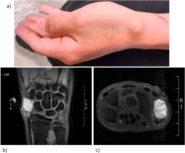
Figure 1
Figure 1a: Pulsatile, tender mass in left anatomical snuffbox.
Figure 1b, 1c: MRI of the left hand revealing a 1.9 cm x 1.2 cm x 1.8 cm pseudoaneurysm of the radial artery within the anatomical snuffbox.
Figure 2
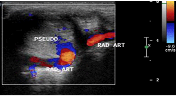
Figure 2
Duplex ultrasonography revealed a partially thrombosed, broadbased radial artery pseudoaneurysm within the anatomical snuffbox.
Figure 3
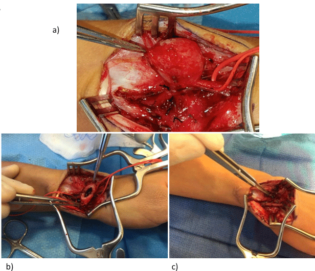
Figure 3
Figure 3a: Proximal and distal control of the radial artery was achieved prior to excision of the pseudoaneurysm sac.
Figure 3b: Incised pseudoaneurysm sac with thrombus.
Figure 3c: End-to-end anastomosis of the affected radial artery with successful re-establishment of pulsatile flow.
Discussion
Radial artery pseudoaneurysms and aneurysms in this unique
location can have a variable pattern of presentation ranging from
an asymptomatic, pulsatile mass to a rapidly expanding, painful
lesion with distal ischemia, paresthesias, overlying skin ulceration,
and bleeding complications [5]. Even the less symptomatic lesions
have the potential for growth over time with subsequent arterial
impingement and potential for thromboembolic complications
[8]. Diagnostic imaging plays an important role in diagnosis and
traditionally includes contrast-enhanced MRI series. The latter
can help identify the location, adjacent arterial involvement, and
associated musculoskeletal abnormalities. Duplex ultrasonography
can play an important role in distinguishing true arterial aneurysms
from pseudoaneurysms and the quality of in-line flow proximal and
distal to the lesion in question. In the case of a pseudoaneurysm, the
so-called “ying-yang” sign is observed via duplex ultrasonography
and is pathognomonic for the alternating pattern of systolic flow into
the pseudoaneurysm and diastolic flow out resulting in the “to-andfro”
spectral waveform signal [9]. In rare situations, pre-operative
angiography can also be a valuable tool to help identify associated
vascular pathologies such as arteritides, arteriovenous fistulas and
malformations, fibromuscular dysplasia, and venous anomalies.
Transcatheter therapeutic options such as covered stent exclusion,
coil and polymerizing agent embolization are extremely limited
and ill advised given the local anatomical restrictions. Percutaneous
thrombin injection and/or ultrasound-guided compression therapy
remain the only viable non-surgical options locally. In the current
case, the broad base and extremely short neck of the pseudoaneurysm
precluded employment of such non-operative maneuvers, as
the risk of distal digital ischemia from non-target embolization
would be extremely high. Careful evaluation and scrutiny of the
pseudoaneurysm morphology on duplex ultrasonography are
paramount in ascertaining patient’s candidacy for percutaneous
therapy.
Of the handful of reported cases of true aneurysms of the
anatomical snuffbox, most have been managed with ligation of the
radial artery followed by surgical resection of the defect [1-8]. A single
report exists of a patient with MRI-confirmed idiopathic radial artery
aneurysm within the anatomical snuffbox, who denied treatment due
to lack of symptoms [10]. The decision to proceed with radial artery
ligation at the time of surgical excision can be based on presence of
collaterals on angiography, a negative Allen’s test, intraoperative
Doppler interrogation, or adequate back bleeding from the distal
stump [3,6,8]. In this particular case, given the dampened pulse
oximeter waveforms noted on the ipsilateral thumb and index fingers
on manual compression of the radial artery, a pre-operative decision
was made to proceed with surgical reconstruction of the radial artery
at the time of pseudoaneurysm excision.
In Behar et al. [7] a radial artery aneurysm was reported in the
right upper extremity from repetitive occupational injury. As a tailor,
the aneurysm caused tenderness and numbness in the patient’s hand
causing difficulty using scissors to cut cloth. The development of the
aneurysm was thought to be caused by back pressure of flow towards
the wrist due to repeated compression of the radial artery from the
handle of the scissors. The aneurysm was confirmed on duplex imaging
and post-op pathology. Pre-op Allen’s test was consistent with ulnar
dominant supply to the palmar arch. The aneurysm was explored
with temporary occlusion of radial artery demonstrating excellent
flow through ulnar artery. Bypass was considered, but rejected due
to high possibility of recurrence from occupation. Therefore, the
aneurysm was excised with no reconstruction necessary.
Dryton et al. [5] reported a case of radial artery pseudoaneurysm
in the right wrist after a cat bite, in a patient who presented with
intermittent pain and discoloration of her right thumb and index
finger. The patient had a history of systemic lupus erythematosus and
deep venous thrombosis on anticoagulation. Bedside pulse oximetry
Allen’s test confirmed good collateralization across the palmer arch,
and uneventful excision of radial artery and pseudoaneursym was
performed.
Poirier et al. [8] reported a mycotic aneurysm of right radial artery.
The patient presented with a pulsatile mass and fever. Arteriogram
was performed consistent with a 1.5cm aneurysm fed by the dorsal
branch of radial artery. He was taken to the operating room and
surgical excision of the mycotic aneurysm and ligation of the feeding
vessel was performed due to presence of adequate collateral flow and
presence of infection.
Finally, two case studies demonstrated traumatic radial artery
aneurysms that were also surgically treated with aneurysm excision
and radial artery ligation. Gabriel et al. [3] described a healthy male
patient with a pulsatile mass of the left hand after sustaining a dog bite
4 months prior with no active signs of infection or skin ulceration.
Doppler arterial interrogation showed dilatation of the left radial artery
consistent with a local aneurysm and decision was made for surgical
treatment. Allen’s test preop was negative, and thus radial artery
was safely ligated. In two case reports by Wenger et al. [4] traumatic
injuries to right anatomical snuffbox were sustained. Both patients
were symptomatic in the area with post-op histopathology consistent
with pseudoaneurysms. Pre-op testing with Allen’s test and Doppler
ultrasound were consistent with acceptable collateral circulation by
ulnar artery, and thus surgical resection of the pseudoaneurysms with
concomitant radial artery ligation was performed in both cases.
Herein, we have described a rare case of symptomatic posttraumatic
radial artery pseudoaneurysm treated by surgical excision
and arterial reconstruction. The location of these rare lesions restricts
transarterial endovascular treatment options. Ultrasound-guided
compression and thrombin injection remain viable non-operative
techniques only for treatment of pseudoaneurysms with appropriate
morphological features such as long, narrow neck. Surgical excision
with ligation remains the gold-standard therapeutic modality.
However, in rare situations such as in the current case, arterial
reconstruction may be necessary. Options include end-to-end
anastomosis, interposition grafting via revered vein or exogenous
conduits. The latter can be compromising in the presence of an
infections etiology and small caliber of the affected artery.
This research did not receive any specific grant from funding
agencies in the public, commercial, or not-for-profit sectors. The
patient consented to the publication of this manuscript.
References
- Sterling AP, Habermann ET. Traumatic aneurysm of the radial artery. Hand. 1975; 7: 294-296.
- Luzzani L, Bellosta R, Carugati C, Talarico M, Sarcina A. Aneurysms of the Radial Artery in the Anatomical Snuff Box. EJVES Extras. 2006; 11: 94-96.
- Gabriel SA, Marcondes de Abreau MF, Simoes CR, Antonio, Chrispim CG, de Camargo O Jr. True posttraumatic radial artery aneurysm. J vasc Bras. 2013; 12: 320-323.
- Wenger DR, Boyer DW, Sandzen SC. Traumatic Aneurysm of the Radial Artery in the Anatomical Snuff Box. Hand. 1980; 12: 266-270.
- Dryton G, Allen KB, Borkon AM, Aggarwal S, Davis JR. Do not bite the hand that feeds you. Ann Vasc Surg. 2015; 29: 362 e3-4.
- Jedynak J, Frydman G. Idiopathic True Aneurysm of the Radial Artery: A Rare Entity. EJVES Extras. 2012; 23: e21-e22.
- Behar JM, Winston JS, Knowles J, Myint F. Radial artery aneurysm resulting from repetitive occupational injury: Tailor's thumb. Eur J Vasc Endovasc Surg. 2007; 34: 299-301.
- Poirier RA, Stansel HC. Arterial Aneuyrsms of the Hand. Am J Surg. 1972; 124: 72-74.
- Rozen G, Samuels DR, Blank A. The to and fro sign: the hall-mark of pseudoaneurysm. Isr Med Assoc J. 2001; 3: 781-782.
- Walton NP, Choudhary F. Idiopathic radial artery aneurysm in the anatomical snuff box. Acta Orthop Belg. 2002; 68: 292 - 294.