Case Report
Acute Type B Dissection in a Bicuspid Aortic Valve Patient During Cardiopulmonary Bypass Associated with Arch Cannulation and the use of an End-Hole Cannula
Schulte K, Attia R* and Young C
Department of Cardiothoracic Surgery, Guy’s and St Thomas’ Hospital, UK
*Corresponding author: Rizwan Attia, Department of Cardiothoracic Surgery, St. Thomas’ Hospital, 6th Floor East Wing, Westminster Bridge Road, London, SE1 7EH, UK
Published: 09 Sep, 2016
Cite this article as: Schulte K, Attia R, Young C. Acute Type B Dissection in a Bicuspid Aortic Valve Patient During Cardiopulmonary Bypass Associated with Arch Cannulation and the use of an End-Hole Cannula. Clin Surg. 2016; 1: 1106.
Abstract
The incidence of acute type B aortic dissection is 2.9 per 100,000. Bicuspid aortic valve diseaese is a known to increase the risk to 3.1 per 10,000. An age adjusted relative risk increase of over 8-fold.
Aortic dissection occurs at an earlier age along with more rapid development of aortic aneurysms.
Cardiac surgery with cardiopulmonary bypass is associated with mechanical manipulation of the
ascending aorta that occassionally leads to type A aortic dissection. This occurs in 0.12-0.16% of
patients with cardiac surgery. The incidence of type B dissection post cardiac surgery is much lower.
The incidence and managment of this condition is not well defined in the literature.
We report the case of a 48-year-old man with bicuspid mixed aortic valve disease and aortopathy
presenting with a 50mm ascending aortic aneurysm who underwent minimally invasive aortic valve
and ascending aortic replacement that led to an acute Stanford type B/Debakey IIIb aortic dissection.
The patient was successfully treated with thoracic endografting and remains well at 2-year followup.
We describe the management of the case and focus on sites of operative trauma at surgery. We
discuss aortic morphology during primary surgery and consider the aortic histopathology at the site
of primary entry tear.
Current guidelines do not provide conclusive information on characteristic features and
management in these patients. We discuss emergent thoracic aortic endografting as a treatment
option for these high-risk patients and discuss all the management strategies that would improve
outcomes for this patient cohort.
Keywords: Bicuspid aortopathy; Aortic valve replacement; Ascending aortic replacement; Complication; Aortic dissection; Thoracic endovascular aortic repair (TEVAR)
Abbreviations
ATBAD: Acute Type B Aortic Dissection; BAV: Bicuspid Aortic Valve; CTA: Computer Tomographic Angiography; TEVAR: Thoracic Endovascular Stent Grafting
Introduction
The incidence of acute type B aortic dissection is 2.9 per 100,000 in the population [1]. Bicuspid aortic valves (BAV) are the commonest congenital cardiac defect with a prevalence estimated
between 0.5 to 2% [2]. Aortic aneurysms associated with BAV are a manifestation of multiple single
defects, for instance mutations in signalling and transcriptional regulators of NOTCH 1 (9q34.3)
that lead to abnormal valve development and de-repression of calcium signalling [3]. Mutations in
GATA 5 have also been reported [4]. There are numerous other gene defects that are described in
extracellular matrix proteins such as FBN1, FN1, FGF8, HOXA1 and TGFBR)1/2 [5]. Histological
abnormalities are observed in bicuspid aortopathy as the result of abnormal regulatory pathways
involving smooth muscle cells within the aortic media affecting the structural integrity and flexibilty
of the aorta [6]. The resulting aortopathy develops early and both children and adults with bicuspid
aortic valve disease have larger aortic annuli, sinuses and proximal ascending aortae than those
with tri-leaflet valves. The reported increased incidence of aortic dissection varies from 0 to 10-fold
compared with patients with aortic stenosis [7].
Iatrogenic aortic dissection is a rare but major complication in invasive vascular interventions
[6]. Whereas the ascending thoracic aorta is usually affected in cardiac surgery [8 and 9] dissection of the descending thoracic aorta is commonly caused by manipulation of intraluminal tools introduced via.the femoral artery for catheter-based interventions.Acute dissection has been described to complicate 0.12-0.16% of cardiac surgery and 0.6% of aortic valve replacements [10 and 11]. In these cases a low-risk elective operation is converted to an emergent to high-risk case with serious morbidity and mortality.
Guidance on management of this case is difficult as current guidelines do not provide conclusive information on characteristic features and management of acute aortic dissections with endovascular grafts. The operative mortality is between 6-66% suggesting substantial differences in management strategies and thus potential for improvements [12]. This case describes acute type B aortic dissection (ATBAD), in the setting of post-operative BAV aortopathy. We discuss the aortic morphology during primary surgery and consider the aortic histopathology at the site of primary entry tear, the importance of timely diagnosis and multi-disciplinary management including endovascular grafting as treatment options in these high-risk patients.
Figure 1
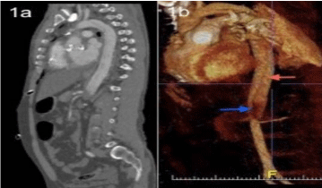
Figure 1
Standford type B/ Debakey Type IIIb aortic dissection extending below the diaphragm. A) CT-Angiography sagittal view, B) 3D reconstruction (flap seen red arrow; occlusive thrombus and flap extending into the region of the visceral vessels seen blue arrow).
Figure 2
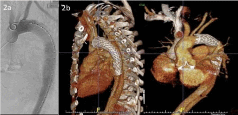
Figure 2
Gore®Tag® thoracic endoprothesis in the proximal descending aorta. A) Fluroscopy demonstrating deployment of the endograft B) 3D reconstruction with an demonstrating the endograft in Mitchell and Ishimaru landing zone 3, sealing the entry tear. The frame of the perimount prosthesis is seen at the LVOT.
Case Presentation
48-year-old man with bicuspid aortopathy presented with a 50mm ascending aortic aneurysm with moderate aortic regurgitation in a setting of preserved left ventricularfunction. He underwent minimally invasive aortic valve and ascending aortic replacement through a mini J-sternotomy. Cardiopulmonary bypass was established cannulating the atrium and aortic arch with an end hole aortic cannula. The aortic cross clamp was applied and the heart arrested with one litre of cold blood cardioplegic solution delivered directly into the coronary ostia. The ascending aorta was excised down to the sino-tubular junction. The aortic valve was excised and replaced with a 27 mm CE Perimount bioprosthesis. The ascending aortawas replaced with a 28 mm Dacron graft. Following routine air drill the aortic cross clamp was released, sinus rhythm regained and at normothermia, bypass was discontinued without inotropic support. Post-operative course was complicated by pericardial tamponade necessitating re-sternotomy. Patient failed to progress with worsening lactatemia and developed acute lower limb ischaemia. Computer tomographic angiography (CTA) demonstrated an acute Standford type B/Debakey type IIIb dissection complicated by coeliac and superior mesenteric compression due to a spiralling flap (Figure 1). The patient underwent thoracic endovascular stent grafting (TEVAR) to seal the entry tear in the proximal descending aorta with a GORETAG® endograft (Figure 2). This led to angiographic resolution of the visceral malperfusion. Due to the previous physiological insult to the viscera in the setting ofcontinued clinical deterioration, the patient underwent a laparotomy, extended subtotal colectomy, ileostomy and mucous fistula formation for ischaemic bowel. Recovery was complicated with multi-organ failure and prolonged neuro-rehabilitation. Patient was discharged well with mild residual weakness in the left leg 42-days post-operatively. He remains well at 2-year follow-up.
Discussion
Aortic dissection can complicate cardiac surgery either
intraoperatively, early or late post-operatively. This converts an
elective low-risk operation to high-risk surgery with a high morbidity
and mortality [12].
Patients with bicuspid aortic disease are at high-risk as the
underlying aorta is structurally abnormal. This pertains tothe gene
defects highlighted [3 and 4], that led to defective extracellular matrix
proteins [5]. As a result matrix disruption, elastin and lamellar
fragmentation is observed accompanied with increased apoptosis of
vascular smooth-muscle cells. These events may lead to an aorta with
weakened structural integrity and reduced elasticity [7]. At these sites
aortic manipulation and trauma are the likely causes of entry tears
namely cannulation site, cross-clamping injury, cardioplegia site and
suture lines. The arterial flow during cardiopulmonary bypass has
to be monitored and high line pressures avoided. In cases of high
cannulation, vessel bifurcation points need to be avoided and the
flow jet directed down the descending thoracic aorta. Use of end hole
cannulae may increase wall shear stress and place the patient at risk
of intimal injury. Altered aortic haemodynamics associated with BAV
have been implicated in development of aortopathy [13]. BAV fusion
has been show on 4D MRI to be associated with changes in systolic
flow uniformity, peak systolic velocities and flow angles. Altered flow
jet patterns towards aortic wall lead to altered wall shear forces and
aortopathy [14]. The structurally abnormal wall may thus be more
likely to develop iatrogenic aortic dissection as in our case.
The appropriate treatment strategy for descending aortic
dissection continues to be a challenge. This case highlights the
importance of high clinical index of suspicion in patients not
progressing post-operatively. The only clinical indication of problem
was high lactate followed by eventual development of lower limbischaemia.
Rapid diagnostic confirmation with CTA, intensive care
monitoring, and aggressive blood pressure with antipulse therapy
involving beta blockers are important [15].
The current indications for intervention in acute type B
dissections involve rupture or signs of impending rupture, rapid
diameter progression, malperfusion of abdominal or peripheral
vessels, persisting pain, and uncontrollable hypertension [16]. Some
authors would argue that all type B aortic dissections should be
considered for endovascular repair [17].
Endografting has been shown to be an effective and feasible
treatment strategy in patients with a complicated ATBAD. It is
dependent on closure of the entry tear, re-expansion of the true lumen,
and thrombosis of the false lumen [16]. It has been shown that the
immediate outcomes are better when compared with open surgical
repair, reducing in-hospital mortality and better 1 and 5-year survival
compared to medical management alone [18]. In symptomatic
patients or patients with visceral or end-organ malperfusion thoracic
endograftingleads to a favorable aortic remodeling during follow-up
after intervention [19]. This is specially the case if there is false lumen
thrombosis following the endografting [20].
Conclusion
Early endovascular stent graft placement should be considered in patients with post-operative ATBAD specially in cases with evidence of end-organ malperfusion and haemodynamic instablity. The use of a dispersion tip cannulae to cannulate the aortic arch should be considered to reduced shear stresses on the aorta. Long-term studies are required to delinate the use of endovascular treatment options in patients with connective tissue disease and genetically tiggered thoracic aortic disease.
References
- Mészáros I, Mórocz J, Szlávi J, Schmidt J, Tornóci L, Nagy L, et al. Epidemiology and clinicopathology of aortic dissection. Chest. 2000; 117: 1271–1278.
- Hoffman JIE, Kaplan S. The incidence of congenital heart disease. J Am Coll Cardiol. 2002; 39: 1890-1900.
- Mohamed SA, Aherrahrou Z, Liptau H, Erasmi AW, Hagemann C, Wrobel S, et al. Novel missense mutations (p.T596M and p.P1797H) in NOTCH1 in patients with bicuspid aortic valve. Biochem Biophys Res Commun. 2006; 345: 1460–1465.
- Padang R, Bagnall RD, Richmond DR, Bannon PG, Semsarian C. Rare non-synonymous variations in the transcriptional activation domains of GATA5 in bicuspid aortic valve disease. J Molec Cell Cardiol. 2012; 53: 277–281.
- Abdulkareem N, Smelt J, Jahangiri M. Bicuspid aortic valve aortopathy: genetics, pathophysiology and medical therapy. Interact Cardiovasc Thorac Surg. 2013; 17: 554–559.
- Michelena HI, Khanna AD, Mahoney D, Margaryan E, Topilsky Y, Suri RM, et al. Incidence of aortic complications in patients with bicuspid aortic valves. JAMA. 2011; 306: 1104–1112.
- Verma S, Siu SC. Aortic Dilatation in Patients with Bicuspid Aortic Valve. N Engl J Med. 2014; 370: 1920–1929.
- Estrera AL, Garami Z, Miller CC, Sheinbaum R, Huynh TTT, Porat EE, et al. Cerebral monitoring with transcranial Doppler ultrasonography improves neurologic outcome during repairs of acute type A aortic dissection. J Thorac Cardiovasc Surg. 2005; 129: 277–285.
- Stanger O, Oberwalder P, Dacar D, Knez I, Rigler B. Late dissection of the ascending aorta after previous cardiac surgery: risk, presentation and outcome. Eur J Cardio-thorac Surg. 2002; 21: 453–458.
- Collins JS, Evangelista A, Nienaber CA, Bossone E, Fang J, Cooper JV, et al. Differences in clinical presentation, management, and outcomes of acute type a aortic dissection in patients with and without previous cardiac surgery. Circulation. 2004; 110: II237–242.
- Kodolitsch von Y, Aydin MA, Koschyk DH, Loose R, Schalwat I, Karck M, et al. Predictors of aneurysmal formation after surgical correction of aortic coarctation. J Am Coll Cardiol. 2002; 39: 617–624.
- Hirose H, Svensson LG, Lytle BW, Blackstone EH, Rajeswaran J, Cosgrove DM. Aortic dissection after previous cardiovascular surgery. Ann Thorac Surg. 2004; 78: 2099–2105.
- Girdauskas E, Borger MA, Secknus M-A, Girdauskas G, Kuntze T. Is aortopathy in bicuspid aortic valve disease a congenital defect or a result of abnormal hemodynamics? A critical reappraisal of a one-sided argument. Eur J Cardio-thorac Surg. 2011; 39: 809–814.
- Mahadevia R, Barker AJ, Schnell S, Entezari P, Kansal P, Fedak PWM, et al. Bicuspid aortic cusp fusion morphology alters aortic three-dimensional outflow patterns, wall shear stress, and expression of aortopathy. Circulation. 2014; 129: 673–682.
- Estrera AL, Miller CC, Safi HJ, Goodrick JS, Keyhani A, Porat EE, et al. Outcomes of medical management of acute type B aortic dissection. Circulation. 2006; 114: I384–389.
- Schoder M, Czerny M, Cejna M, Rand T, Stadler A, Sodeck GH, et al. Endovascular repair of acute type B aortic dissection: long-term followup of true and false lumen diameter changes. Ann Thorac Surg. 2007; 83: 1059–1066.
- Nienaber CA, Kische S, Rousseau H, Eggebrecht H, Rehders TC, Kundt G, et al. Endovascular repair of type B aortic dissection: long-term results of the randomized investigation of stent grafts in aortic dissection trial. Circ Cardiovasc interv. 2013; 6: 407–416.
- Fattori G, Saito N, Seregni M, Kaderka R, Pella A, Constantinescu A, et al. Commissioning of an Integrated Platform for Time-Resolved Treatment Delivery in Scanned Ion Beam Therapy by Means of Optical Motion Monitoring. Technol Cancer Res Treat. 2014; 13: 517-528.
- Andacheh ID, Donayre C, Othman F, Walot I, Kopchok G, White R. Patient outcomes and thoracic aortic volume and morphologic changes following thoracic endovascular aortic repair in patients with complicated chronic type B aortic dissection. J Vascular surg. 2012; 56: 644–650.
- Trimarchi S, Tolenaar JL, Jonker FHW, Murray B, Tsai TT, Eagle KA, et al. Importance of false lumen thrombosis in type B aortic dissection prognosis. J Thorac Cardiovasc Surg. 2013; 145: S208–212.