Case Report
Skull Base Metastasis of a Breast Carcinoma
Ferri GG*,Castellucci A, Vecchio VD and Brandolini C
1Department of Experimental, Diagnostic and Specialty Medicine, S. Orsola-Malpighi University Hospital, Italy
*Corresponding author: Gian Gaetano Ferri, Department of Experimental, Diagnostic and Specialty Medicine, S. Orsola-Malpighi University Hospital, Via Massarenti 9, 40138 Bologna, Italy
Published: 09 Sep, 2016
Cite this article as: Ferri GG, Castellucci A, Vecchio VD, Brandolini C. Skull Base Metastasis of
a Breast Carcinoma. Clin Surg. 2016; 1: 1105.
Abstract
Skull base metastases from primary malignant tumors are relatively uncommon lesions. They usually originate from the breast, lung, prostate, skin, liver and cervix. We report a case of a 76 years old female complaining of dizziness, mild left hearing loss, tinnitus and recurring left facial paresis. A computed tomography scan of the temporal bones documented a much extended osteolytic lesion of the left temporal bone. A left mastoidectomy evidenced a soft-tissue etheroplastic mass involving diffusely the middle ear. Several bioptic specimens were obtained and the definite histopathological response was breast lobular carcinoma metastasiss.
Case Presentation
A 76 years old woman referred to our clinical institution complaining of dizziness, mild
left hearing loss, tinnitus and recurring left facial paresis. Ten years before, after a right breast
carcinoma had been diagnosed, she had been submitted to surgery (right quadrantectomy) followed
by radiotherapy and hormone therapy.
Vestibular symptoms (objective vertigo sometimes accompanied by nausea and vomiting)
started several months before diagnosis and disappeared during a two-month period. At the
moment of our neurotologic examination, no kind of nystagmus was observed. Tone audiometry
evidenced a bilateral presbyacusis, slightly worse at the left side where a conductive component
was present, only partially affecting the patient who also complained of omolateral tinnitus. Two
months before, the patient had been affected by a left facial nerve paresis (treated with vitamin B
complex and regressed after four days) that recurred a few weeks later.
A previous brain and temporal bone MRI investigation had not evidenced radiologic signs of
disease. An accurate high-resolution computed tomography (HRCT) scan of the temporal bones
documented a much extended osteolytic lesion of the left temporal bone, comprehending bony
erosion of the mastoid, tegmen tympani and petrous apex (Figure 1). Anteriorly, the metastatic
lesion also caused a lytic erosion of the clivus, while posteriorly a suspected involvement of middle
fossa dura and sigmoid sinus region was recorded (Figure 2). Finally, coronal scans evidenced the
erosion of the first cervical metamers and the involvement of the vertical tract of the facial canal
(Figure 3). Instead, the ossicular chain and the otic capsule appeared to be preserved.
Therefore, a left mastoidectomy was performed under general anesthesia: a soft-tissue
etheroplastic mass involved the sinodural angle and the perisinusal mastoid cells. The tegmen antri
wall appeared infiltrated. Several bioptic specimens were obtained; the histopathological response
reported signs of osteofibrous tissue infiltrated by neoplastic cells.
Immunohistochemical investigations also evidenced a significant
positivity for estrogen and progesterone hormones in the neoplastic
cells. The definite response was breast lobular carcinoma metastasis.
Finally, due to both the radiologic and histopathologic findings,
the patient was submitted to the oncologic section of our hospital.
Actually, about one year after the surgical procedure, the patient feels
better and vestibular symptoms have decreased.
Figure 1
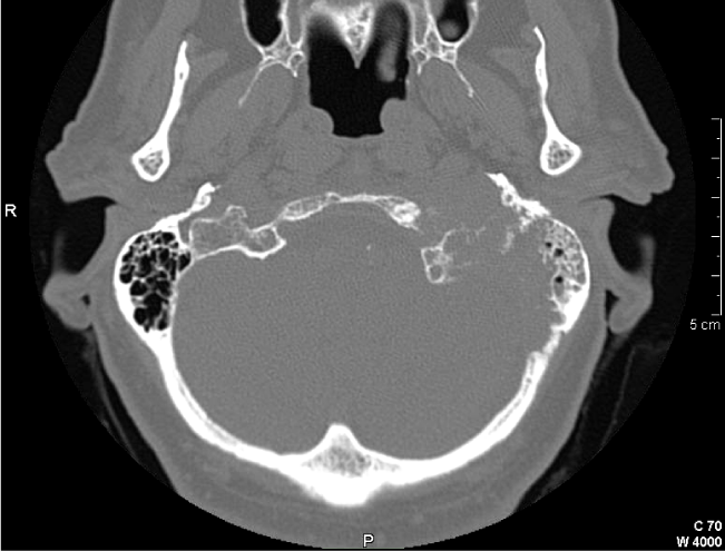
Figure 1
Axial high-resolution CT scan of the temporal bones shows an extended osteolytic erosion of the left temporal bone, including the mastoid and petrous apex.
Figure 2

Figure 2
Axial high-resolution CT scan of the temporal bones evidences the metastatic lytic lesion of the clivus (arrow) and posteriorly a suspected involvement of left middle fossa dura and sigmoid sinus region (open arrow).
Figure 3
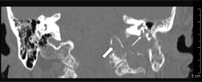
Figure 3
Coronal high-resolution CT scan of the temporal bones demonstrates the involvement of the vertical tract of the left facial nerve canal (arrow) and the erosion of the first left cervical metamers (open arrow).
Discussion
Skull base metastases from primary malignant tumors are
relatively uncommon lesions. They often originate from the breast,
but also lung, prostate, skin, liver and cervix are cited [1]. Generally,
malignant cells are supposed to follow the hematogeneous route of
dissemination to the bone marrow [1 and 2] and within the temporal
bone, petrous apex, mastoid and internal auditory canal are the most
commonly involved sites [1]; in particular, petrous apex appears the
first temporal bone area affected by neoplastic cells before mastoid
involvement [1].
Skull base metastases may be occult and asymptomatic, and
postmortem findings in patients with possible secondary lesions
confirm that their incidence is greater than previously reported [1].
Otherwise, hearing loss, otorrhea, tinnitus, vertigo and facial palsy are
some of the most frequently reported symptoms [1 and 3].
Under a radiographic point of view, osteolytic lesions are often
revealed [4], but they can be found in cholesteatoma, primary
neoplasms and paragangliomas as well, even if a destructive action
to bone appears more characteristic of tumoral diseases [3]. On the
contrary, in other cases, soft tissue density masses in middle ear and
mastoid cavities can be observed [3], leading to an initial suspect of
chronic otitis media [5].
References
- Gloria-Cruz TI, Schachern PA, Paparella MM, Adams GL, Fulton SE. Metastases to temporal bones from primary non-systemic malignant neoplasms. Arch Otolaryngol Head Neck Surg. 2000; 126: 209-214.
- Berlinger NT, Koutroupas S, Adams G, Maisel R. Patterns of involvement of temporal bone in metastatic and systemic malignancy. Laryngoscope. 1980; 90: 619-627.
- Lan MY, Shiao AS, Li WY. Facial paralysis caused by metastasis of breast carcinoma to the temporal bone. J Chin Med Assoc. 2004; 67: 587-590.
- LJones HM. Case of metastasis in the temporal bone from a carcinoma of the breast. J Laryngol Otol. 1969; 83: 293-298.
- Maddox HE. Metastatic tumors of the temporal bone. Ann Oto Rhinol Laryngol. 1967; 76: 149-165.