Case Report
Simplified Approach to Repair Early Pseudoaneurysm of the Left Coronary Button Following Composite Graft Because of Acute Type A Aortic Dissection
Thierry Carrel*, Samuel Hurni and Christoph Huber
Clinic for Cardiovascular Surgery, University Hospital Bern and University of Bern, Switzerland
*Corresponding author: Thierry Carrel, Clinic for Cardiovascular Surgery University Hospital, CH-3010 Bern, Switzerland
Published: 04 Jul, 2016
Cite this article as: Carrel T, Hurni S, Huber C. Simplified Approach to Repair Early Pseudoaneurysm of the Left Coronary Button Following Composite Graft Because of Acute Type A Aortic Dissection. Clin Surg. 2016; 1: 1057.
Abstract
We present a simplified surgical technique performed in a 37-yr old man who presented with a
pseudoaneurysm of the left coronary ostium 2 months after repair of acute type A aortic dissection
by composite graft. Intraoperatively, the surgical situs showed extreme adhesions. The leakage at the
level of the coronary suture line was exposed from inside the aortic graft. Repair was performed by
7.0 polypropylene sutures and the postoperative course was uneventful. The patient was discharged
on postoperative day 6.
Keywords: Aortic dissection; Composite graft; Coronary button pseudoaneurysm
Introduction
Pseudo-aneurysm is a rare but challenging complication that may occur at the level of aortic anastomoses or coronary re-implantation suture lines following composite graft replacement [1]. Etiology includes weak tissue secondary to aortic dissection or connective tissue disease or technical failure during construction of the anastomosis [2,3]. Repair of pseudo-aneurysm has to be performed surgically in the vaste majority of cases and exceptionally through interventional technique. Aim of this brief report is to present a simplified approach to treat a pseudo-aneurysm of the left coronary orifice in the early postoperative period following composite graft replacement of the aortic root because of type A aortic dissection.
Case Presentation
The 37-yr old patient had undergone composite graft repair because of acute type A aortic
dissection 2 months before in another University Hospital in this country. At initial presentation,
he suffered from pericardial tamponade, had to be intubated during transportation and operated as
emergency because of unstable hemodynamics. Initial repair was performed without complication
and consisted in the implantation of a composite graft with a mechanical valve. The distal anastomosis
was performed in the open aortic arch technique during a brief period of moderate hypothermic
circulatory arrest using selective antegrade cerebral perfusion for brain protection. Re-institution of
the cardiopulmonary bypass was achieved through cannulation of the side-arm of the aortic graft
(Vascutek© Anteflow, Renfreshire, UK). Coronary reimplantation was performed in the modified
button technique with running polypropylene suture 6.0 using a pericardial strip as reinforcement.
Intraoperative Transo Esophageal Echocardiography (TEE) at the end of the procedure was normal.
The immediate postoperative evolution was uneventful and the patient discharged at postoperative
day 8. A first CT-scan was performed 4 weeks postoperatively and demonstrated an extravasation
of contrast medium (8mm in size) behind the aorta, close to the left coronary artery origin. It was
decided to treat conservatively and lower the pressure at systolic values between 80 and 100 mm Hg.
However, at second CT-scan 4 weeks later, the pseudo-aneurysm had increased in size with 17 mm
(Figure 1a).
Indication to cure the pseudo-aneurysm as soon as possible was confirmed, although the
patient was asymptomatic. Prior reopening the sternotomy, the external iliac artery and vein were
exposed. Preparation of the mediastinal and intrapericardial situs was horribly difficult because of
extremely strong adhesions between the vascular prosthesis and the adjacent structures (mainly the
pulmonary artery but also the right atrium and the superior vena cava. For this reason we decided to
cannulate the femoral artery and vein through Seldinger technique and cooled the patient to a core
temperature of 28°C. Shortly before hypothermic arrest, Pentothal was administred and thereafter
cardiopulmonary bypass was interrupted without prior clamping
the prosthesis: this would have considerably prolonged the time on
bypass. The previous anastomosis between the arch prosthesis and
the composite graft was opened (Figure 2), then the distal graft was
prepared from inside to separate it from the adjacent structures.
Thereafter it was clamped (hypothermic arrest was 5 minutes) and
cardiopulmonary bypass was re-instituted and the patient rewarmed.
During this period, inspection of the left coronary orifice from inside
the aortic prosthesis was performed. At the lower part of the suture
line, a small leakage could be identified. It was fixed with 2 separate
U-stitches from inside of the coronary button anastomosis and in
addition the previous suture line was stretched with an additional
stitch. Cross-clamp time was 22 minutes only. Intraoperative TEE
was available before and after repair (Figure 1b and C). Immediate
postoperative evolution was uneventful and the patient could be
discharged at postoperative day 6. CT-scan at 4 weeks did not show
any perfusion in the shrinked pseudo-aneurysm cavity (Figure 1d).
Figure 1a
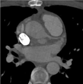
Figure 1a
CT scan performed 2 months after initial surgery showing a
pseudoaneurysm at the level of the left coronary button anastomosis
posterior to the aortic root.
Figure 1b
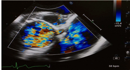
Figure 1b
Preoperative echocardiography showing the perfused
pseudoaneurysm of the left coronary button anastomosis.
Figure 1c
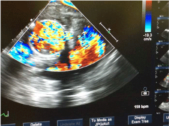
Figure 1c
Intraoperative transesophageal echocardiography after
exclusion of the pseudoaneurysm.
Figure 1d
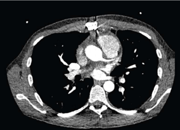
Figure 1d
Postoperative CT-scan one month after repair of the
pseudoaneurysm showing normal situs.
Figure 2
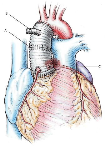
Figure 2
Technique of repair: the previous anastomosis between the
composite graft and the hemi-arch protheses is open during a short period
of circulatory arrest. A. Then the distal graft is controlled and clamped and
cardiopulmonary bypass restarted. B. Thereafter, the pseudoaneurysm is
fixed from inside the composite graft. C.
Discussion
We described a simplified technical approach to pseudo-aneurysm of the left coronary orifice in the early course following composite graft replacement because of type A acute aortic dissection (Figure 2). This complication is a rare but challenging event that always need surgical repair even though the dense adhesions behind the aorta most would probably prevent rupture. Another potential complication might be compression of the left main coronary artery due to expansion of the aneurysm. Diagnosis is made by echocardiography and/or CT-scan which should always be performed in the first 3 months following repair of acute type A aortic dissection. TEE may help to localize the exact position of the leakage and to intraoperatively control that the problem has been fixed. In the present case, moderate hypothermic cardiopulmonary bypass helped to save time because the control the aortic graft was greatly facilitated. After incision of the graft in its anterior part, the transection was performed under visual control and circular preparation of the graft was possible without injury to the pulmonary artery or the superior vena cava. Once the cranial part of the prosthesis was clamped, CPB could be restarted and repair of the aneurysm was performed from inside during rewarming. Complete re-confection of the anastomosis of the left coronary artery ostium to the composite graft is rarely necessary since the leakage can usually be fixed by one or several single stitches from inside the aortic graft. This technique is safe since it does not require exposure of the coronary button from outside the graft (behind the aorta) and can be applied in the early but also late course following composite graft repair to fix a dehiscence of the coronary artery button anastomosis. In addition, this case emphasizes the necessity of follow-up imaging after aortic surgery in order to detect unexpected changes that could endanger the patient.
References
- Carrel T, Tkebuchava T, Turina M. Reoperations after previous surgery on the thoracic aorta: etiology, techniques and results. Ann Thorac Surg. 1993; 56: 259-268.
- Luciani N, De Geest R, Lauria G, Farina P, Luciani M, Glieca F, et al. Late reoperations after acute aortic dissection repair: Single-center experience. Asian Cardiovasc Thorac Ann. 2015; 23: 787-794.
- Mattesini A, Porto I, D'Alfonso MG, Cecilia A, Gensini GF, Valente S. A rapidly growing coronarypseudoaneurysm. Cardiovasc Revasc Med. 2015; 16: 320-321.