Case Report
Calcifying Pseudotumor of the Spine: A Case Report
Giardina F*, Guerra G, Righi A and Bertoni F
Department of Orthopaedics, Istituto Ortopedico Rizzoli, Italy
*Corresponding author: Federico Giardina, Department of Orthopaedics, Istituto Ortopedico Rizzoli, Via Di Barbiano 1/10, 40136, Bologna, Italy
Published: 12 Jul, 2016
Cite this article as: Giardina F, Guerra G, Righi A, Bertoni F. Calcifying Pseudotumor of the Spine: A Case Report. Clin Surg. 2016; 1: 1042.
Abstract
Objective: To describe a case of calcifying pseudotumor of the spine and to review and discuss the significant literature above this rare clinical entity.
Summary of Background Data: Calcifying pseudotumor of the spine has been rarely reported. They are non-neoplastic, calcified lesions of the central nervous system believed to be reactive in nature. They are encountered both intracranially and spinally with a virtually equal prevalence.
Methods: We present the case of a 68-year-old man with a L4-L5 radiculopathy without an history
of trauma, infection or neoplastic process referred to the spine. CT and MRI scans showed a L4-L5
calcified intradural mass with an iso-ipointense signal to the spinal cord on T1- and T2-weighted
sequences. Patient underwent a laminectomy to obtain complete resection of the lesion. The mass
was round and hard and the histological examination revealed the typical features of calcified
pseudoneoplasm of the neural axis (CAPNON).
Results: At five years long follow-up the patient had complete resolution of his symptoms.
Conclusions: This clinical entity should be taken into consideration in the differential diagnosis of
intracranial and intraspinal calcified lesions. The surgical resection is necessary to obtain symptom’s relief and proper histological validation.
Keywords: Pseudotumor; Spine
Introduction
Calcifying pseudoneoplams of the neuraxis (CAPNON) are rare, non-neoplastic, calcified lesions of the central nervous system (CNS) believed to be reactive in nature [1,2]. Since their first description by Rhodes and Davis in 1978 [3] only 39 cases have been reported in the literature, using also the name of “fibro-osseous lesions of the central nervous system” or “calcifying pseudotumour of the neuraxis” [4]. We report the case of a 68 year-old man with a calcifying pseudotumor of the spine and review the available English literature indexed in MEDLINE.
Case Report
A 68-year-old man with a history of six months low back pain radiating into his right leg was admitted to our institution in 2007. There was no previous history of back pain, spinal injury or infective process. Neurologic findings were consistent with a L4-L5 radiculopathy. An MRI and CT scans were obtained demonstrating a L4-L5 intradural mass. The CT demonstrated a calcified mass located at L4-L5 space which was initially judged as calcified synovial cysts (Figure 1); on MRI the mass was iso-ipointense to the spinal cord on both T1- and T2-weighted sequences, without surrounding edema (Figure 2). The patient underwent a laminectomy and gross total resection of the lesion. The mass was round and hard; the histological examination of the specimen showed the typical features of chondromyxoid matrix organized in a nodular pattern associated with fibrousstroma and calcifications (Figure 3). After resection of the mass, the patient had no local recurrence and a subsequent MRI showed no residual mass; so, at the actual five years follow-up, there is complete relief by symptoms.
Discussion
TCalcifying pseudoneoplasms are timorous lesions of the CNS with a low prevalence. There is no clear age predominance, although a male preference was noted [1,2,4-9]. They are encountered both intracranially and spinally with a virtually equal prevalence [4]. The clinical presentation generally depends on the location and size of the lesion. Symptoms are related to the “mass effect” on the adjacent tissue. In spinal affections patients complain of local or radiating pain, while in cases with intracranial CAPNON seizures were the most common finding, followed by recurrent headache. Dense calcifications are the recurring radiological characteristic of CAPNON as seen in plain radiographs. These lesions appear like hyper dense masses in all CT scans performed in all known cases, with a peripheral calcification and less frequently bone erosion by the calcifying mass [6,9-14]. MRI imaging can be variable: according to Stienen et al. [4], who reviewed all reported cases of CAPNON, in the 18 cases of patients presented in their study who underwent MRI, 87.5 % of the spinal lesions appeared hypo intense in T1- as well asT2-WI, while in the group of intracranial lesions, 77.8 % showed hypo intensity in T1- and 88.9 % in T2-WI. Overall, after contrast administration, most lesions showed rim or inhomogeneous internal linear enhancement. On the basis of previous reports the clinico-radiographic differential diagnosis that must be considered are meningioma, chordoma, chondrosarcoma, vestibularschwannoma, vascular lesions such as cavernous malformation, infections such as tuberculosis, calcified herniated disks and calcified synovial cysts [1,15]. By review of the literature, basic points of histopathological diagnosis include chondromyxoid matrix in a nodular pattern, palisading spindle to epithelioid cells, variable amounts of fibrous stroma, calcification or osseous metaplasia and foreign-body reaction with giant cells [1,11]. A complete surgical resection of the neoplasm should be attempted, otherwise partial resection or “debulking” should be performed when there are technical difficulties. Considering the particular nature of the lesion, when a proper histopathological diagnosis is obtained, there are no recommendations concerning adjuvant medical or radiation therapy [4].
Figure 1
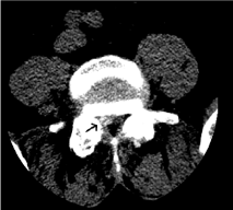
Figure 1
CT scans showed intradural calcified mass in the lower lumbar
spine (arrow).
Figure 2
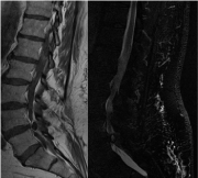
Figure 2
MRI imaging of the L4-L5 intradural mass evidenced the typical
features of spinal CAPNON.
Figure 3
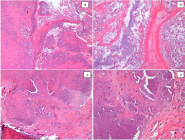
Figure 3
Histopathologic features of this case of calcifying pseudoneoplasms
of the neuraxis include chondromyxoid matrix organized in a nodular pattern
(A) at low power field and B at high power field) associated with fibrous
stroma and calcifications (C). Spindle and epithelioid cells bordered the
chondromyxoid matrix nodules (D).
Conclusion
Calcifying pseudoneoplasms are rare and benign lesions of the CNS believed to be reactive in nature [2]. This clinical entity should be taken into consideration in the differential diagnosis of intracranial and intraspinal calcified lesions. The more common findings are dense calcification on the CT and T1-WI and T2-WI hypo intensity in the MRI, however definitive diagnosis cannot be confirmed before surgery. Complete resection of the lesion should be attempted to relieve the patient’s symptoms and to obtain pathologic validation, which is important to avoid wrong diagnosis of a mimic neoplastic malignancy that changes therapeutic behavior.
References
- Bertoni F, Unni KK, Dahlin DC, Beabout JW, Onofrio BM. Calcifying pseudoneoplasms of the neural axis. J Neurosurg. 1990; 72: 42-48.
- Mohapatra I, Manish R, Mahadevan A, Prasad C, Sampath S, Shankar SK. Calcifying pseudoneoplasm (fibro osseous lesion) of neuraxis (CAPNON)- a case report. Clin Neuropathol. 2010; 29: 223-226.
- Rhodes RH, Davis RL. An unusual fibro-osseous component in intracranial lesions. Hum Pathol. 1978; 9: 309-319.
- Stienen MN, Abdulazim A, Gautschi OP, Schneiderhan TM, Hildebrandt G, Lücke S. Calcifying pseudoneoplasms of the neuraxis (CAPNON): clinical features and therapeutic options. ActaNeurochir (Wien). 2012 Oct 3.
- Chang H, Park JB, Kim KW. Intraosseous calcifying pseudotumor of the axis: a case report. Spine (Phila Pa 1976). 2000; 25: 1036-1039.
- Garen PD, Powers JM, King JS, Perot PL Jr. Intracranial fibro-osseous lesion. Case report. J Neurosurg. 1989; 70: 475-477.
- Hodges TR, Karikari IO, Nimjee SM, Tibaleka J, Friedman AH, Cummings TJ, et al. Calcifying pseudoneoplasm of the cerebellopontine angle: case report. Neurosurgery. 2011; 69: E117-120.
- Jun C, Burdick B. An unusual fibro-osseous lesion of the brain. Case report. J Neurosurg. 1984; 60: 1308-1311.
- Liccardo G, Lunardi P, Menniti A, Floris R, Pastore FS, Fraioli B. Calcifying pseudo-tumor of the spine: description of a case and review of the literature. Eur Spine J. 2003; 12: 548-551.
- Mayr MT, Hunter S, Erwood SC, Haid RW Jr. Calcifying pseudoneoplasms of the spine with myelopathy. Report of two cases. J Neurosurg. 2000; 93: 291-293.
- Qian J, Rubio A, Powers JM, Rosenblum MK, Pilcher WH, Shrier DA, et al. Fibro-osseous lesions of the central nervous system: report of four cases and literature review. Am J Surg Pathol. 1999; 23: 1270-1275.
- Shrier DA, Melville D, Millet D, Qian J, Millet D, Nelson C, et al. Fibroosseous lesions involving the brain: MRI. Neuroradiology. 1999; 41: 18-21.
- Tatke M, Singh AK, Gupta V. Calcifying pseudoneoplasm of the CNS. Br J Neurosurg. 2001; 15: 521-523.
- Aiken AH, Akgun H, Tihan T, Barbaro N, Glastonbury C. Calcifying pseudoneoplasms of the neuraxis: CT, MR imaging, and histologic features. AJNR Am J Neuroradiol. 2009; 30: 1256-1260.
- Moser FG, Tourje EJ, Pressman BD, Blinderman EE. Calcifying pseudotumor of the cervical spine. AJNR Am J Neuroradiol. 1994; 15: 580.